
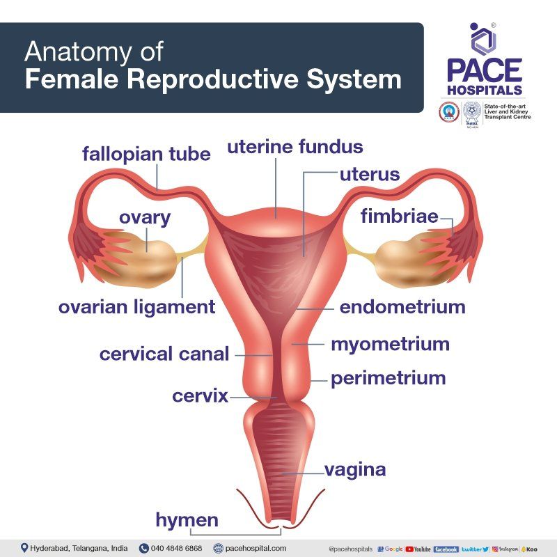
Internal anatomy of female reproductive system
There are numerous varieties of operations in hysterectomy and various factors influencing the surgeon to opt the specific type of hysterectomy, but before delving deeper, a brief understanding of the anatomy and physiology of the female reproductive system is necessary.
The following are the female reproductive organs:
- Vagina: also, known as the “birth canal” vagina is a canal joining the cervix (the lower part of the uterus) to the outside of the body.
- Uterus: often known as the womb, the uterus is a hollow, pear-shaped organ that accommodate a growing foetus. The cervix, which is the lower portion of the uterus that enters into the vagina, and the corpus, which is the larger portion of the uterus, are separated anatomically. A developing baby can be held by the corpus with ease. Sperm enters, and menstrual blood exits through a canal in the cervix.
- Ovaries: On either side of the uterus are two tiny, oval-shaped glands called the ovaries, in which eggs and hormones are produced.
- Fallopian tubes:
The ova (egg cells) move through these tiny tubes, which are connected to the upper section of the uterus, to reach the uterus. In the fallopian tubes, an egg and a sperm are often fertilised. Once within the uterus, the fertilised egg implants to the uterine lining.


