Interventional Radiology Hospital in Hyderabad for Interventional Treatment
PACE Hospitals is one of the best Interventional Radiology Hospital in Hyderabad, offering effective minimally invasive treatment for disorders related to blood clots, blockages, tumours and chronic pain. The team of skilled and experienced interventional radiologists have vast expertise in managing a wide array of disorders requiring image-guided interventional treatment, including:
- Uterine Fibroids
- Ovarian Cysts
- Kidney Stones
- Biliary Obstruction
- Ureteral Obstruction
- Pelvic Congestion Syndrome
- Carotid Artery Disease
- Varicose Veins
- Deep Vein Thrombosis (DVT)
- Arteriovenous Malformations (AVMs)
- Aneurysms (aortic, cerebral)
- Tumours (liver, kidney, lung)
- Abscess Drainage (liver, kidney)
- Gastrostomy Tube Placement
- Portal Vein Thrombosis
- Cancer Pain Management
- Biopsies
Why choose PACE Hospitals?
Extensive IR Care
Providing treatment to a wide range of disorders related to tumours, blood clots, blockage and chronic main managements with high precision and lesser recovery time.
Advanced State-of-the-art Facility
Equipped with advanced and cutting edge diagnostic equipment, robotic and minimally invasive surgical facilities for interventional Procedures.
Skilled Interventional Radiology Doctor
Team of experienced interventional radiologist with vast experience in minimally invasive and image guided procedures.
Advanced Centre for Interventional Radiology in Hyderabad, Telangana

PACE Hospitals is one of the best hospital for interventional radiology in Hyderabad. The department is staffed with a team of experienced interventional radiologists who coordinate closely with the multidisciplinary team of another department specialist to provide precise diagnostics and minimally invasive treatment for vascular disorders, oncological conditions, gastrointestinal conditions, urological issues and chronic pain management, to deliver the patient-centric and highest standard of care to the patients. Interventional radiology doctors are highly skilled in a comprehensive range of image-guided interventional and minimally invasive procedures including angioplasty, radiofrequency ablation, vascular stenting, embolization and more.
The Department of Interventional Radiology at PACE Hospitals is equipped with cutting-edge state-of-the-art diagnostic facilities, including CT scans, MRIs, and ultrasound-guided imaging, to evaluate and perform minimally invasive procedures to manage complex conditions. These procedures are intended to minimize hospital stay and discomfort and provide faster recovery.
3,28,338
99,825
684
2011
Best Interventional Radiology Doctor in Hyderabad
A team of the best interventional radiology doctor in Hyderabad, India, have extensive expertise in image-guided minimally invasive procedures such as Angioplasty and Stenting, Embolization, Thrombectomy, Endovenous Laser Therapy (EVLT), Radiofrequency Ablation (RFA), Chemoembolization, Biliary Drainage and Stenting, Gastrostomy Tube Insertion (PEG), Nephrostomy, Ureteral Stenting, Abscess Drainage and Biopsies for the wide range of complex conditions like peripheral artery disease (PAD), varicose veins, liver and kidney tumours, uterine fibroids, deep vein thrombosis (DVT), Aneurysms (aortic, cerebral), Tumours (liver, kidney, lung), Abscess Drainage (liver, kidney) and more. The specialized team of interventional radiologists are highly skilled and apt with the latest minimally invasive treatment modalities to provide the utmost treatment care with precision, minimal complications and a high success rate.
Dr. Lakshmi Kumar Chalamarla
MBBS, DNB (Radiology), PDCC fellowship (Abdominal Imaging), PDCC fellowship (Interventional Radiology), EBIR
Experience : 11+ years
Senior Interventional Radiologist and Abdominal Imaging Specialist
Interventional Radiology Conditions Explained by Drs
Need Help?
Struggling with conditions like chronic pain, fibroids, blocked arteries, vascular blockages and clots conditions like peripheral artery disease (PAD), deep vein thrombosis (DVT), varicose veins, varicocele, arteriovenous malformations (AVMS), aneurysms, biliary obstruction, carotid artery disease, kidney stones, urethral strictures or seeking minimally invasive treatment for tumours and cancers of liver, lung, digestive system, uterus and kidney, we offer minimally invasive treatment tailored to your need with less discomfort and shorter recovery time.
What we treat?
We specialize in treating various critical conditions of blood clots, tumours, and blockages affecting the vascular system and other organs like the liver, kidney, lungs, uterus, digestive system, breast, prostate and bone & joints. From vascular conditions like Peripheral Artery Disease (PAD), Deep Vein Thrombosis (DVT), Varicose Veins, Aneurysms, Pulmonary Embolism to all kinds of cancer requiring radiofrequency ablation (RFA) and chemoembolization and conditions like Uterine Fibroids, Adenomyosis, Gastrointestinal Bleeding, Biliary Obstruction, Kidney Stones, Aortic Aneurysms, Coronary Artery Disease (CAD) requiring embolization and stent placement and solutions to the chronic pain management, our team of interventional radiology doctor is committed to delivering effective, precise and compassionate treatments with minimal recovery time.

Gastrointestinal and Hepatobiliary Conditions
Biliary obstruction
Biliary obstruction is the blockage of the bile duct, which transports the bile from the liver to the intestine. Bile is a liquid containing bile salts, bilirubin, and cholesterol released by the liver. Bile duct blockage leads to the accumulation of bile in the liver and causes jaundice.
It is caused by multiple factors such as gallstones, bile duct cysts, enlarged lymph nodes, tumours of the pancreas or bile ducts, and inflammation. Symptoms include fever, dark urine, yellowing of skin cancer(jaundice), abdominal pain in the upper right side, pale stools, nausea and vomiting. Biliary obstruction can cause serious complications such as hepatic damage, kidney failure, bleeding problems, infections and nutritional deficiencies.
Gastrointestinal bleeding
GI bleeding is a symptom of any medical condition or disease rather than a single condition. It includes any bleeding that occurs in the gastrointestinal tract (digestive tract). Acute is short-term, which comes suddenly and becomes severe. Chronic can last long and be characterised by slight bleeding.
Many conditions can cause GI bleeding, and a healthcare professional can identify the cause by finding its underlying condition. Possible causes of GI bleeding include colitis, esophagitis, gastritis, haemorrhoids or anal fissures, colon polyps, peptic ulcers etc. Symptoms of GI bleeding may include black stool, dizziness, paleness, anaemia, and a drop in blood pressure.
Liver abscesses
A liver abscesses is a pus-filled mass that occurs in the liver due to infection or injury. Most abscesses are classified as either pyogenic or amoebic, with a minority caused by parasites and fungi.
Diabetes, being male, older age, liver cirrhosis, immunocompromised state and use of PPIs (proton-pump inhibitors) can increase the risk of developing liver abscesses. Signs and symptoms include fever, tenderness and hepatomegaly. Some people can experience non-specific symptoms such as fever with weight loss, malaise and chills. If not treated, it can rupture and cause shock and peritonitis.
Ascites
Ascites is a medical condition characterised by the collection of fluid in the cavities of the abdomen. Cirrhosis is a common cause of developing ascites. It is also caused by elevated (high) blood pressure in the specific veins of the liver, decreased albumin levels, liver diseases, heart diseases, kidney failure, and certain cancers, such as stomach, pancreatic, liver and lung.
Accumulated fluid in the abdomen can cause discomfort and swelling, worsen in a few days, and exert pressure on other organs, leading to bloating, back and abdomen pain, constipation, breathlessness, fatigue and frequent urination.
Encephalopathy
Encephalopathy describes any condition that affects the functioning, structure and alters the mental function of the brain. It is not a single condition and can have various underlying causes, such as brain tumours, kidney failure, bacterial or viral infections, autoimmune disorders, etc.
There are many kinds of encephalopathy, including hepatic encephalopathy, chronic traumatic encephalopathy, hypoxic ischaemic, and Wernicke encephalopathy. Hepatic encephalopathy is caused by long-term conditions such as an infection, cirrhosis, or bleeding in the digestive tract. Symptoms include sleep problems, slurred speech, anxiety, mood or personality changes and cognitive impairment.
Portal hypertension
Portal hypertension is an increased blood pressure in the veins of the portal venous system. The portal vein is the primary vein that leads to the liver.
When something obstructs or slows the blood flow through the portal vein, it can cause elevated pressure in the portal venous system. Symptoms of this condition include oedema, jaundice, gastrointestinal bleeding, and ascites. The most common cause of portal hypertension is liver cirrhosis.
Arterial Conditions
Arteriovenous malformations (AVMs)
Arteriovenous malformations (AVMs) are abnormal blood vessel tangles that lead to irregular and unusual connections between the arteries and veins. They commonly occur during a baby's development before birth and can develop anywhere in the body, including the spinal cord, any part of the brain, or its surface.
Many people do not show any symptoms. However, in some cases, a weakened blood vessel may rupture, resulting in blood leakage into the brain, called a haemorrhage. Other symptoms include seizures, headache, pain, visual problems, etc. Surgery is one of the treatment options to cure this condition.
Aneurysms
Aneurysm is characterised by a weakened wall of blood vessels, which leads to an abnormal bulge (enlargement). It is commonly seen in arteries, even though it can occur in any blood vessel of the brain (central aneurysm), aorta, kidney, intestine, neck, spleen and legs. Most often, it affects the arteries of the brain and heart.
The exact cause of aneurysm is idiopathic (unknown), but having conditions such as older age, being male, having a family history, obesity, hyperlipidaemia, genetic factors, smoking and hypertension (major risk factors) can increase the risk of developing this condition. The symptoms that a person may experience depend on which artery is affected by the aneurysm.
Hereditary haemorrhagic telangiectasia (HHT)
It is also called as Osler-Weber-Rendu syndrome. Hereditary haemorrhagic telangiectasia (HHT) is a congenital(inborn) genetic disorder affecting blood vessels. It is characterised by the improper development of blood vessels. Sometimes, it causes bleeding called arteriovenous malformations (AVMs).
In the HHT condition, a person may present without capillaries (tiny blood vessels that connect the arteries and veins for passing the blood), which easily leads to the rupture of the arteries and veins. It is a genetic condition caused by a faulty gene (inherited from parents). Symptoms of this condition include regular nosebleeds and visible red spots in some places on the body.
Peripheral arterial disease (PAD)
Peripheral arterial disease (PAD) is characterised by the buildup of fatty (cholesterol) deposits in the arteries of arms or legs, resulting in decreased blood supply to the leg muscles. It is commonly caused by the blockages of arteries in the heart (atherosclerosis) or plaque buildup.
A common symptom of this illness is pain, discomfort, or cramps in the legs while walking or exercising, which goes away after a few minutes of rest. Other symptoms include a colour change (paler than usual or blue), numbness or weakness in the legs, and colder feet. Older age, family history and genetics, smoking, stress, metabolic syndrome, thrombocytosis, obesity, and high blood pressure can increase the risk of developing PAD.
Pain Management in Certain conditions
Chronic back pain
Back pain is considered the most common medical issue and is regarded as chronic if it lasts three or more months. Chronic back pain is the most common medical condition lasting three or more months.
It will come and suddenly give relief, ranging from a dull, continuous pain to a sudden, sharp pain that may radiate down to the legs.
Sometimes, it comes from a fall, accident, or lifting something very heavy or due to age-related degenerative changes in the spine, some medical conditions can also cause this condition. Treatment options depend on underlying causes and symptoms.
Sciatica
The sciatic nerve arises in the lower back and runs down the back side of each leg, controlling the muscles of the lower leg and back of the knee. Sciatica is a medical condition characterised by weakness, pain, tingling or numbness in the leg. Sciatica is usually not dangerous and can be managed with self-care treatments, but severe cases may require surgery.
This condition is caused by the damage or pressure on the sciatic nerve. Common causes include spinal stenosis, tumours, pelvic fracture or injury and slipped or herniated disk.
Joint pain
Joint pain is a common medical condition in older people. Joints are the parts where the bones will meet and allow the bones to move. These include hips, shoulders, knees, and elbows. Pain in any joint can be referred to as joint pain.
It can be caused by several reasons, such as injury or arthritis, cancer, osteoporosis, gout, infections, rickets, etc. Symptoms may vary based on the cause. For example, one may experience gout symptoms such as hot, swollen toe joint pain suddenly.
Cancer-related pain
Pain is one of the most common symptoms experienced by cancer patients. It may cause by the cancer disease or its treatment or due to a combination of the factors. Tumours, surgeries, radiation, targeted, intravenous chemotherapy, supportive care therapies and diagnostic procedures may cause pain in cancer patients.
Younger patients have a higher probability of developing cancer pain and pain flares than older patients. Patients with advanced (progressed) cancer can have more pain. Pain from the cancer is caused by the compression of the tumours on the bones, nerves, or organs. Some diagnostical tests and procedures are painful, and some drugs may be used to feel calm or fall asleep. Imagery or relaxation therapies can also help manage pain and anxiety related to treatment.
Compression fractures
Compression fractures are tiny breaks in the vertebrae (bones in the spine). Vertebrae are the bones of the spine. They are common in women who are over 50. As they age, they are more likely to break due to osteoporosis, a condition defined by weak and brittle bones. It can be commonly caused by osteoporosis, and other causes may include tumours in the spine (multiple myeloma), trauma to the back, and bone tumours.
One can experience symptoms ranging from mild to severe. Some people may not have symptoms. Compression fractures can happen suddenly and can cause severe back pain. Treatment can lower the risk of more fractures.
Musculoskeletal Conditions
Osteoporotic fractures
Osteoporotic fractures can occur due to a medical condition called osteoporosis. This condition causes bones to become weak and break (fragile and brittle) easily due to low bone density or mass, leading to a greater risk of fractures.
Bone loss can happen silently, without any apparent symptoms, until an actual fracture occurs. Osteoporotic fractures can start with minimal trauma such as strain, fall or bump. Even minor falls or routine activities like twisting and bending can cause fractures in individuals with osteoporosis.80% of osteoporosis cases are seen in women and older individuals, while men can also be affected at any age.
Bone metastases
If the cancer (malignant) cells develop in the bone, it is called primary bone cancer, but if they spread to bone from elsewhere in the body, it is called bone metastasis or secondary bone cancer. This bone metastasis can cause problems such as broken bones, pain, spinal cord compression, and high blood calcium levels.
Almost all cancers have the ability to spread to the bones. Still, the most common cancers that spread to bones are lung, breast, kidney, prostate, thyroid, ovarian and melanoma, and the most common site for bone metastases is the spine. Other sites are ribs, skull, hip bone, femur (upper leg bone) and humerus (upper arm bone).
Gynaecological Conditions
Uterine fibroids
Uterine fibroids are the growths that form in the uterus. A Woman's uterus is made up of muscle. Fibroids originate from the muscle, bulging from outside or inside the uterus. They are prevalent and non-cancerous, but they may become cancerous.
The exact cause is idiopathic (unknown). Hormones and genetics might be the cause. Most fibroids are minor (small in size) and do not cause any symptoms. However, some women may show symptoms such as increased menstrual bleeding, pelvic pressure and pain, menstrual clots that last longer than usual, increased urination and painful sex.
Pelvic congestion syndrome
Pelvic congestion syndrome (PCS) is characterised by long-term (chronic) pelvic pain that lasts for three-six months, which is not related to pregnancy or menstruation in a woman of reproductive age.
Women in the age group (20 to 45) and women who have given birth more than once and have varicose veins (these conditions) can increase the risk of developing PCS. Symptoms of this condition include varicose veins on the upper thighs or butt, pain during sex, bulging veins in front of the vagina, stressed or irritable bladder-which makes it hard to control the urine and dull aching pain in the pelvic area and lower back.
Ovarian cysts
Ovarian cysts are fluid-filled sacs that can develop in one or both ovaries. These sacs can contain gas, liquid, or semi-solid material. It can be caused by hormonal factors, endometriosis and infections.
It is estimated that 10 out of 100 women present with this condition. Usually, they do not cause any symptoms. Ovarian cysts show signs when it ruptures; in these cases, symptoms may include pelvic pain, painful sex, urinary urgency, bloating, swollen tummy, heavy periods or irregular periods or lighter periods, weight gain and difficulty conceiving.
Neurovascular Conditions
Stroke
A stroke is characterised by the blockage of blood flow to the brain or blood vessel bursts in the brain. Stroke is classified into two types. Stoppage of blood supply to the brain is called ischemic stroke, which causes a shortage of oxygen and nutrient supply. A sudden haemorrhage (bleeding) in the brain is known as a haemorrhagic stroke, which causes pressure on brain cells and leads to damage.90% of the strokes are ischemic, and the rest are haemorrhagic.
Signs of the stroke include weakness, paralysis or numbness on one side of the face or body. Other signs and symptoms include sudden weakness, severe headache, vision problems and trouble speaking and understanding.
Carotid artery stenosis
Carotid artery stenosis is characterised by the narrowing or blockage of the carotid arteries. These arteries supply the oxygenated blood to the brain from the heart. It can be caused by atherosclerosis. Having diabetes, a family history of stroke, trauma to the neck, high blood pressure, smoking, high cholesterol, and older age can increase the risk of developing carotid artery stenosis.
If left untreated, it can lead to a stroke or stroke symptoms. It is the reason for one-third of all strokes.
Symptoms of this condition include memory loss, confusion, problems with thinking, reasoning and speaking, and numbness or weakness in part of the body or one side. Management of carotid artery stenosis can reduce the risk of stroke.
Intracranial haemorrhage
Intracranial bleeding is a neurological condition referred to as one of the types of strokes; intracranial bleeding is characterised by the bursting of the artery in the brain and causes localised bleeding in the surrounding tissues. Bleeding (haemorrhage) occurs within the brain tissue or between the brain and membranes that cover it or between the skull and the covering of the brain.
It can be caused by several factors, such as head trauma, aneurysm, blood and bleeding disorders, etc. Symptoms may vary based on the location, severity, and amount of tissue affected. Some symptoms include vomiting and nausea, weakness, headache, and paralysis of the leg, arm or face.
Cancers
Liver cancer
The liver is one of the largest organs in the body, has two lobes and is located at the upper right side of the abdomen inside the rib cage. It helps in digesting the food and removal of toxins. Primary Liver cancer is a condition characterised by the formation(growth) of cancerous cells in the tissues of the liver. Secondary liver cancer is a separate condition where the cancer initially develops (grows) in other parts and spreads to the liver.
Hepatocellular carcinoma is the main type of liver cancer. It is the third leading cause of death (cancer-related) worldwide, and another main type is called cholangiocarcinoma (bile duct cancer). Signs and symptoms of this cancer include a swollen abdomen and easy bruising, loss of appetite, a hard lump on the right side below the ribcage, weight loss and fever.
Kidney cancer
Kidneys are a pair of bean-shaped organs attached to the upper back side wall of the abdomen, protected by a lower rib cage. They act as excretory organs by removing the excess amounts of salt, water and waste products from the body. Kidney cancer, also known as renal cancer, characterised by the development of cancer cells in the tissues of the kidneys. Symptoms include blood in urine, a lump or swelling in the back, tiredness, sweating, loss of appetite and weight loss.
Some conditions increase the risk of kidney cancer, such as smoking, a family history of kidney cancer, obesity, high blood pressure and long-term dialysis for chronic kidney disease.
Lung cancer
Cancer is referred to as the unlimited growth of cells. If it occurs in the lungs (bronchi or alveoli), it is called lung cancer, and this type of growth is usually seen in bronchi or alveoli. It starts in the lungs and may spread to the other organs in the body or lymph nodes, and cancer from other organs may also come to the lungs.
Mainly lung cancers are divided into two types: non-small cell (more common) and small cell lung cancer; they grow in different manners. So, lung cancer treatment and symptoms depend on the spreading and stages. Symptoms include coughing up phlegm, wheezing, weakness, loss of appetite and weight loss, chest pain and recurrent respiratory infections.
Bone tumours
A bone tumour is a collection (bunch)of abnormal cells in the bone. Bone tumours can be noncancerous (benign) or cancerous (malignant). Cancerous tumours attack and destroy the body's tissues and spread to other parts, causing new tumours, called metastasis, representing cancer that has advanced to a late stage. Most tumours originating in the bones are benign (they don't spread to other locations). Types of benign bone tumours: Osteochondromas, giant cell tumours, non-ossifying fibroma unicameral, enchondroma, fibrous dysplasia.
Bone cancer is an uncommon cancer that develops in the bones. It can affect any bone, but in most cases, it affects the long bones of the legs or upper arms. Common symptoms include persistent bone pain that worsens over time. Other symptoms are swelling and inflammation over a bone, leading to difficulty in movement if the cancer cells affect near a joint, a bone lump and a weak bone that fractures (breaks) easily than normal bone. Common types of bone cancer include Osteosarcoma, Ewing sarcoma and chondrosarcoma.
Pancreatic cancer
Pancreas is a six inches long gland that is pear-shaped that lies between the stomach and the spine. It breaks down food and controls blood sugar levels by making certain hormones such as insulin and glucagon. Pancreatic cancer is a medical condition where cancerous (malignant) cells form in the pancreas. Smoking, being overweight, having a family history of pancreatic cancer, and having a history of diabetes or chronic pancreatitis can increase the risk of developing pancreatic cancer. Signs and symptoms include pain, jaundice and weight loss, but it can be challenging to diagnose early and often requires medical tests and procedures.
Vascular Conditions
Deep vein thrombosis (DVT)
DVT (Deep vein thrombosis) is characterised by a blood clot in the deep veins, generally in the leg, but can also occur in the veins of arms, pelvis, thigh, brain and intestines. DVT is caused by multiple factors, such as cancer and its treatment, smoking, obesity, pregnancy, older age, not moving for a long time (sitting), injury to a deep vein, varicose veins, etc.
Symptoms of this condition include swelling, pain, tenderness in the leg (while walking or standing), veins on the skin surface that look larger than normal, discoloured skin and severe headache.
Varicose veins
Varicose veins are twisted, swollen, and dilated(enlarged) veins that lie under the skin, usually in the legs. They can also develop in other parts of the body, such as in the rectum, as haemorrhoids (a type of varicose vein).
Symptoms include swelling, pain, feeling heaviness in the legs, bluish veins, itching around veins, and nighttime leg cramps.
It can be caused by damaged or weakened walls of veins and valves, increased blood pressure in veins (due to pregnancy, obesity, constipation, tumour) Being older, sitting or standing for longer, and having a family history of varicose veins or deep vein thrombosis (DVT) can increase the risk of developing varicose veins.
Venous malformation (VM’s)
Veins are the part of the human circulatory system that distributes blood throughout the body.
There are three types of blood vessels in the body:
- Arteries (moves oxygenated blood from the heart to other body parts).
- Veins (transport deoxygenated blood from tissues to lungs and heart).
- Capillaries (tiny blood vessels that connect the arteries and veins).
Venous malformations are an improper(enlarged) formation of veins and are not part of the veinous system. It is a genetic condition present at the time of birth. VMs are most commonly seen in the head and neck and appear in bluish, purple spots on the skin, growth under the skin or bruises on the skin. Pain and swelling are the most common symptoms of VMs.
Lymphatic malformations (LM)
The lymphatic system is part of the human immune system, which helps protect the body from infections. Lymph vessels are tubes that move lymph to the bloodstream, where lymph nodes filter it.
Lymphatic malformations (LM) are rare vascular abnormalities that can arise anywhere on the body and are characterised by the abnormal formation of lymph nodes. They are formed during the baby's development before birth. LM can be seen on the skin's surface with tiny bumps filled with clear, bluish-purpled liquid or originate as swelling under the skin. LMs will infect, swell, and bleed.
Urological Conditions
Benign prostatic hyperplasia (BPH)
Benign prostatic hyperplasia is a non-cancerous(benign) condition where the prostate can enlarge. It is prevalent among men and is more common among those aged 50 and above. The exact cause of this condition is idiopathic (unknown). Researchers have suggested that benign prostatic hyperplasia may happen due to increased estrogen activity because of decreased testosterone in the blood as men age.
Symptoms include unusual urine colour or smell, frequent urination (eight or more times a day), urinary urgency (inability to delay urination), painful urination or ejaculation, etc. Men with older age, erectile dysfunction, obesity, heart and circulatory disease, type 2 diabetes and family history of benign prostatic hyperplasia can have a higher chance of developing BPH.Treatment options include medications, surgery and minimally invasive procedures.
Urinary tract obstructions (obstructive uropathy)
Obstructive uropathy is characterised by blockages (obstructions) occurring in one or both ureters. Ureters are the tubes that transport the urine from the kidneys to the bladder. An obstructed ureters prevents urine from moving to the urinary bladder and out of the body. If untreated, it can damage the kidneys, which causes pain and risk of infection. It can cause sepsis, kidney failure, or death in severe cases.
It is common in men aged over 60 due to the enlarged prostate (BPH), and other causes include tumours or cysts in the abdominal area that compress the ureter, blood clots, gastrointestinal problems and kidney stones. Symptoms include pain in the abdomen and lower back, fever, nausea or vomiting, difficulty urinating, swollen legs, and recurring urinary tract infections (UTIs).
Kidney stones
A kidney stone is a solid object, a pebble-like substance consisting of tiny crystals that can form in one or both kidneys. Usually, substances such as phosphate, oxalate and calcium that dissolve in urine, can become too concentrated and can isolate as crystals. A kidney stone forms when these crystals attach, collecting into a small mass or stone. Kidney stones are in various mineral types, such as calcium, struvite, uric acid, and cystine stones.
Sharp pain in the groin, lower abdomen or in the back or urine in blood are the common symptoms of this condition. If the stone is small enough, it can easily pass through the urinary tract.
Renal artery stenosis
Renal artery stenosis (RAS) is characterised by the narrowing of one or both arteries that carry the blood to the kidneys. Renal arteries are the vessels that transport blood from the aorta to the kidneys. One of the major causes of this condition is atherosclerosis. Another cause is the abnormal growth of cells on the walls of the renal artery, known as fibromuscular dysplasia.
Having diabetes, obesity, high blood pressure, a family history of cardiovascular disease, high cholesterol, and older age can increase the risk of developing renal artery stenosis. In most cases, RAS has no symptoms until it progresses, and signs include hypertension, decreased kidney function or both. The major complications of this condition are chronic kidney disease and end-stage renal disease (kidney failure).
Varicocele
Varicocele is termed as abnormal dilated (enlarged) veins in the scrotum. Veins distribute blood from multiple organs back to the heart. Usually, the veins have a valve for ensuring the movement of blood in the proper direction. But when the valves of the testicular vein don't work properly, the blood goes back in the opposite direction to the scrotum due to gravity, leading to varicocele.
It is not a severe problem, but it can cause three main issues: scrotal discomfort, impaired fertility or reduced testosterone production by the testis. For this reason, this condition is not usually treated unless one may have one of these problems. It is commonly seen on the left side, and most patients do not show any noticeable discomfort, but it can rarely hurt.
Pulmonary Conditions
Pulmonary embolism
A pulmonary embolism is a blood clot (blockage) that forms in the blood vessels in the lungs. It occurs due to the movement of a chunk (blood clot) from another body part, such as the leg or arms, into the veins of the lung. It restricts the blood flow to the lungs, leading to decreased oxygen levels and increased blood pressure in pulmonary arteries. Symptoms of this condition include sudden shortness of breath, chest pain (which worsens during breathing), anxiety, irregular heartbeat, sweating, and coughing.
Causes of pulmonary embolism include injury to a vein, cardiovascular disease, changes in blood clotting factors (elevated clotting factors due to hormone replacement or some cancers), and blood accumulation due to inactivity from more extended periods such as bed rest or surgery.
Pleural effusion
Pleural Effusion, or water on the lung, occurs due to fluid collection in the space(cavity) between the chest cavity or pleural cavity and the lungs. Pleura, called thin membranes, cover the inside of the chest cavity or pleural cavity and outside of the lungs. Usually, some fluid is present to lubricate the lungs, which helps expand during breathing. But in some medical conditions, the excess liquid is going to accumulate. This condition can affect breathing. Symptoms of pleural effusion include shortness of breath, fever, dry cough and chest pain (especially when the patient is deep breathing).
It can be caused due to result of some medical treatments and other health conditions such as cirrhosis, congestive heart failure, pancreatitis, tuberculosis, pulmonary embolism, congestive heart failure, open-heart surgery, radiation therapy, and ovarian hyperstimulation.
Pulmonary Arteriovenous Malformations (AVMs)
Pulmonary Arteriovenous Malformations (PAVMs) is an abnormality in which arteries and veins in the lung get directly connected without the involvement of the capillary system, leading to the direct flow of blood from arteries to venins impairing the oxygen exchange, causing the decreased level of oxygen in the blood. This abnormality usually manifests with symptoms like shortness of breath, cyanosis (bluish skin), recurrent nosebleeds, and fatigue. The treatment strategy usually includes Embolization to block abnormal blood vessels in the lungs.
Pulmonary Hypertension
Pulmonary hypertension is a progressive lung disease in which blood pressure in the lungs' arteries becomes abnormally elevated. This rise in blood pressure could happen due to the blockage or narrowing in the blood vessels, making it hard for the right side of the heart to pump blood. Some early symptoms can include breathlessness, dizziness, and swelling in the extremities.
Effective treatment options for pulmonary hypertension may include minimally invasive procedures to improve blood flow, such as balloon angioplasty or stent placement in pulmonary arteries.
Airway Obstructions
Airway obstructions is a condition which indicates the impairment of the flow of air into the lung due the the blockages in the bronchi or treaches, this blockakes can cause due the reasons like build up od scar tissues, infections or the formotion of tumours. some of the early symptoms are shortness of breath to wheezing and coughing.
Management of airway obstructions includes interventional radiology and a minimally invasive procedure called airway stenting, which relieves blockages in the trachea or bronchi.
Portal hypertension
Portal hypertension is an increased blood pressure in the veins of the portal venous system. The portal vein is the primary vein that leads to the liver.
When something obstructs or slows the blood flow through the portal vein, it can cause elevated pressure in the portal venous system. Symptoms of this condition include oedema, jaundice, gastrointestinal bleeding, and ascites. The most common cause of portal hypertension is liver cirrhosis.
Paediatric Conditions
Congenital vascular malformations
Congenital vascular malformation is a medical condition that includes inborn vascular anomalies of veins, lymph vessels, or both arteries and veins.
• Lymph vessels: Lymphatic malformations
• Veins and lymph vessels: Venolymphatic malformations (VLM)
• Arteries and veins (without capillaries): Arteriovenous malformation (AVM)
It may appear as birthmarks on a baby's skin. Vascular birthmarks occur in approximately 1% of all births. It can occur in any part of the body, but if it appears in the brain or spine, it can cause serious problems. It can cause different symptoms based on the location.
Paediatric cancer
It is also called Childhood cancer and is a top cause of cancer-related deaths in children and adolescents. Cancer is an abnormal development of cancer (malignant) cells that occur anywhere in the body, including lymph nodes and blood systems. Cancer is a genetic disease caused by changes or mutations to genes that control the function of cells, leading to abnormal and irregular behaviour, which spreads into the other parts of the body and causes damage and death if it is not treated. Most of the time, the cause is idiopathic (unknown) in childhood cancers, but few occur due to lifestyle or environmental factors.
Common cancers that are diagnosed in children under the age of 15 are leukaemia (28%), brain and spinal cord cancers (27%), neuroblastoma and non-Hodgkin lymphoma (6%), Hodgkin lymphoma (3%), Wilms tumour (5%).
Palliative Care
Palliative treatment of tumours
Palliative therapy includes medications to relieve symptoms and side effects of tumours and its treatment. It aims to enhance (improve) the quality of life in cancer patients and individuals with severe medical conditions. The goal is to prevent and treat the symptoms as early as possible and the side effects of the disease and its treatment while also addressing social and psychological issues as early as possible.
Patients can receive palliative therapy in various settings, including hospitals, homes, outpatient clinics, and long-term care facilities, under the direction of licensed healthcare professionals.
Pain management for advanced cancer patients
Cancer causes pain to most of the cancer patients in their treatment. Approximately 80% of cancer patients have two or three episodes of pain, and some patients may experience pain with advanced disease. This pain will disturb the quality of life in cancer patients.
In this situation, a healthcare professional may perform palliative surgery to relieve them from pain, which involves the removal of painful primary or metastatic tumour growths. Palliative surgery is often a part of treatment for advanced cancer. Other operations that may be included are:
- Reduce and stop the bleeding
- Relieve the nerve pain or pressure
- Prevent broken bones
- Remove a block in the digestive system or other area
- Relieve nerve pain or pressure.
Patient Testimonials
Interventional Radiology Diagnostic Tests and Procedures
We focus on providing precise interventional radiology diagnosis and treatment through minimally invasive techniques, offering patients an alternative to traditional surgeries. Our skilled and experienced interventional radiologist uses advanced, cutting-edge and high-precision imaging equipment like CT, MRI, and ultrasound to perform minimally invasive procedures, image-guided targeted treatment like angioplasty, ablations, tumour embolization, and catheter-based procedures for conditions including blood clots, tumours, and blockages without major surgery ensuring exceptional outcomes with quicker recovery and minimal discomfort.

1. CT scan (Computed Tomography): A CT scan utilizes an X-ray machine linked to a computer system to take a series of images of organs from different angles, which helps create detailed 3D pictures of the inside of the body. Before the scan, dye or contrast material can be used, or gives a needle into a vein, gives dye to swallow, which helps produce the images by highlighting specific areas in the body. The CT machine moves around the patient during the CT scan by creating the images. It will help diagnose multiple conditions and cancers such as colorectal cancer and lung cancer.
2. MRI scan (Magnetic Resonance Imaging): An MRI is an imaging instrument that uses powerful radio waves and magnets to take images of the body in slices. These slices are merged to create detailed cross-sectional pictures of the inside of the body to distinguish between normal and diseased tissue, which can show the places where the tumours are located and also reveal the metastases. It is often used for imaging of the connective tissue, brain, muscle, spine and inside of bones.
During the process, the patient has to lie on a table and is pushed into a long chamber, where the MRI makes loud thumping noises and rhythmic beats. A special dye (contrast agent) was injected into a vein, which shows the pictures more brightly.
3. Ultrasound: An ultrasound, also known as ultrasonography or sonography, utilizes high-energy sound waves to produce images of the internal organs of the body. When sound waves contact the body's organs, they are sent back by bouncing off them. A tool called a transducer turns the sound waves into images.
It is used to guide the needle placement to take sample tissue or fluid from joints, tendons, organs, muscles, cysts or fluid collections. An ultrasound is helpful in finding the tumour to healthcare professionals by showing the exact location of the tumour in the patient's body. The healthcare professional can see tumours by examining the echoes produced by sound waves as they pass through the body's tissues.
4. Angiography: Angiography is a medical imaging method that uses X-rays to check the blood vessels and heart chambers for anatomical and structural examination. A catheter is inserted into an artery through a small cut (incision) along with a contrast agent. The catheter is guided to an area that is being examined, and a series of X-rays are taken to visualize the blood vessels and to find the abnormalities. The created images of X-rays during this technique are called angiograms. It can help to examine and identify the multiple problems that affect the blood vessels, including Peripheral arterial disease, aneurysm (brain), blood clots or pulmonary embolism, atherosclerosis, etc.
5. Angioplasty (Percutaneous transluminal coronary angioplasty) (PTCA): Angioplasty is a minimally invasive procedure to open (widen) the clogged narrowed coronary arteries to restore the regular blood flow to the heart muscle without opening the heart.
It uses a balloon to open the blocked or narrowed artery. In this procedure, a catheter (long, thin tube) is inserted into an artery, a thin wire is guided to the narrowed coronary artery, delivering a tiny balloon to the affected artery, which is inflated to open (widen) the artery which presses the blood clot or plaque against the sides of the artery, which makes more space for blood flow. It is used to treat coronary heart disease (CHD), heart attacks, atherosclerosis, and angina to improve the blood flow to the heart.
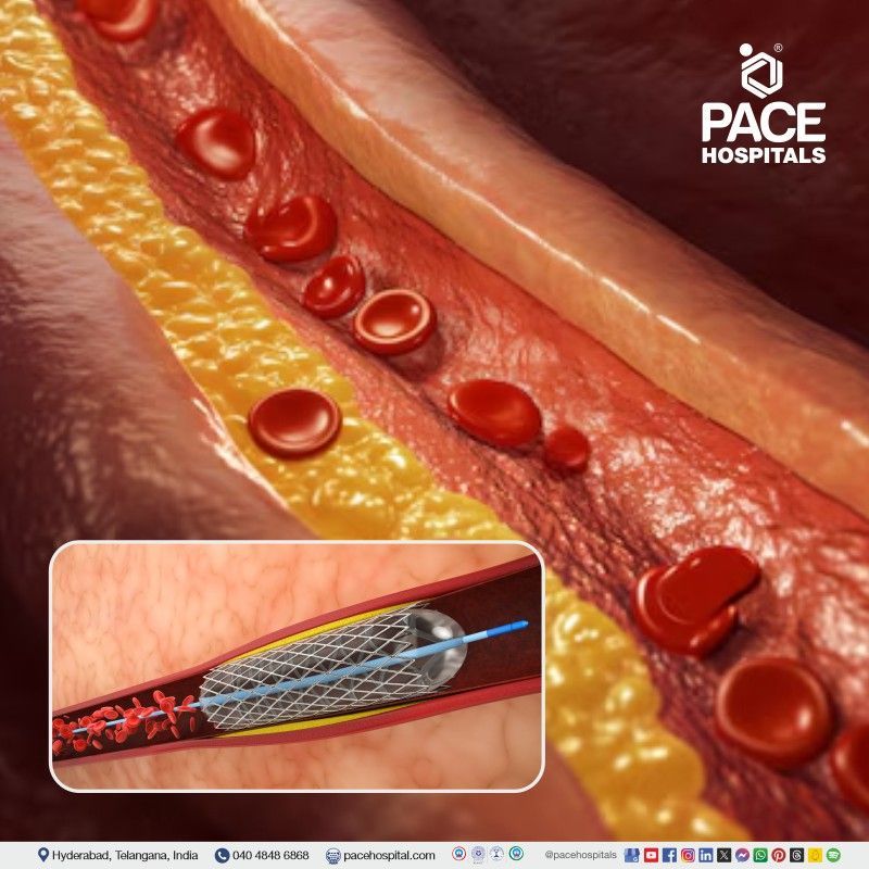
6. Stenting: Stenting is a minimally invasive technique made with fabric, metal, silicone mesh or materials, usually used to open and hold the open passages of the narrowed arteries. Stents treat narrowed coronary arteries (provides oxygenated blood to the heart muscle), aneurysm (bulge in the artery's wall) and narrowed airways in the lungs. Angioplasty is a medical procedure to stretch and widen (open) the clogged or narrowed artery. In modern angioplasty procedures, a stent is used to open and hold the passage permanently to treat the narrowed coronary artery and prevent the further blockage or narrowing of the artery. The combination of angioplasty with stenting is usually called percutaneous coronary intervention (PCI).
7. Embolization: Embolization is a minimally surgical treatment that helps close or block the blood vessels or abnormal blood channels. This is performed by introducing a substance called an embolic agent into the blood vessel to obstruct the flow of blood by using catheters.
It can be used to treat many conditions such as Arteriovenous malformations (AVM), brain aneurysms, cancers (tumour bleeding), uterine fibroids, varicocele, vascular malformations, heavy menstrual bleeding, epistaxis (frequent nosebleeds), etc. Embolic agents include balloons, liquid glue, gelatin form, metallic coils, particulate agents and liquid sclerosing agents.
8. Chemoembolization: Chemoembolization is a minimally surgical treatment for liver cancer that can be performed when the tumour cannot be treated by surgery or radio frequency ablation (RFA). During this process, chemotherapy drugs and embolic agents are injected into the tumour to block the arteries that supply blood (tumour). It also treats cancers that spread to the liver, such as sarcoma, breast, and colon cancers.
A healthcare professional uses X-ray imaging guidance to insert a small catheter through the femoral artery in the groin into the blood vessels that supply the liver tumour. Catheter ensures the treatment of the tumours while minimizing the harm to healthy areas in the liver and the rest of the body.
9. Biopsy: In most cases, healthcare professionals must perform a biopsy to confirm the cancer. A biopsy is a minor procedure of removal of a sample of abnormal tissue, which is examined under the microscope and other examinations on the cell sample. Biopsy samples may be removed in several ways.
- With a Needle: Healthcare professional uses a needle to withdraw fluid or tissue. This method is performed to take some breast, prostate and liver biopsies.
- With an Endoscopy: Healthcare professionals insert a thin, lighted tube known as an endoscope into a mouth or anus (opening of the body) and remove some or complete abnormal tissue through the endoscope.
10. Fine-needle Aspiration: FNA (Fine-needle aspiration) is a procedure that healthcare professionals use to get a cell sample from an abnormal area or suspicious lump. It may also perform during a bronchoscopy or endoscopy.
It uses a thin needle and syringe to pull (take) out the abnormal cells, tissue and fluids. It will identify the masses (tumours) in the breast, skin, lymph nodes and thyroid.
11. Uterine Fibroid Embolization (UFE): Uterine Artery Embolization, or Uterine Fibroid Embolization (UFE), is a procedure to treat or reduce the size of symptomatic uterine fibroids (shrink non-cancerous tumours in the uterus) by blocking the blood supply.
Monitoring through fluoroscopy (a form of real-time x-ray), the embolic agents (agents for blocking blood flow) are delivered to the uterus to treat the fibroids. The duration of recovery and hospitalization are minimal as this procedure is not a major surgery. Uterine artery embolization is also a safe and reliable procedure for post-partum haemorrhage (blood loss by the mother during birth).
12. Endometrial Ablation: Endometrial ablation is a minimally invasive procedure that destroys the endometrial tissue lining of the uterus to treat abnormal uterine bleeding. It can be caused by several reasons, such as hormonal changes, fibroids and polyps in the uterus. It is typically recommended for women who don't wish to become pregnant in the future.
Gynaecologists don't make any incisions (cuts). Instead, they use a thin tool inserted through the vagina to reach the uterus and destroy the uterine lining.
13. Foreign Body Removal: If someone has an inhaled foreign object lodged in their airway, bronchoscopy is the preferred procedure to remove it. A healthcare professional may need to use rigid or flexible bronchoscopy to remove the object safely.
14. IVC filters: An inferior vena cava filter (IVC) is a tiny medical metal device placed in the inferior vena cava to prevent blood clots from travelling to the lungs. It is used to treat pulmonary embolisms. Healthcare professionals may recommend an IVC filter if the people have blood clots and cannot take blood thinners.
The vena cava filter has two types. One is the inferior vena cava (IVC) filter, which prevents the blood clots from the lower body and from travelling into the lungs, and the superior vena cava filter, which prevents the blood clots from the upper body from travelling into the lungs.
15. Cryotherapy: Cryotherapy is a medical procedure that utilizes extreme cold to freeze and eliminate abnormal cells. Healthcare professionals often use this technique to treat certain types of cancers, including lung cancer, by inducing tissue necrosis or cellular death. This is accomplished using a specialized cryoprobe attached to a bronchoscope or administering a cryotherapy spray. Cryotherapy is also referred to as cryoablation.
Bronchoscopic cryotherapy can address various medical conditions, such as foreign body removal, endobronchial biopsy, transbronchial biopsy, and the treatment of benign and malignant central airway obstruction.
16. Vertebroplasty: When a vertebra fractures or breaks, it leads to the development of bone fragments. Pain will occur if they slide or rub against each other or push into the spinal cord. Vertebroplasty is a medical procedure that involves injecting the affected bone with a cement mixture, which is helpful to fuse the fragments or breaks, strengthen the vertebra and provide relief from the associated pain or discomfort.
It involves injecting a fractured vertebra with a special orthopaedic cement called polymethylmethacrylate (PMMA). A healthcare professional slowly injects the cement into the vertebra. This process gives relief from spinal pain and restores mobility. A healthcare professional takes a complete medical, medication history and performs a physical examination to determine the exact location and nature of vertebra-related pain by taking X-rays, MRIs or CT scans.
17. Kyphoplasty: Vertebroplasty and kyphoplasty are medical procedures used to relieve the painful vertebral compression fractures in the spinal column, which can be caused by osteoporosis. A healthcare professional may use the imaging guidance for injecting the special cement mixture into fractured bone (known as vertebroplasty), or a balloon is inserted into the fractured or affected bone to create a space, which can then be filled with cement (known as kyphoplasty).
During the process, the skin is numbed locally. Then, a balloon is inserted through (a hollow needle or tube) a trocar into the fractured vertebra, where the balloon is inflated to create a space for cement injection. The balloon is removed before giving the injection.
18. Nephrostomy: Kidneys produce urine, which flows through the ureter to the urinary bladder. Sometimes, urine flow is obstructed due to kidney stones, infections, cancers, trauma, etc.
A nephrostomy is a medical procedure to clear out (drain) the urine from the kidney using a catheter (nephrostomy tube). Usually, urine drains from the kidneys to the urinary bladder through the ureters. But, when there is a blockage or obstruction, a nephrostomy tube (thin plastic tube) is inserted through the back of the kidney to the point(area) where the urine collects, to temporarily drain the blocked urine. This procedure allows the kidney to function properly and protects it from further damage.
19. Cholangiography: Cholangiography is a procedure that involves the examination of the bile ducts and gallbladder structures by using a contrast dye to show them up on an X-ray. A healthcare professional sends the endoscope through the throat by numbing it. The tube has a light and camera at the end, is guided to the targeted area and injects the dye to see the gall bladder and bile duct structures on the monitor screen.
This procedure typically takes 30 to 60 minutes to complete. It diagnoses the size and extent of gallbladder cancer and finds the blockages in the bile duct. If the patient has a blockage, the healthcare professional will put a stent (small tube), allowing the bile to clear out of the body into a drainage bag to relieve the jaundice symptoms and reduce the risk of infection.
20. Fluoroscopy: Fluoroscopy is an imaging method that visualises continuous X-ray images on the monitor screen. It studies moving body structures, much like an X-ray movie. Fluoroscopy can be used alone as a diagnostic tool or in combination with other procedures. It is used in many techniques, such as barium X-rays, intravascular ultrasound, biopsies, etc. It is helpful to identify the heart or intestinal disease, guide the treatments such as injections, implants, or orthopaedic surgery, and place the catheters, stents, or other devices inside the body (in the heart or blood vessels).
21. Catheters Insertions: An interventional radiologist uses a catheter into different parts of the body to deliver the medication, drain fluids, and perform various therapeutic procedures. For example, healthcare professionals use a catheter inserted into a large vein to provide chemotherapy medicines or haemodialysis. Additionally, angiographic catheters are crucial in any vascular intervention. After insertion into the blood vessel, catheters can conduct diagnostic angiography and deliver stents.
22. Thrombolysis: Thrombolysis is a process of using medications or minimally surgical procedures to break the blood clots and prevent further clotting. A healthcare professional can inject clot-dissolving drugs through an IV (intravenous) line or use a catheter to deliver the drugs directly to the area (site) of the blockage in the patient.
Thrombolysis is often used to treat blood clots in the legs (Deep vein thrombosis) and pelvic area; if untreated, it can break off and travel to the lungs (pulmonary embolism). In catheter-directed thrombolysis, healthcare professionals use an endovascular (minimally invasive) procedure to reach and dissolve blood clots.
23. Radiofrequency Ablation: Radiofrequency ablation (RFA) is a minor surgical technique that uses heat to shrink or destroy the tissue of the tumours, nodules or other growths in the patient body. It treats benign and malignant tumours, chronic venous insufficiency in the legs, chronic neck and back pain.
- Nerve Ablation: This process treats chronic pain by disabling the nerve fibres that carry the pain signals to the brain through the spinal cord.
- Chronic venous insufficiency: Venous insufficiency is an inadequate flow of blood from one or more diseased veins in the legs to the heart, which can result in the pooling of blood in the legs, ankles and feet with pain. RFA can help to treat this condition by sealing off the diseased vein and diverting the blood flow to healthier veins located in the legs.
24. Gastrointestinal Interventions: The primary purpose of gastrointestinal intervention is to provide treatments to patients who are unable to drink orally or to consume food and who may be at a more increased risk of aspiration and treat various conditions in the digestive tract, such as draining abscesses, managing gastrointestinal bleeding, and placing feeding tubes such as gastrostomy and gastrojejunostomy.
25. Hysterosalpingogram: Hysterosalpingogram is an X-ray imaging technique to examine potential causes of pregnancy problems or infertility in women. It uses X-rays to provide a picture of the fallopian tubes and uterus. It can detect scar tissue, structural abnormalities, fibroids, tumours, polyps, or blockages in the tubes. Obstetrician-gynaecologist may recommend this procedure if a patient has had difficulty conceiving or has experienced multiple miscarriages.
26. Intravascular ultrasound (IVUS):
Intravascular ultrasound (IVUS) is an imaging technique used to create(give) pictures of the blood vessels and heart from inside the body. A healthcare professional inserts a catheter in the blood vessel in the wrist or groin by using a catheter with an ultrasound probe. Healthcare professionals navigate a catheter to the heart using the ultrasound probe, which uses sound waves to visualize the heart's artery walls.
It is a type of cardiac catheterization (group of procedures) that uses catheters to identify and treat heart conditions. It is used during specific procedures such as angioplasty or atherectomy.
Why choose PACE Hospitals?
- A Multi-Super Speciality Hospital.
- NABH, NABL, NBE & NABH - Nursing Excellence accreditation.
- State-of-the-art Liver and Kidney transplant centre.
- Empanelled with all TPA’s for smooth cashless benefits.
- Centralized HIMS (Hospital Information System).
- Computerized health records available via website.
- Minimum waiting time for Inpatient and Outpatient.
- Round-the-clock guidance from highly qualified super specialist.
- Standardization of ethical medical care.
- 24X7 Outpatient & Inpatient Pharmacy Services.
- State-of-the-art operation theaters.
- Intensive Care Units (Surgical and Medical) with ISO-9001 accreditation.


