Chronic Calcific Pancreatitis Treatment in Hyderabad, India
PACE Hospitals stands as the best hospital for chronic calcific pancreatitis treatment in Hyderabad, Telangana, India. Our experienced team of gastroenterologists, endocrinologists, surgeons, and dietitians provides a comprehensive and patient-centered approach to managing this complex condition. Using advanced diagnostic tools, we accurately assess pancreatic damage and its complications, including chronic pain, malabsorption, and diabetes.
We offer personalized treatment plans that may include pancreatic enzyme therapy, pain management, nutritional support, and surgical interventions—tailored to slow disease progression and restore function.
Book Appointment for Chronic Calcific Pancreatitis Treatment
Chronic Calcific Pancreatitis Treatment Online Appointment
Thank you for contacting us. We will get back to you as soon as possible. Kindly save these contact details in your contacts to receive calls and messages:-
Appointment Desk: 04048486868
WhatsApp: 8977889778
Regards,
PACE Hospitals
HITEC City and Madeenaguda
Hyderabad, Telangana, India.
Oops, there was an error sending your message. Please try again later. Kindly save these contact details in your contacts to receive calls and messages:-
Appointment Desk: 04048486868
WhatsApp: 8977889778
Regards,
PACE Hospitals
HITEC City and Madeenaguda
Hyderabad, Telangana, India.
Why Choose PACE Hospitals for Chronic Calcific Pancreatitis Treatment?

Advanced Diagnostic Facilities: CT scan, MRI with MRCP, EUS, Pancreatic Function Tests (PFTs)
Expert Gastroenterologists, Endocrinologists & GI Surgeons in Hyderabad
Minimally Invasive and Surgical Treatment Options
Affordable & Transparent Pancreatitis Treatment with Insurance & Cashless Options
Recognized for excellence in healthcare delivery with
NABH, NABL, NBE/DNB, NABH Nursing Excellence, and ISCCM accreditations.
Diagnosis of Chronic Calcific Pancreatitis (CCP)
The diagnosis of chronic calcific pancreatitis (CCP) can be challenging, especially in the early stages, as the clinical manifestations may mimic other gastrointestinal conditions. Hence, a thorough clinical, biochemical, and radiological assessment is essential.
Diagnosing CCP involves a thorough clinical evaluation, history taking, and confirmatory imaging and laboratory studies. Cross-sectional imaging, especially CT scans or MRI, can confirm the diagnosis by revealing pancreatic calcifications, ductal dilatation, and atrophy.
The gastroenterologist can make the initial diagnosis and treatment plan based on the severity of the disease, and evaluate the following before selecting the appropriate tests to confirm CCP:
- The presented signs and symptoms
- Age, family history, and general health of the suspected patient
- Medical and medication history
- Results of previous medical tests
- Physical Examination
The presented signs and symptoms
Patients typically present with chronic or recurrent episodes of epigastric abdominal pain radiating to the back, worsened by eating or drinking. Other common symptoms include malabsorption (manifested by steatorrhea or oily stools), weight loss, nausea, vomiting, and signs of
diabetes mellitus due to loss of endocrine function.
In advanced stages, symptoms of exocrine and endocrine pancreatic insufficiency become more pronounced. In some cases, painless CCP may be detected incidentally during imaging.
Age, Family History, and General Health of the Suspected Patient
Chronic calcific pancreatitis (CCP) typically affects adults between the ages of 30 and 50, although it can occur at any age. One of the most significant risk factors is chronic alcohol consumption, particularly among males. Other notable contributors include smoking, poor nutritional status, and a history of recurrent acute pancreatitis. Hereditary forms of the disease, often linked to genetic mutations, may present earlier in life and are commonly associated with a positive family history. In evaluating a patient's risk for CCP, gastroenterologists consider demographic details, lifestyle habits, and familial predispositions.
Medical and Medication History
An in-depth medical history is essential in identifying underlying causes or risk factors for chronic calcific pancreatitis (CCP). Common contributing factors include recurrent episodes of acute pancreatitis,
cystic fibrosis, hypercalcemia, hyperlipidaemia, autoimmune conditions, and previous pancreatic duct obstruction or trauma. Evaluating for these conditions helps determine the etiology and guide appropriate management strategies.
Reviewing the patient’s medication history is equally important, as certain drugs, such as diuretics, anticonvulsants, immunosuppressants, and corticosteroids, have been associated with pancreatic inflammation. In some instances, drug-induced pancreatic injury can progress to fibrosis and calcification. Additionally, gastroenterologists assess for comorbidities like diabetes mellitus and liver disease, which frequently coexist with CCP and may complicate the clinical course.
Results of Previous Medical Tests
Doctors evaluate previous medical test reports such as abdominal
ultrasound, CT scan, MRI, or
ERCP to assess the presence and extent of pancreatic calcification, ductal irregularities, and structural fibrosis. These imaging studies provide valuable information on the chronicity and anatomical changes associated with chronic calcific pancreatitis.
They also review pancreatic function tests, including faecal elastase-1 and the 72-hour faecal fat test, to determine the degree of exocrine insufficiency. In addition, blood test results are examined for abnormalities in pancreatic enzyme levels, blood glucose, and nutritional markers, all of which help in assessing disease severity and associated metabolic complications.
Physical Examination
After assessing the patient's clinical symptoms, risk factors, and medical history, the gastroenterologist conducts a focused physical examination to identify abdominal abnormalities and systemic complications associated with chronic calcific pancreatitis. This examination is structured into the following key areas:
- Abdominal Examination
- Inspection and Palpation: May reveal epigastric tenderness, abdominal distention, and in some cases, a palpable mass suggestive of a pancreatic pseudocyst.
- Vital Signs: Abnormalities such as fever (in the presence of infection), tachycardia, or hypotension may indicate complications like bleeding or infected collections.
- Nutritional Assessment
- Signs of Malnutrition: Observations may include visible weight loss, muscle wasting, dry skin, glossitis, and clinical features of fat-soluble vitamin deficiencies (e.g., night blindness or easy bruising).
- Stool Findings: Positive tests for steatorrhea can indicate significant exocrine insufficiency.
- Endocrine Evaluation
- Signs of Diabetes Mellitus: These may include dry mouth, polyuria, persistent fatigue, and peripheral neuropathic symptoms, reflecting endocrine dysfunction due to pancreatic damage.
Diagnostic Tests
Following the physical examination, doctors may recommend a combination of the following diagnostic tests to confirm chronic calcific pancreatitis, evaluate pancreatic damage and function, and detect complications based on the patient’s symptoms and clinical findings:
Imaging Tests
- Abdominal X-ray
- Ultrasound (USG)
- Computed Tomography (CT) Scan
- Magnetic Resonance Cholangiopancreatography (MRCP)
Laboratory Blood Tests
- Serum Amylase and Lipase
- Fasting Blood Glucose and HbA1c
- Serum Trypsinogen
Oxygen Saturation and Respiratory Function Tests
- Pulse Oximetry
- Arterial Blood Gas (ABG) Analysis
Microbiological Tests
- Pancreatic Fluid Culture (via EUS or ERCP-guided aspiration)
- Stool Elastase Test
Minimally Invasive Procedures
- Endoscopic Ultrasound (EUS)
- Endoscopic Retrograde Cholangiopancreatography (ERCP)
- Fine Needle Aspiration (FNA)
Imaging Tests
- Abdominal X-ray: Abdominal X-rays can reveal dense, coarse calcifications in the pancreas, which are hallmark indicators of CCP. This test is useful in detecting advanced stages of the disease. Although not very sensitive in early stages, it is a quick, accessible, and low-cost initial imaging modality.
- Ultrasound (USG): Ultrasound can detect hyperechoic foci with acoustic shadowing, suggesting pancreatic calcifications. It is also useful in evaluating pancreatic size, ductal dilatation, and parenchymal atrophy. While operator-dependent, USG is a non-invasive and widely available tool.
- Computed Tomography (CT) Scan: CT scan offers high-resolution images that clearly identify pancreatic calcifications, ductal dilatation, pseudocysts, and gland atrophy. It is the preferred imaging modality for diagnosing CCP and assessing complications. CT also helps rule out pancreatic malignancy when features overlap.
- Magnetic Resonance Cholangiopancreatography (MRCP): MRCP provides detailed images of the pancreaticobiliary ducts without contrast injection. It identifies strictures, dilated ducts, and intraductal stones associated with CCP. Being non-invasive and free from ionizing radiation, it’s ideal for repeated follow-up in chronic cases.
Laboratory Blood Tests
- Serum Amylase and Lipase: These pancreatic enzymes are usually elevated during acute inflammation, but may be normal or low in chronic cases. Persistently low levels in CCP suggest long-standing exocrine dysfunction. These tests help differentiate acute-on-chronic pancreatitis episodes from active inflammation.
- Fasting Blood Glucose and HbA1c: These tests evaluate pancreatic endocrine function by assessing blood sugar control. Chronic inflammation damages insulin-producing cells, leading to secondary diabetes. Detecting hyperglycaemia or abnormal HbA1c helps confirm endocrine insufficiency in CCP.
- Serum Trypsinogen: Low levels of serum trypsinogen are indicative of pancreatic exocrine insufficiency, a key feature of CCP. It is particularly useful in diagnosing early or subclinical disease stages. This test also complements stool-based assessments of enzyme output.
Oxygen Saturation and Respiratory Function Tests
- Pulse Oximetry: Pulse oximetry measures peripheral oxygen saturation, which can be affected in patients with severe abdominal pain or systemic complications. While not specific for CCP, it is important for monitoring patients during acute flare-ups or procedural sedation.
- Arterial Blood Gas (ABG) Analysis: ABG assesses oxygenation, ventilation, and acid-base balance. It is especially relevant in severe CCP cases with systemic manifestations or when surgery is required. It also helps evaluate respiratory compromise in patients with multi-organ involvement.
Microbiological Tests
- Pancreatic Fluid Culture (via EUS or ERCP-guided aspiration): This test identifies bacterial infections in pancreatic collections, such as abscesses or infected pseudocysts. Infection is a known complication of CCP. Culturing fluid helps guide appropriate antibiotic therapy and drainage strategies.
- Stool Elastase Test: This non-invasive test measures pancreatic elastase levels in stool, which are reduced in exocrine insufficiency. It provides indirect confirmation of the CCP when low values are found. It’s particularly useful in early or non-calcific presentations of the disease.
Minimally Invasive Procedures
- Endoscopic Ultrasound (EUS): EUS allows high-resolution visualization of the pancreas from the gastrointestinal tract. It detects early parenchymal changes, ductal irregularities, and small calcifications. It also enables tissue sampling via fine-needle aspiration, especially when cancer is suspected.
- Endoscopic Retrograde Cholangiopancreatography (ERCP): ERCP is both diagnostic and therapeutic, offering direct visualization and access to the pancreatic ducts. It identifies strictures, intraductal stones, and irregular duct morphology typical of CCP. Interventions like stent placement or stone extraction can be performed during the procedure.
- Fine Needle Aspiration (FNA): FNA is typically performed under EUS guidance to collect pancreatic tissue or fluid samples. It helps distinguish CCP from pancreatic cancer when imaging is inconclusive. Cytological analysis confirms inflammatory vs malignant etiology in indeterminate lesions.
Differential Diagnosis
A differential diagnosis is a list of possible medical conditions or diseases that can present with similar symptoms in a person. The CCP has an extensive differential diagnosis, outlined as follows:
Pancreatic Ductal Adenocarcinoma
A malignant tumor of the pancreas that can mimic CCP symptoms like weight loss and abdominal pain; imaging and biopsy are essential for differentiation.
Autoimmune Pancreatitis (AIP)
An immune-mediated inflammation of the pancreas presenting with similar imaging findings; elevated IgG4 levels and steroid responsiveness help in diagnosis.
Hereditary Pancreatitis
A genetic condition causing recurrent pancreatitis episodes from a young age; family history and genetic testing aid in identification.
Tropical Pancreatitis
A form of chronic pancreatitis prevalent in tropical regions, often associated with malnutrition and diabetes, distinguished by geographic and demographic factors.
Cystic Fibrosis
A genetic disorder affecting exocrine glands, leading to thick secretions and pancreatic insufficiency; diagnosed through sweat chloride tests and genetic analysis.
Pancreatic Pseudocyst
A fluid-filled sac arising from pancreatitis that can cause abdominal discomfort; imaging helps differentiate it from CCP.
Chronic Alcoholic Pancreatitis
Long-term alcohol abuse leading to pancreatic inflammation and calcification; a history of alcohol intake is a key differentiator.
Idiopathic Chronic Pancreatitis
Chronic pancreatitis with no identifiable cause; diagnosis is made after excluding other etiologies.
Treatment for chronic calcific pancreatitis (CCP) typically begins once the diagnosis is confirmed and symptoms such as persistent abdominal pain, malabsorption, or diabetes become evident. The choice of treatment depends on the severity of symptoms, extent of pancreatic damage, and presence of complications like ductal obstruction or pseudocysts. Here are some approaches that can be chosen for the treatment of CCP:
Conservative Management
- Pain Management
- Enzyme Replacement Therapy (PERT)
- Nutritional and Vitamin Support
- Diabetes Control (Type 3c)
Endoscopic Interventions
- ERCP (Endoscopic Retrograde Cholangiopancreatography)
- Pancreatic Ductal Stone Removal
- Pancreatic Stenting
Surgical Management
- Lateral Pancreaticojejunostomy (Modified Puestow)
- Frey Procedure
- Beger Procedure
- Distal Pancreatectomy
- Total Pancreatectomy
Conservative Management
It is indicated to control symptoms, manage pain, and optimize digestion through enzyme supplementation, dietary modifications, and alcohol cessation, without the need for surgery unless complications arise. The following are the conservative management approaches:
1. Pain Management
Pain is managed using a step-wise approach, starting with NSAIDs. If ineffective, opioid analgesics or low-dose opioids may be added. Antioxidants such as vitamin C may reduce inflammation and oxidative stress, improving pain control. Avoidance of alcohol and smoking is essential to prevent exacerbations.
2. Enzyme Replacement Therapy (PERT)
Pancreatic enzyme supplements (lipase ≥25,000 units/meal) aid fat digestion and reduce post-meal pancreatic stimulation. This helps in alleviating postprandial pain and correcting malabsorption. Preparations typically contain lipase, amylase, and protease and are taken with every meal and snack.
3. Nutritional and Vitamin Support
A high-protein, low-fat diet is advised, with fat restriction adjusted based on steatorrhea severity. Supplementation of fat-soluble vitamins (A, D, E, K), calcium, magnesium, and vitamin B12 is essential to counter malabsorption and prevent deficiencies. Nutritional support helps prevent weight loss and complications.
4. Diabetes Control (Type 3c)
Endocrine insufficiency due to pancreatic damage is treated with insulin therapy. Oral hypoglycaemics are generally avoided in malnourished patients or those with erratic glucose absorption. Regular blood sugar monitoring and education about hypoglycaemia are crucial in long-term management.
Endoscopic Interventions
Endoscopic therapy is used when there's main duct obstruction, ductal hypertension causing severe pain, or complications like inflammatory masses and pseudocysts. It helps relieve obstruction and avoid or delay surgery in select patients.
1. ERCP (Endoscopic Retrograde Cholangiopancreatography)
ERCP allows visualization and dilation of strictures in the pancreatic duct. It enables stent placement to relieve obstruction and is a minimally invasive alternative for ductal drainage. It can also help in the diagnosis of pancreatic ductal abnormalities.
2. Pancreatic Ductal Stone Removal
Stones causing ductal obstruction are treated with extracorporeal shockwave lithotripsy (ESWL) followed by mechanical clearance via ERCP. This reduces ductal pressure and alleviates pain. It’s a preferred approach before surgical options in stone-related CCP.
3. Pancreatic Stenting
Plastic or metal stents are placed to relieve ductal obstruction and manage strictures. This helps in reducing ductal pressure and providing symptomatic relief. Stents may require replacement or removal after several weeks, depending on response.
Surgical Management
Surgery is indicated when medical and endoscopic approaches fail, or when complications such as persistent pain, ductal obstruction, suspected cancer, or pseudocysts arise. It is especially considered in patients with dilated ducts or head masses.
1. Lateral Pancreaticojejunostomy (Modified Puestow)
It is performed when the main duct is dilated (>7 mm), involving opening the duct along its length and connecting it to the jejunum (Roux-en-Y). It offers durable pain relief by improving ductal drainage and decompressing the gland.
2. Frey Procedure
This combines local resection of the pancreatic head with ductal drainage. It is suited for patients with a bulky inflammatory mass in the head and dilated ducts. It preserves pancreatic function while relieving pain and obstruction.
3. Beger Procedure
This is a duodenum-preserving pancreatic head resection. It is indicated for isolated head disease while maintaining duodenal and biliary continuity. It provides symptom relief and preserves digestive tract anatomy.
4. Distal Pancreatectomy
It is performed when the disease is limited to the pancreatic body and tail. It involves surgical removal of the distal pancreas and may require splenectomy. This option may compromise endocrine and exocrine function in the long term.
5. Total Pancreatectomy
This is the last resort for intractable pain or diffuse disease unresponsive to other treatments. It removes the entire pancreas, resulting in permanent diabetes and exocrine insufficiency. It is reserved for selected patients with severe disease.
Post-Treatment Monitoring and Follow-Up
Patients require lifelong follow-up to monitor glycaemic control (e.g., HbA1c), nutritional status, and vitamin levels. Imaging may be repeated if symptoms recur or complications are suspected. Continued use of enzymes and dietary guidance is essential for long-term health maintenance.
Long-Term Outlook and Patient Education
CCP is a progressive and debilitating disease; early intervention improves quality of life. Abstinence from alcohol and smoking is non-negotiable to slow progression. Patient education should focus on medication adherence, diet, enzyme use, glucose monitoring, and psychological support for chronic illness.
Chronic Calcific Pancreatitis Treatment Cost in Hyderabad, India
The cost of Chronic Calcific Pancreatitis Treatment in Hyderabad generally ranges from ₹65,000 to ₹2,40,000 (approximately US $780 – US $2,890).
The exact treatment price varies depending on factors such as the severity of pancreatic calcification, presence of complications (ductal obstruction, pseudocyst), whether endoscopic or surgical intervention is needed, the requirement for ERCP or lithotripsy, surgeon expertise, and the hospital facilities chosen — including cashless treatment options, TPA corporate tie-ups, and assistance with medical insurance approvals wherever applicable.
Cost Breakdown According to Type of Treatment
- Medical Management & Pain Control – ₹65,000 – ₹95,000 (US $780 – US $1,145)
- Endoscopic Treatment (ERCP + Stone Extraction / Stenting) – ₹90,000 – ₹1,60,000 (US $1,080 – US $1,925)
- Extracorporeal Shock Wave Lithotripsy (ESWL) for Pancreatic Stones – ₹1,20,000 – ₹1,90,000 (US $1,445 – US $2,290)
- Surgical Drainage Procedure (Puestow / Lateral Pancreaticojejunostomy) – ₹1,40,000 – ₹2,20,000 (US $1,690 – US $2,650)
- Complex / Recurrent Calcific Pancreatitis with Complications – ₹1,60,000 – ₹2,40,000 (US $1,930 – US $2,890)
Frequently Asked Questions (FAQs) on Chronic Calcific Pancreatitis (CCP)
What is the relation between chronic calcific pancreatitis and diabetes?
Chronic calcific pancreatitis leads to progressive destruction of pancreatic tissue, including insulin-producing beta cells, resulting in impaired glucose metabolism. This increases the risk of developing type 3c diabetes mellitus (pancreatogenic diabetes).
Which Is the best hospital for Chronic Calcific Pancreatitis Treatment in Hyderabad, India?
PACE Hospitals, Hyderabad is considered one of the most trusted centres for treating chronic calcific pancreatitis and complex pancreatic disorders.
Our team of gastroenterologists, endoscopists, and pancreatic surgeons specialises in minimally invasive and advanced interventional techniques to relieve pain, remove ductal stones, restore pancreatic drainage, and manage long-term pancreatic function.
With state-of-the-art endoscopy suites, advanced lithotripsy systems, modern OT infrastructure, and dedicated post-treatment monitoring, PACE Hospitals ensures safe, comprehensive, and long-lasting relief — supported by cashless facility options, TPA corporate tie-ups, and assistance with medical insurance processing for eligible patients.
What are the common symptoms of chronic calcific pancreatitis?
Common symptoms of chronic calcific pancreatitis include persistent upper abdominal pain, steatorrhea (fatty stools), weight loss, and signs of diabetes such as increased thirst and fatigue. Symptoms result from impaired digestive and endocrine functions of the pancreas.
What are the causes of CCP?
Chronic calcific pancreatitis (CCP) is commonly caused by chronic alcohol consumption, genetic mutations, and tropical pancreatitis. Other causes include recurrent acute pancreatitis, autoimmune conditions, and ductal obstructions such as strictures or stones.
What is the difference between chronic calcific pancreatitis and pancreatitis?
The main difference between chronic calcific pancreatitis and general pancreatitis is that chronic calcific pancreatitis involves long-term inflammation with irreversible calcification and damage to the pancreas. In contrast, pancreatitis refers to any inflammation of the pancreas, which can be acute or chronic and may not necessarily involve calcification.
What Is the cost of Chronic Calcific Pancreatitis Treatment at PACE Hospitals, Hyderabad?
At PACE Hospitals, Hyderabad, the cost of chronic calcific pancreatitis treatment typically ranges from ₹60,000 to ₹2,20,000 and above (approximately US $720 – US $2,650), depending on:
- Type of treatment required (medical, endoscopic, lithotripsy, surgical)
- Severity of calcification and duct obstruction
- Presence of pseudocysts or complications
- Specialist expertise and procedure complexity
- Duration of hospital stay and ICU requirement
- Diagnostic tests such as CT/MRCP/Endoscopic Imaging
- Need for repeat ERCP or multiple sessions of therapy
For medical management and basic endoscopic interventions, costs fall at the lower end, while lithotripsy or major pancreatic surgery falls toward the higher end.
After a complete gastroenterology evaluation, imaging studies, and treatment planning, our specialists will provide a personalised and transparent cost estimate based on your condition.
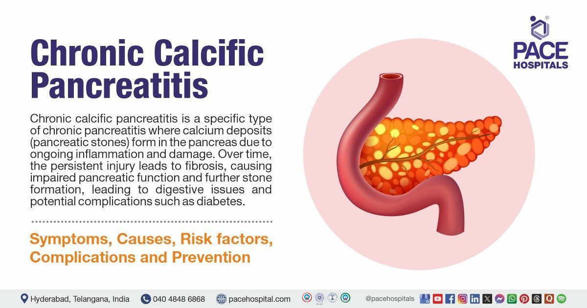
What is chronic calcific pancreatitis?
Chronic calcific pancreatitis is a long-standing inflammation of the pancreas characterized by irreversible damage, fibrosis, and calcification of the pancreatic tissue. It leads to progressive loss of both exocrine and endocrine functions, often causing pain, malabsorption, and diabetes.
How does calcification occur in the pancreas during chronic pancreatitis?
Calcification in chronic pancreatitis occurs due to prolonged inflammation and ductal obstruction, leading to protein-rich plug formation within the pancreatic ducts. Over time, these plugs calcify, resulting in intraductal and parenchymal calcifications.
Can chronic calcific pancreatitis cause hypoglycaemia?
Yes, chronic calcific pancreatitis can cause hypoglycaemia, especially in the early stages of pancreatic diabetes (type 3c), where there is inconsistent insulin secretion. As beta cell function declines irregularly, patients may experience unpredictable blood glucose swings, including hypoglycaemia.
What is the diet recommended for chronic calcific pancreatitis?
The diet for chronic calcific pancreatitis should be low in fat, high in protein, and rich in fruits and vegetables to reduce pancreatic stimulation. Small, frequent meals and pancreatic enzyme supplements are recommended to aid digestion and nutrient absorption.
Why gall bladder not removed in chronic calcific pancreatitis?
The gallbladder is not routinely removed in chronic calcific pancreatitis unless gallstones or biliary obstruction are present. The condition primarily affects the pancreas, and gallbladder removal is unnecessary unless it contributes to symptoms or complications.
What is non-chronic calcific pancreatitis?
Non-chronic calcific pancreatitis typically refers to recurrent pancreatitis without long-term calcification or permanent damage. It involves sudden inflammation that may resolve completely, unlike chronic calcific pancreatitis, which causes irreversible changes.