Breast Cancer Treatment in Hyderabad | Surgery and Cost
At PACE Hospitals, team of best breast cancer doctor, medical oncologist - breast cancer specialist, surgical oncologist - breast cancer surgeon are experienced in handling even the most complex and complicated cases of breast cancer, and having expertise providing breast cancer treatment through medical management and / or performing breast cancer surgery with using advanced 3D HD laparoscopic and robotic surgery techniques with minimal time and high success rate.
Request Appointment for Breast Cancer Treatment
Breast Cancer Treatment - appointment
Why Choose PACE Hospitals for Breast Cancer Treatment?

5,000+ Breast Cancer Screening Performed
Team of the Best Breast Cancer Doctor with 35+ years of expertise
Cost-effective treatment with 99.9% success rate
All insurance accepted with No-cost EMI option
Breast Cancer Stages
In total there are 5 stages of breast cancer which are based on TNM classification. TNM refers to Tumour, Nodes and metastasis respectively.
| Stage | T (Tumour) | N (Nodes) | M (Metastasis) |
|---|---|---|---|
| 0 | Tis | N0 | M0 |
| I (1) | T1 | N0 | M0 |
| II (2) | T0–T3 | N0–N1 | M0 |
| IIIA (3A) | T0–T3 | N1–N2 | M0 |
| IIIB (3B) | T4 | N0–N2 | M0 |
| IIIC (3C) | Any T | N3 | M0 |
| IV (4) | Any T | Any N | M1 |
The knowledge of stage of breast cancer is important as it can provide a clarity to the breast cancer surgeon / surgical oncologist about the prognosis and complications of the cancer, thereby preparing the oncologist to opt for the best treatment modality.
Diagnosis of Breast Cancer
A visit to a Hospital for Breast Cancer is typically recommended by the primary care physician. The patients can get either a physical breast exam, or an ultrasound and x-ray (mammogram) or all the three depending upon the complexity of the disease.
Before commencing imaging tests, the oncologist (cancer specialist) performs a physical breast exam during which a keen and thorough checking for any abnormal growth/protrusion, any signs of disease, such as lumps etc is performed. The doctor may ask questions about the patients’ health, habits, past illnesses, their treatments. Questions could also include the parentage of the patient. All these questions are intended to find a connection between the patient and the disease.
In case the doctor finds any abnormalities or irregularities, the patient is then subjected to imaging tests to understand and confirm the disease. The imaging tests include:
- Optical imaging tests
- Mammogram - Contrast-enhanced mammography
- Breast ultrasound - Elastography
- Biopsy
- Fine needle aspiration cytology
- Core needle biopsy
- Punch biopsy
- Vacuum assisted biopsy
- Wire guided excision biopsy
- Breast MRI - Abbreviated breast MRI (fast breast MRI)
- Nuclear medicine tests (radionuclide imaging)
- Molecular breast imaging
- Positron emission tomography
- Positron emission mammography
- Electrical impedance tomography
Optical imaging tests: These examinations involve shining a light into the breast and then monitoring any light reflection or transmission. This method does not involve radiation or painful breast compression. Preliminary research is currently looking into the efficacy of combining optical imaging with other tests, such as breast magnetic resonance imaging (MRI), ultrasound, or 3D mammography, to aid in the search for breast cancer.
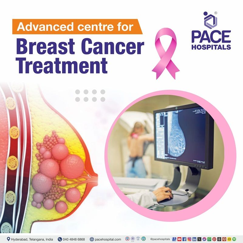
Mammogram: A special kind of breast x-ray in which high-energy rays are used in x-ray machines to capture images of the body's interior. The duration of a mammogram is usually a few minutes. A newer mammography procedure, CEM is recently introduced.
- Contrast-enhanced mammography (CEM): Also called contrast-enhanced spectral mammography (CESM), it involves injecting a contrast dye containing iodine into the bloodstream just before taking two sets of mammograms (at different energy levels). The abnormalities in the breasts can be more visible due to the contrast. Comparative studies are being performed for its potential use in screening women with dense breasts. CEM has the potential to replace MRI if it proves to be as effective.
Breast ultrasound: Diagnosing breast cancer and other breast conditions can be performed through ultrasound waves, which creates a picture of the breast tissue. This is one of the most common diagnostic methods and it requires no special preparation. In few hospitals, elastography is also done as a part of ultrasound exam.
- Elastography: Performed as part of ultrasound exam, elastography comprises detecting the firmness of a suspicious area through ultrasonography when the the breast is compressed slightly. Usually, the breast cancer tumours tend to be firmer and stiffer than the surrounding breast tissue. Through this exam, the differentiation between malignant or benign tissues can be done easily.
Biopsy: It comprises the taking of cells or tissues from the breast and having them examined under a microscope to find signs of cancer. Biopsy is mainly done if the above explained diagnostic tools reveal an abnormal finding in the breast tissue. There are 5 types of biopsy examinations:
- Fine needle aspiration cytology (FNAC): Breast tissue cells can be sampled via a fine needle and syringe. After that, a microscope examination of the samples is possible. This examination could be performed in the hospital's outpatient clinic.
- Core needle biopsy (CNB): A breast tissue sample can be taken via needle biopsy and examined under a microscope. A hollow needle equipped with a precision cutting instrument is utilized for taking samples from the part of the breast that has shown abnormalities on a mammogram or in an ultrasound scan. The needle's handle is equipped with a precision cutting instrument. Under a microscope, the samples are analysed. Conditions like ductal carcinoma in situ or cancer can be detected.
- Punch biopsy: Non-cancerous skin conditions like eczema, inflammatory breast cancer, and Paget's disease can be diagnosed with a punch biopsy, in which a small cutting device is used to remove a sample that can be examined under a microscope. This examination may be performed in the hospital's outpatient clinic.
- Vacuum assisted biopsy (VAB): The mode of diagnosis involves the process of obtaining a tissue sample from the site of an abnormal growth by inserting a vacuum-powered instrument through a small incision in the skin. Instead of having to withdraw the probe after each sample, as in core needle biopsy, vacuum pressure can be used to remove the abnormal cells and tissue.
- Wire guided excision biopsy: This method is used in the cases where an abnormal area on a mammogram or breast ultrasound has been detected which the oncologist was unable to detect it during a physical examination. It involves the insertion of a thin wire into the breast in order to remove the abnormal tissue.
- Excision biopsy: A small size of the palpable lump in the breast is surgically removed from the body. In most cases, only the affected lymph nodes are taken out. The tissue is then examined under a microscope.
Breast magnetic resonance imaging (MRI) scan: Cross-sectional images of the body are created by using magnetism and radio waves. It takes 360-degree images of the body and reveals soft tissues in stunning detail. An MRI is a type of imaging procedure that is typically performed in the radiology (x-ray) department on an outpatient basis. This is performed as mammograms and breast ultrasounds are not particularly effective at detecting lobular breast cancer.
The extent of the cancer and the surgical options available can be determined with the help of a breast MRI scan. A newer type of MRI called fast breast MRI is recently developed.
- Abbreviated breast MRI (fast breast MRI): Although this newer MRI requires fewer scans than the conventional breast MRI, enhanced images are developed due to the administration of gadolinium (contrast material) through an IV line before the exam. It is currently being determined if it can diagnose cancer in women with denser breasts.
Nuclear medicine tests (radionuclide imaging): Organ and tissue function can be investigated with nuclear imaging. A small amount of a radioactive substance (called radioactive tracer or simply tracer) is administered into the body during the procedure. The tissue in the body takes up the tracer and its presence can be detected by the tests. Various types of tracers are such as technetium, thallium, gallium, iodine, and xenon are utilised for different types of examinations. The various types of radionuclide imaging tests are:
• Molecular breast imaging (MBI)
• Positron emission tomography (PET)
• Positron emission mammography (PEM)
- Molecular breast imaging (MBI), also known as scintimammography or breast-specific gamma imaging (BSGI): The tracer technetium-99m sestamibi is injected into the blood and imaged using a special camera while the breast is gently compressed. Monitoring of breast issues (like a lump or an abnormal mammogram) or to aiding in the staging of breast cancer can be feasible with this test. It is also being researched as a potential screening test to be used alongside mammograms in the detection of breast cancer in women with dense breasts. One potential disadvantage is the exposure of the entire body to radiation; due to which annual screenings using this method are probably not feasible.
- Positron emission tomography (PET) scan: In order to perform a PET scan, a different kind of radioactive tracer is injected into the bloodstream. If there is reason to suspect that the breast cancer has spread to other parts of the body, a standard PET scan using a form of radioactive sugar (known as FDG) may be performed. Some advanced oestrogen receptor (ER)-positive breast cancers can now be monitored for metastasis using fluoroestradiol F-18, a novel type of tracer.
- Positron emission mammography (PEM): This imaging tests combines the aspects of both PET scans and mammograms. Tracers are injected into the bloodstream for both PET and PEM. Similar to a mammogram, the breast is gently compressed while images are taken. PEM has the potential to be more sensitive than conventional mammography at detecting clumps of cancer cells within the breast. This exam is currently studied to find is use in staging breast cancer. Since PEM subjects the entire body to radiation, it probably won't be used annually for breast cancer screening.
Electrical impedance tomography (EIT): Abnormal electrical conductance is seen in cancer cells when compared to healthy cells. By taping small electrodes to the skin, extremely weak electrical currents are sent through the breast. Through EIT their effects are read, thus differentiating a malignant from a benign growth. In this exam, the breasts are not compressed or subjected to radiation. A potential application of this test is in the categorization of mammographically detected tumours. Nevertheless, more clinical trials are needed to determine its efficacy in screening for breast cancer.
Staging of Breast Cancer
The size and the extent of the cancer spread are the deciding factors of staging a breast cancer. Detecting the stage of breast cancer is necessary for the oncologist to treat the patient by:
- Finding the severity of the breast cancer and how likely is the patient can beat it.
- Considering all the options before settling on a course of treatment.
- Checking out any potential treatment clinical trials
Regardless of whether or not a cancer progresses, its stage is always referred to as the one assigned at diagnosis. Any additional details about a cancer's evolution over time are added to the original stage. This means that even though the cancer may progress, the stage remains constant.
Staging of breast cancer can be done by the amalgamation of x-rays, lab tests, and other diagnostic tests or procedures. Other factors affecting the staging of breast cancer are:
- Location of tumour
- The cell type (adenocarcinoma (cancer developed in gland) or squamous cell carcinoma)
- The size of the tumour
- Metastasis of cancer (spread of cancer)
Advanced Cancer Centre for Breast Cancer Treatment in Hyderabad
PACE Hospitals is one of the Advanced and Best Breast Cancer Hospital in Hyderabad backed up with wide expertise and vast experience team of breast cancer specialist including breast cancer surgeon (surgical oncologist), breast cancer doctor (medical onologist), nursing, and paramedical staff. Oncology department at PACE Hospitals equipped with state-of-the-art facility and robotic surgery technology offering comprehensive treatment for breast cancer.
Considerations and goals of an oncologist before treating a breast cancer patient
Before starting the necessary therapy, the oncologist primarily looks up at the medical history and her risk factors to understand in which group of breast cancer patients does she fit in:
- High risk group or
- Low risk group
Once the proper risk group is determined, the goals of the oncologist are planned.
The goal of therapy for breast cancer in patients who do not have obvious evidence of distant metastases (meaning outside the breast, chest wall, and regional lymph nodes) is cure, or at least substantial survival prolongation.
For these patients, treatment strategies are divided into primary and systemic considerations.
- Primary therapies consist of surgical and radiation treatments directed toward the breast and loco regional lymph nodes which are designed to minimize the incidence of loco regional recurrence while maintaining quality of life and cosmesis (the practice of preserving or restoring physical appearance, usually after an operation conducted for non-cosmetic reasons) as much as possible by excising the cancer and sterilizing unaffected breast tissue as appropriate.
- Adjuvant systemic treatments, consisting of endocrine, anti-HER2, and/or chemotherapies, are given to treat micrometastases that may have already escaped to distant sites but are not yet detectable.
Treatment of Breast Cancer
The treatment of breast cancer includes multiple modalities and is determined based on the stage of disease.
Surgery, radiation therapy, hormonal therapy, chemotherapy, and biologic therapy can all be used in many different combinations based on a patient’s specific disease.
- Neoadjuvant therapy: If the therapy is given before surgery, it is called neoadjuvant therapy
- Adjuvant therapy: If the treatment is administered after surgery, it is termed adjuvant therapy
Breast Cancer Surgery
Surgery involves mastectomy (removal of breast) or breast-conserving surgery plus radiation therapy.
- Skin-sparing mastectomy: Allows for easier breast reconstruction by preserving the pectoral muscles and a sufficient amount of skin to cover the wound.
- Nipple-sparing mastectomy: Similar to the above, but the areola and nipple are also preserved.
- Simple mastectomy: Spares the pectoral muscles and axillary lymph nodes
- Modified radical mastectomy: Cutting into the pectoral muscles is avoided, but takes out some lymph nodes in the armpits.
- Radical mastectomy: Removal of axillary lymph nodes and the pectoral muscles. This is rarely done unless the cancer has invaded the pectoral muscles.
- Breast-conserving surgery (BCS): The size of the tumour and the necessary margins are calculated (based on the tumour’s size in relation to the volume of the breast), and the affected area is then surgically removed. Depending on how much breast tissue is removed, the procedure may be referred to as a lumpectomy (part of the affected breast or the lump is removed surgically), wide excision, or quadrantectomy (cutting off the tissue in a breast quadrant, as a type of partial mastectomy).
When the entire tumour can be removed, the survival and recurrence rates of patients with invasive cancer are not so different between mastectomy and breast-conserving surgery plus radiation therapy.
So, within reasonable bounds, patient preference can direct treatment selection.
- Less invasive surgery and the possibility of retaining the breasts are the main benefits of breast-conserving surgery combined with radiation therapy.
- Removing the tumour completely with a margin free of cancerous tissue is more important than preserving the patient's appearance.
- Patients with ptosis (sagging) breasts may benefit from consulting a plastic surgeon about oncoplastic surgery in order to achieve good resection margins.
When combined with radiation therapy, neoadjuvant chemotherapy allows some patients who would have otherwise needed a
mastectomy to instead undergo breast-conserving surgery.
Radiation Therapy
High doses of radiation are used in radiation therapy (also called radiotherapy) to kill cancer cells and reduce the size of tumours.
- Local recurrence in the breast and regional lymph nodes are greatly reduced by radiation therapy following breast-conserving surgery, and overall survival of the patient may be improved.
- Adding radiation therapy to lumpectomy plus tamoxifen does not significantly reduce the rate of mastectomy for local recurrence or the occurrence of distant metastases or increase the survival rate, so it may not be necessary for patients aged greater than 70 and up with early oestrogen receptor–positive (ER+) breast cancer.
- Radiation therapy side effects (such as fatigue and skin changes) are typically mild and short-lived.
- There is less of a chance of developing lymphedema (localized swelling of the body caused by an abnormal accumulation of lymph), brachial plexopathy (condition marked by numbness, tingling, pain, weakness, or limited movement in the arm or hand), radiation pneumonitis (radiation-induced lung injury characterized by the presence of acute diffuse alveolar damage), rib damage, secondary cancers, or cardiac toxicity.
Neoadjuvant Therapy
The recent years have seen a rise in the popularity of neoadjuvant chemotherapy, which is administered before tumour removal surgery. The patients suffering with locally advanced breast cancer or those who could benefit from size reduction prior to conservation therapy are currently offered neoadjuvant chemotherapy.
There is now sufficient evidence that shows a patient will have a better outcome if neoadjuvant chemotherapy results in a complete pathologic response. Therefore, the degree of response to neoadjuvant chemotherapy is a crucial factor in determining patient outcome and has a significant impact on both patient selection and follow-up management.
Adjuvant Chemotherapy or Endocrine Therapy
Endocrine therapy: Hormone therapy (also called hormonal therapy, hormone treatment, or endocrine therapy) slows or stops the growth of hormone-sensitive tumours by blocking the body's ability to produce hormones or by interfering with effects of hormones on breast cancer cells.
- Adjuvant endocrine therapy is indicated for nearly all patients with a diagnosis of ER-positive breast cancer and never for those with ER-negative disease.
- The duration of adjuvant endocrine treatment depends upon the stages of the cancer.
- The standard recommendation was at least 5 years of therapy, which clearly reduces the risk of recurrence during that time and for a few years after discontinuation.
- However, the annual risk of distant recurrence during the subsequent 15 years is 0.5–3%, depending on the initial T and N status.
Chemotherapy: Administration of anti-cancer drugs either by intravenous route or oral route is chemotherapy. Typically, multiple-agent adjuvant chemotherapy is more effective than single-agent chemotherapy.
- Although chemotherapeutic agents are usually delivered in combination, sequential single-agent chemotherapy is as effective, and may be slightly less toxic, although it requires longer total duration to deliver.
- Chemotherapy is associated with nausea, vomiting, and alopecia (loss of hair) in nearly 100% of patients.
- Nausea and vomiting are usually well controlled with modern antiemetics.
- Convincing studies have suggested that the strategy of constricting blood flow to the scalp with various means of cooling is commonly effective in sparing hair loss, without evidence of increased scalp metastases.
- More importantly, chemotherapy causes potential life-threatening or life-changing toxicities in 2–3% of all treated patients.
Biological therapy: (also called immunotherapy) treats the patient by either attacking cancer cells directly or by stimulating the immune system of the patient to attack cancer cells.
Biologic targeted therapy: (also called molecularly targeted drugs/therapies, or precision medicine or just targeted therapy) Targeted therapy specifically attacks the proteins which are overproduced in greater quantities in cancer cells thus disabling their function by preventing cancer cells from receiving growth signals.
- Targeted therapy is more preferable as it allows normal and healthy cells to survive unlike chemotherapy which can kill healthy cells during the elimination of cancer cells, causing side effects such as hair loss and low blood counts.
- Targeted therapy is mostly given in patients with high level of HER2. HER2 is a protein that promotes cell growth which is present in 20% of all breast cancers and is the most prevalent abnormal protein in the disease.
Complications of Breast Cancer Treatment
The complications of breast cancer can arise from its treatment, whether chemotherapy, radiation, hormonal therapy, or surgery.
Surgical complications include:
- Infection
- Pain
- Bleeding
- Cosmetic issues
- Permanent scarring
- Alteration or loss of sensation in the chest area and reconstructed breasts
Chemotherapy complications include:
- Nausea/vomiting and diarrhoea
- Alopecia (Hair loss)
- Memory loss ("chemo brain")
- Xerovagina (Vaginal dryness)
- Menopausal symptoms / fertility issues
- Neuropathy (damage/dysfunction of nerves resulting in numbness, tingling, muscle weakness and pain in the affected area.
Hormonal therapy complications include:
- Hot flashes
- Vaginal discharge dryness
- Fatigue
- Nausea
- Impotence in males with breast cancer
Complications of radiation therapy:
- Pain and skin changes
- Fatigue
- Nausea
- Hair loss
- Heart and lung issues (long-term)
- Neuropathy
Survival Rate for Breast Cancer
Early-stage breast cancer is highly curable, whereas metastatic breast cancer is not.
- The 5-year survival for localized, early stage breast cancer is approximately 98%.
- In patients with stage 2 or 3 breast cancer, the 5-year survival rate is approximately 83%
- In patients with stage 4 breast cancer, the 5-year survival rate is approximately 26%.
- Patients who present with metastatic disease typically present with symptoms of their disease based on the metastatic site. In patients with metastatic disease, the goal is palliation of symptoms (relief of symptoms and suffering caused by cancer and other life-threatening diseases) and improvement in quality of life.
Take Home Message
We would like to advocate is that in a percentage of breast cancer patients a breast conserving surgery (BCS) is possible, and the affected breast need not always be removed and this breast conserving surgery (BCS) has no bearing on the outcome of the treatment i.e. the cure of the cancer.
Like everything comes with a rider in life, the breast conserving surgery (BCS) in breast cancer is not the total treatment in itself and this has to be followed with next line of treatment.
Breast cancer treatment consists of surgery which is the main stay of treatment with or without additional (adjuvant) modalities like radio therapy and chemo therapy. The surgery is of twofold as described -- total removal of breast or BCS with radiation.
So BCS and radiotherapy forms one method of surgical treatment. Most of the time, the patient fails to understand that radiotherapy to the remaining part of the breast is needed following a BCS operation. Even after whole breast removal, sometimes radiation to the chest wall and axilla is given depending on the local extent of the tumour originally-- LABS (locally advanced breast cancer).
Whereas surgery in whatever form and radio therapy are essentially a kind of local treatment i.e the tumour site of origin is treated and whenever it is discovered that cancer is spreading or likely to spread to other organs via blood stream chemo therapy in various forms is advocated.
Breast cancer is a very serious problem to the epidemiologists all over the globe and the magnitude is huge (No of patients is large). While one in 8 women has some kind of breast "problem" it is estimated that one in 160 women of all ages put together develops one of the cancers in that "axis"--breast, ovary, endometrium (uterus) and colon.
- This axis is genetically induced, and specific germ line mutations are cause of the cancers.
- The germ line mutations are inherited, meaning these cancers run in families.
The dilemma of recurrence of cancer even after treatment also plagues the mind of the unfortunate breast cancer patient. The recurrence and involvement of other organs (metastasis) depends on the original stage of the disease at the time of initial treatment. So every treated breast cancer patient is kept on follow up for about 5 years post treatment and asked to visit the cancer clinic at regular intervals of time every 3 months in the 1st year; every 4 months in the 2nd year; every 6 months in the 3rd and 4th years after 5 years of follow up she is asked to have annual check as per the protocol to detect any recurrence.
Breast Cancer Treatment Cost in Hyderabad, India
The cost of Breast Cancer Treatment in Hyderabad generally ranges from ₹1,20,000 to ₹6,50,000 (approximately US $1,445 – US $7,820).
The exact breast cancer treatment cost varies depending on factors such as the stage of cancer, type of treatment required (surgery, chemotherapy, radiation therapy, targeted therapy, immunotherapy), tumor size and location, diagnostic work-up, oncologist expertise, and the hospital facilities chosen — including cashless treatment options, TPA corporate tie-ups, and assistance with medical insurance approvals wherever applicable.
Cost Breakdown According to Type of Breast Cancer Treatment
- Breast Lumpectomy (Breast-Conserving Surgery) – ₹1,20,000 – ₹1,80,000 (US $1,445 – US $2,165)
- Simple / Total Mastectomy – ₹1,40,000 – ₹2,20,000 (US $1,690 – US $2,650)
- Modified Radical Mastectomy (MRM) – ₹1,80,000 – ₹2,80,000 (US $2,165 – US $3,315)
- Breast Reconstruction Surgery (Implants or Flap-Based) – ₹2,50,000 – ₹5,00,000 (US $3,010 – US $6,020)
- Chemotherapy (Per Cycle) – ₹18,000 – ₹55,000 (US $215 – US $660)
- Radiation Therapy (Complete Course) – ₹1,20,000 – ₹2,50,000 (US $1,445 – US $3,010)
- Targeted Therapy (Per Cycle) – ₹65,000 – ₹2,00,000 (US $785 – US $2,410)
- Immunotherapy (Per Cycle) – ₹1,50,000 – ₹3,20,000 (US $1,810 – US $3,850)
Frequently Asked Questions (FAQs) on Breast Cancer Treatment
What causes breast cancer?
While each case of breast cancer usually has an unknown cause, on the other hand, many of the potential causes of these malignancies are already well understood. Hormones appear to contribute to many instances of breast cancer, though the precise mechanisms by which this occurs is not yet well understood.
Which Is the best Hospital for Breast Cancer Treatment in hyderabad, India?
PACE Hospitals, Hyderabad is regarded as one of the most trusted cancer care centres for early-stage to advanced breast cancer management.
Our multidisciplinary oncology team—comprising surgical oncologists, medical oncologists, radiation oncologists, oncoplastic surgeons, and breast-care specialists—provides comprehensive, evidence-based cancer treatment tailored to each patient’s medical and emotional needs.
With advanced oncology infrastructure, high-precision radiotherapy systems, dedicated chemotherapy units, and 24/7 cancer care support, PACE Hospitals ensures safe, effective, and patient-centric breast cancer treatment — supported by cashless facility options, TPA corporate tie-ups, and assistance with medical insurance processing for eligible patients.
What are the symptoms of breast cancer?
Breast cancer signs or symptoms vary from person to person and in a few people, it could be asymptomatic (no presentable symptoms). The usual symptoms include:
- Breast swelling or thickening
- General pain in any area of the breast/nipple area
- Redness or skin changes in one or both breast/nipple area
- Discharge from nipple other than breast milk, including blood
- Any change in the shape, size or colour of the breast
- New nodes and lumps felt inside / on the breast or underarm (armpit)
- Flaking or peeling of the nipple skin or the breast
- Irritation or itching on one or both breast
- The nipple that turns inward
How to prevent breast cancer?
There have been significant breakthroughs in the study of breast cancer. In recent years, there has been a decline in breast cancer deaths.
Unfortunately, among women aged 20-59, breast cancer remains the most common cancer-related cause of death. Screening, chemoprevention, and biological prevention are just a few of today's more direct and effective approaches to cancer prevention.
Reducing risk factors and taking chemoprevention are two main measures to prevent breast cancer.
How do you know if you have breast cancer?
There are various symptoms of breast cancer. The patients must be cautious of any new lump developing in the breast or underarm. The thickening or swelling of part of the breast as well as irritation or dimpling of breast skin can also be considered breast cancer. Often redness or flaky skin in the nipple area or the breast is seen.
What Is the cost of Breast Cancer Treatment at PACE Hospitals, Hyderabad?
At PACE Hospitals, Hyderabad, the cost of breast cancer treatment typically ranges from ₹1,10,000 to ₹6,20,000 and above (approximately US $1,325 – US $7,460), depending on:
- Type of surgery required (lumpectomy, mastectomy, MRM)
- Need for reconstruction (implant-based or flap-based)
- Stage and type of breast cancer
- Requirement for chemotherapy, radiation therapy, targeted therapy, or immunotherapy
- Oncologist expertise and multidisciplinary care needs
- Duration of hospital stay (day-care / inpatient)
- Additional imaging, pathology, and follow-up assessments
For early-stage cancers treated with breast-conserving surgery, costs fall toward the lower end, while advanced or multi-modal treatments may fall toward the higher end.
After a detailed oncological evaluation, imaging, and histopathology review, our oncology specialists provide a personalised treatment plan and transparent cost estimate based on the patient’s clinical condition and treatment goals.
How many types of breast cancer are there?
The different types of breast cancers include:
- Ductal Carcinoma in Situ (DCIS)
- Invasive ductal carcinoma (IDC)
- Invasive lobular carcinoma (ILC)
- Medullary Carcinoma of the Breast
- Tubular Carcinoma of the Breast
- Papillary Carcinoma of the Breast
- Mucinous Carcinoma of the Breast
- Cribriform Carcinoma of the Breast
- Lobular Carcinoma in Situ (LCIS)
- Triple-negative breast cancer
- Recurrent breast cancer
- Inflammatory breast cancer
- Paget's disease of the nipple
- Male breast cancer
- Phyllodes tumours
- Metastatic breast cancer
Where is breast cancer located?
Cells in the milk ducts are the usual starting point for breast cancer (invasive ductal carcinoma). It is also possible for breast cancer to begin in cells or tissues outside of the ducts and lobules.
Is breast cancer genetic?
Yes, breast cancer could be genetic and inherited from parents. Although the majority of breast cancers cannot be attributed to environmental factors, about 5-10% of them are linked to gene mutations passed down through families.
Breast cancer risk can be increased by a number of inherited mutations in genes. The risk of developing breast and ovarian cancer is greatly increased in carriers of the most well-known of these genes, BRCA1 and BRCA2.
An oncologist may recommend a blood test to detect mutations in BRCA or other genes if the patient has a strong family history of breast or other cancers.
What causes triple negative breast cancer?
Researchers don't know what causes triple negative breast cancer, but they think BRCA1 genetic mutation might play a part. The BRCA1 gene is meant to prevent cancer. When it mutates, however, the gene reverses course and makes your cells more vulnerable to cancer.
Though the exact cause of triple-negative breast cancer is unknown, researchers and oncologists suspect the role of BRCA1 in genetic mutation. The BRCA1 gene is meant to prevent cancer. However, when this gene undergoes a mutation, it stops being functional and instead makes you the patient more susceptible to cancer.
How fast does breast cancer grow?
Breast cancers usually progress slowly, taking about 280 days to double in volume. If it is assumed that every cancer starts with a single cell and divides every 280 days, then a tumour measuring 2 millimetres in diameter (the smallest size at which a mammogram can detect it) will have been present for more than 18 years. A tumour that can be seen by a doctor will have been there even longer.
hat is usually the first sign of breast cancer?
A new lump or mass is the first typical sign of breast cancer (although most breast lumps are not cancer).
Breast cancers are most often characterised by a painless, hard mass with irregular edges; however, these tumours can also be soft, round, tender, or even painful.
Can dense breast tissue turn into cancer?
The risk of developing breast cancer is higher in women with dense breasts compared to those with more fatty tissue. The difficulty in interpreting a mammogram due to dense breasts is unrelated to this increased risk.
What are the 5 warning signs of breast cancer?
The five signs of breast cancer include Fatigue, Changes in skin (moles and freckles), Unexplained weight loss, Constant pain and the Presence of lumps.
The ABCDEs of skin changes in moles and freckles include:
- Asymmetry: two halves of the mole don’t match
- Borders: the edges of moles are irregular or uneven
- Colour: moles have different or changing shades of brown, tan, black, red, blue or pink
- Diameter: moles are usually (but not always) bigger than 6 mm
- Evolution: Moles changes in appearance (size, shape or colour), or symptoms (bleeding, oozing or itching)
What percentage of women get breast cancer?
About 25.8 per 100,000 women (age-adjusted rate) and mortality rates of 12.7 per 100,000 women place breast cancer as leading cancer in Indian women. With an expected 2.3 million new cases in 2020 (11.7% of all cancer cases), it is the leading cause of global cancer incidence. Between 1965 and 1985 there was a nearly 50% increase in breast cancer incidence in India.
Can smoking cause breast cancer?
Premenopausal women who start smoking have an increased risk of developing breast cancer. Extreme second-hand smoke exposure has also been linked to an increased risk of breast cancer in postmenopausal women, according to the findings of some studies.
The risks of radiation therapy to the lungs, blood clots from hormonal therapy medications, and poor wound healing after breast reconstruction are all exacerbated by smoking.
Is nipple pain a sign of breast cancer?
No, not all nipple pain could be linked up with breast cancer. Apart from breast cancer, the differential diagnosis of nipple pain could be of the following conditions:
- Sore nipples: commonly seen in the early days after birth, the most frequent issue with breastfeeding. This is temporary and once the baby is held in the right position it should go away.
- Nipple pain, redness, and swelling can indicate a number of medical issues, such as Raynaud's syndrome, eczema of the nipple/areola and scaling dermatitis at times.
Can breast biopsy cause cancer to spread?
Yes, there is a complication of tumour cells dispersal during biopsy procedures, either into the lymphatic system via the interstitial fluid or into the bloodstream via the veins draining the tissue. A biopsy carries the additional risk of spreading cancer by dragging cells along the surgical incision or needle track.
What percent of stage 4 breast cancer patients survive?
As per the research data the 5-year survival rate is only 28% in patients diagnosed with stage 4 breast cancer after diagnosis. Comparatively, this fraction is much lower than in the earlier stages.
The 5-year survival rate across all stages is 90%. Despite the odds, with early detection and the right treatment life can be extended with improved quality of life for patients suffering from stage 4 breast cancer.
What are 5 ways to prevent breast cancer?
The 5 ways of preventing breast cancer which doesn’t involve the intervention of the oncologist are:
- Avoiding the usage of tobacco,
- Avoiding too much ionising radiation
- Maintaining a healthy body mass index,
- Exercising and limiting alcohol intake
- Breastfeeding
The patient can initiate the primary prevention factors for herself. Although they are widely available, high-risk women often fail to take advantage of chemoprevention drugs.
What type of collagen causes breast cancer?
Studies demonstrated the increased expression of collagen XIII in human breast cancer tissue when compared with normal mammary glands. Increased chances of recurrence were associated with higher collagen XIII mRNA levels in breast cancer tissue. It was found that collagen XIII expression enhanced and promoted invasive tumour growth.
How to reduce risk of breast cancer?
A woman's risk of developing the disease is increased by a number of clinical and genetic factors. Risk of breast cancer reduction can be done by:
- Avoiding the usage of tobacco, exogenous hormones and not getting too much ionising radiation are the primary prevention factors which also include maintaining a healthy body mass index, exercising, breastfeeding and limiting alcohol intake.
- The patient can initiate the primary prevention factors for herself.
- Although they are widely available, high-risk women often fail to take advantage of chemoprevention drugs.
What age is breast cancer most common?
Typically, breast cancer diagnoses in women in their twenties and thirties have been uncommon. Only 5% of all cases have been reported among people at this age.
- Women between the ages of 65 and 74 have the highest incidence of being diagnosed with breast cancer. The average age at diagnosis is 63.
- A 2021 analysis of the latest available data confirms that breast cancer is the most common cancer in young adults (ages 15-39), accounting for 30% of all cancers in this age group.
- Invasive breast cancer was diagnosed in 5.6% of women younger than 40 in the United States in 2017, according to data compiled by the Surveillance, Epidemiology, and End Results (SEER) database.
- According to recent statistics, Indian women are more likely to be diagnosed with the disease at a younger age than their Western counterparts. Cancer registry data were analysed by the National Cancer Registry Program to determine trends in cancer incidence from 1988-2013. There is a rising incidence of breast cancer, as evidenced by all population-based cancer registries.
Can bra cause breast cancer?
No, the usage of a bra does not cause cancer. Studies were specifically performed to bust this myth. The researchers demonstrated that none of the characteristics of bra use, such as cup size, frequency of use, time spent in a bra per day, whether or not the bra had an underwire, or when regular bra use first began, were associated with increased risks of invasive ductal carcinoma or invasive lobular carcinoma.
Among postmenopausal women too, the findings did not support the hypothesis of the association of wearing a bra with an increased risk of breast cancer.


