Gallbladder Stones Treatment | Diagnosis, Stages, Surgery & Cost
PACE Hospitals is recognized as the Best Hospital for Gallstones Treatment in Hyderabad, offering advanced care for patients with gall bladder stones. Our expert team provides comprehensive cholelithiasis treatment, including lifestyle guidance, medications, and minimally invasive gallstone surgery.
With specialized diagnostics and experienced gallstone specialists, we ensure safe and effective cholelithiasis surgery, helping patients relieve pain, prevent complications, and restore digestive health.
Book an Appointment for Gallstones Treatment
Gallbladder Stones Treatment Appointment
Thank you for contacting us. We will get back to you as soon as possible. Kindly save these contact details in your contacts to receive calls and messages:-
Appointment Desk: 04048486868
WhatsApp: 8977889778
Regards,
PACE Hospitals
HITEC City and Madeenaguda
Hyderabad, Telangana, India.
Oops, there was an error sending your message. Please try again later. Kindly save these contact details in your contacts to receive calls and messages:-
Appointment Desk: 04048486868
WhatsApp: 8977889778
Regards,
PACE Hospitals
HITEC City and Madeenaguda
Hyderabad, Telangana, India.
Why Choose PACE Hospitals for Gallbladder Stones (Cholelithiasis) Treatment?
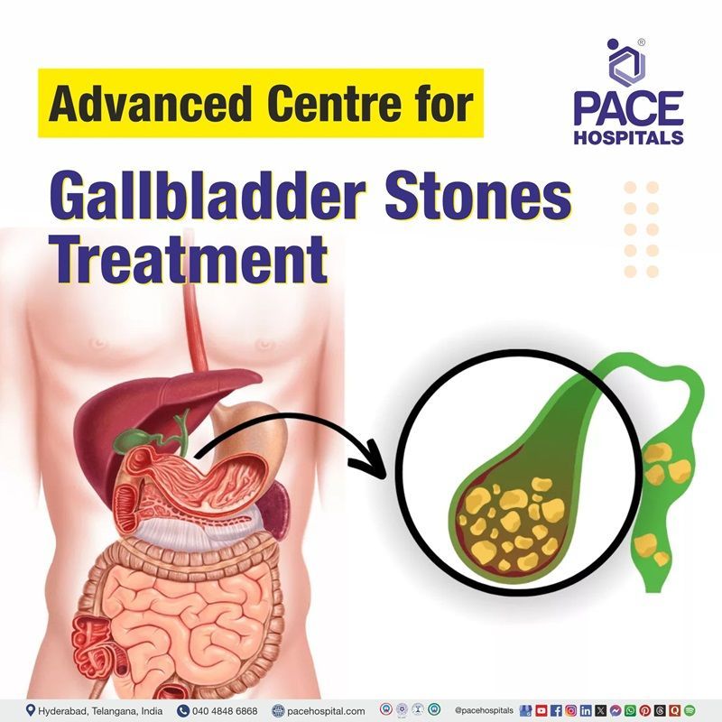
Advanced Diagnostic Facilities: Ultrasound Abdomen, CT Scan, MRCP, ERCP for Gallbladder & Biliary Tract Evaluation
Specialized Gallbladder Stone Treatment by Leading Gastroenterologists in Hyderabad
Advanced ICU Support with Laparoscopic Cholecystectomy & Bile Duct Exploration for Gallbladder Stone Care
Affordable & Transparent Gallbladder Stone Treatment at PACE Hospitals with Insurance & Cashless Options
Gallstones Diagnosis
The diagnosis of gallstones is often based on a combination of symptom history and physical examination findings, which help the gastroenterologist in concluding the diagnosis. Gallstones are managed depending on whether they cause noticeable symptoms such as abdominal pain, bloating, or nausea.
To determine the appropriate diagnostic approach, the gastroenterologist takes into account the following factors before selecting the tests to diagnose gallstones:
- Medical history
- Physical examination
Medical history
Medical history plays an important role in diagnosing gallstones by helping clinicians identify general symptoms and risk factors that suggest their presence.
- Patients usually report episodic right upper quadrant or epigastric abdominal pain, often described as severe and crampy, which occurs after eating fatty meals.
- This pain, known as biliary colic, may radiate to the back or right shoulder and can last from minutes to several hours. Associated symptoms such as fever, nausea, vomiting, or jaundice provide essential clues about potential complications like cholecystitis or bile duct obstruction.
- Gathering information about risk factors such as female sex, obesity, age, rapid weight loss, and certain medical conditions further supports the suspicion of gallstones.
- A detailed history also helps differentiate gallstone-related pain from other causes of abdominal discomfort. Thus, the medical history serves as a foundation for additional physical examination and diagnostic imaging, especially abdominal ultrasound, which supports the diagnosis.
Physical examination
The physical examination of gallbladder stones allows the doctor to examine for indicators of problems with the gallbladder directly.
- During the exam, the doctor gently presses on the abdomen, especially the upper right side, where the gallbladder is located. If pressing this area causes sudden pain or discomfort, it may indicate irritation or inflammation related to gallstones.
- The doctor also looks for other signs, such as tenderness, abdominal bloating, or muscle stiffness in that area. In some cases, yellowing of the skin or eyes (jaundice) may be noticed, which can occur if a gallstone blocks the bile duct.
- Although a physical examination alone cannot confirm the presence of gallstones, it provides important clues about the condition and helps the doctor determine whether further imaging tests, such as an ultrasound, are necessary. In simple words, the exam helps link the patient's pain and symptoms to possible gallbladder disease.
Diagnostic Tests for Gallstones
Based on the above information, a gastroenterologist advises diagnostic tests for gallstone to confirm their presence and evaluate for possible complications such as infection, obstruction, or inflammation.
The following tests might be recommended to diagnose gallstones and related conditions:
- Laboratory tests
- Complete blood count (CBC) with differential
- Liver function panel
- C-reactive protein (CRP)
- Amylase
- Serum bilirubin
- Alkaline phosphatase (ALP)
- Lipase
- Imaging studies
- Abdominal radiography
- Ultrasonography (USG)
- Computed tomography (CT)
- Magnetic resonance cholangiopancreatography (MRCP)
- Hepatobiliary iminodiacetic acid (HIDA) scan
- Endoscopic evaluations
- Endoscopic ultrasonography (EUS)
- Endoscopic retrograde cholangiopancreatography (ERCP)
- Other invasive imaging tests
- Percutaneous transhepatic cholangiography (PTC)
Laboratory tests
Laboratory tests are important for diagnosing gallstones because they help identify inflammation, infection, and bile duct blockage, as well as evaluate liver and pancreatic involvement, thereby aiding in accurate diagnosis and treatment. Lab tests for gallstones include the following:
- Complete blood count (CBC) with differential: A CBC checks the levels of white blood cells, red blood cells, and platelets. In gallstone disease, a rise in white blood cells often indicates infection or inflammation of the gallbladder (cholecystitis) or bile ducts (cholangitis). This helps doctors determine whether abdominal pain is associated with a disease.
- Liver function panel: This panel contains enzymes such as Aspartate Aminotransferase (AST), Alanine Aminotransferase (ALT), and gamma-glutamyl transferase (GGT). If gallstones obstruct bile flow, these enzymes increase, indicating liver stress or injury. This allows doctors to determine if the pain is caused by liver involvement or bile duct blockage. It also helps rule out primary liver diseases such as viral hepatitis or alcohol-related liver injury, since gallstones primarily cause obstruction, not direct liver inflammation.
- C-reactive protein (CRP): CRP is a sensitive marker of inflammation. When gallstones lead to infection of the gallbladder or severe inflammation in the bile ducts, CRP levels increase sharply. High CRP values point toward acute conditions, such as acute cholecystitis, rather than simple or silent gallstones. It also helps rule out mild, non-inflammatory causes of abdominal pain like indigestion or functional bowel problems.
- Amylase: The pancreas releases amylase to aid in the digestion of food. If gallstones block the pancreatic duct, enzymes like amylase rise in the blood. This suggests gallstone-induced pancreatitis, which can be a serious complication.
- Serum bilirubin: Bilirubin builds up in the blood when bile ducts are blocked. High bilirubin levels may cause yellowing of the eyes and skin (jaundice), which is common if gallstones obstruct the common bile duct. This test measures the extent to which bile flow is affected. It also helps differentiate between gallstone-related jaundice and jaundice caused by liver disease or breakdown of red blood cells (haemolytic anaemia).
- Alkaline phosphatase (ALP): Alkaline phosphatase is often elevated when gallstones block the bile ducts. It increases more specifically in cases of bile duct obstruction compared to other liver enzymes. If ALP is high, along with bilirubin, it strongly indicates a blockage in bile flow.
- Lipase: Lipase is another pancreatic enzyme, more specific than amylase. A rise in lipase strongly suggests pancreatitis, often caused by gallstones that enter and block the pancreatic duct. This test helps doctors confirm if the pancreas is involved in addition to gallbladder disease.
Imaging studies
Imaging studies are critical for accurately diagnosing gallstones, as they allow for the confirmation of their presence of gallstones, assessment of their location, and detection of complications. This includes:
- Abdominal radiography:Plain X-rays of the abdomen are sometimes utilized as an initial test, although their use in gallstone diagnosis is limited. Most gallstones are composed of cholesterol and are radiolucent (not visible on X-ray), which means only about 10–15% of stones that contain calcium can be seen. While not the most reliable tool for gallstones, radiography may help rule out other abdominal conditions or detect complications such as bowel obstruction caused by gallstones.
- Ultrasonography (USG): Ultrasound is the gold standard first-line test for suspected gallstones. It is non-invasive, widely available, inexpensive, and safe (no radiation exposure). USG can directly visualize gallstones within the gallbladder as echogenic foci that cast acoustic shadows. It can also detect gallbladder wall thickening, pericholecystic fluid, or sonographic Murphy's sign, all of which suggest acute cholecystitis. Additionally, ultrasound can detect bile duct dilatation if stones are obstructing the biliary tree. Ultrasound may miss very small stones or stones in the bile duct; hence, other diagnostic tests may be used.
- Computed tomography (CT): Computed tomography (CT) is valuable in identifying calcified stones, assessing the anatomy in complicated cases, and evaluating complications such as pancreatitis, perforation, abscess, or gangrenous cholecystitis. CT offers detailed cross-sectional imaging that is especially useful when US findings are inconclusive or when other intra-abdominal pathologies are suspected.
- Magnetic resonance cholangiopancreatography (MRCP): MRCP is a specialised MRI technique that provides excellent visualisation of the pancreatic and biliary ducts. It is beneficial for detecting stones within the common bile duct (choledocholithiasis) and for mapping out the biliary anatomy before surgery. MRCP has very high sensitivity and specificity for biliary tract obstruction, making it the preferred non-invasive tool when ultrasound cannot conclusively detect bile duct stones.
- Hepatobiliary iminodiacetic acid (HIDA) scan : This procedure is primarily used to assess gallbladder function and biliary patency rather than to visualise gallstones directly. It is beneficial when acute cholecystitis is suspected, but the USG is inconclusive. In the presence of a gallstone obstructing the cystic duct, the radiotracer will not enter the gallbladder, resulting in non-visualization of the organ, a hallmark finding in acute cholecystitis. A HIDA scan can also assess gallbladder ejection fraction and diagnose functional disorders or biliary leaks following surgery.
Endoscopic evaluation
Patient with symptoms of right upper quadrant pain and abnormal liver function tests (LFTs). Endoscopic evaluation is indicated to assess for choledocholithiasis (common bile duct stones) as a possible cause.
The following are the endoscopy methods for gallstone diagnosis:
- Endoscopic Ultrasonography (EUS): EUS is a minimally invasive technique that combines endoscopy with ultrasound, allowing detailed images of the gallbladder and bile ducts from inside the stomach or duodenum. It is highly accurate for detecting even small gallstones and common bile duct stones, especially when other imaging tests are inconclusive.
- Endoscopic Retrograde Cholangiopancreatography (ERCP): ERCP involves inserting an endoscope into the duodenum and injecting contrast to visualize the bile ducts under X-ray. It is mainly used for both diagnosis and treatment. Stones in the bile duct can be directly removed during the procedure.
Other invasive imaging tests
This imaging test may be indicated for the diagnosis of gallstones when non-invasive methods are inconclusive or when intervention is planned. This includes:
Percutaneous Transhepatic Cholangiography (PTC)
PTC is done by inserting a needle through the liver into the bile ducts and injecting contrast to outline the biliary system. It helps diagnose stones causing obstruction and can also be used to remove stones or place drainage tubes if ERCP is not possible.
Stages of Gallstone Formation
Gallstone disease is often described in stages, based on whether stones are present, whether they cause symptoms, and if complications develop. They include the following:
- Lithogenic state (pre-stone stage)
- Asymptomatic gallstones (Silent stage)
- Symptomatic gallstones (Biliary colic stage)
- Complicated cholelithiasis
Stage 1: Lithogenic state (pre-stone stage)
The lithogenic state is the earliest phase in gallstone formation. At this stage, bile becomes abnormal in composition, usually containing too much cholesterol or too few bile salts. This imbalance makes bile prone to crystallisation. Although no stones are present yet, microscopic crystals may begin to form. The lithogenic state itself produces no symptoms and is often undetectable on routine imaging.
Stage 2: Asymptomatic gallstones (silent stage)
In this stage, actual gallstones are present but do not cause pain or digestive symptoms. Most cases were discovered incidentally during imaging for other reasons. About 70–80% of people with gallstones remain in this stage throughout life without developing complications. Because the risk of progression is low, no treatment is needed unless specific high-risk features are identified, such as very large stones or certain underlying conditions.
Stage 3: Symptomatic gallstones (biliary colic stage)
When gallstones temporarily obstruct the cystic duct, they cause pain and discomfort known as biliary colic. This ache is commonly felt in the upper right abdomen, especially after consuming fatty foods, and can persist for minutes to hours. Once symptoms occur, recurrence is common, and surgical removal of the gallbladder is generally recommended.
Stage 4: Complicated cholelithiasis
At this stage, gallstones persistently block ducts or move into the common bile duct, and they may cause serious complications. These include acute cholecystitis (inflammation of the gallbladder), gallbladder stone pancreatitis (inflammation of the pancreas due to a stone blocking the duct opening), choledocholithiasis (stones in the common bile duct), and cholangitis (infection of the bile ducts). These conditions require urgent medical or surgical treatment.
Gallstones Differential Diagnosis
The gallstones symptoms, especially upper abdominal pain, nausea, vomiting, and jaundice, are not unique and can overlap with many other medical conditions. As a result, doctors consider a wide range of possible causes before confirming gallstone disease. Evaluating these different conditions helps avoid misdiagnosis and ensures the correct treatment. Below are some important conditions that can mimic or resemble gallbladder stone symptoms:
- Appendicitis: It can mimic gallstone pain because both may cause abdominal pain. However, appendicitis usually starts around the belly button and shifts to the lower right abdomen, whereas gallstone pain is more in the upper right abdomen.
- Renal calculi (kidney stones): These can cause severe flank or abdominal pain similar to gallstones. The pain often radiates to the groin, and blood may be found in urine, which helps distinguish it from gallstones.
- Cholangiocarcinoma: This is cancer of the bile ducts. It can produce symptoms similar to gallstones, such as jaundice, abdominal pain, and bile duct blockage. Imaging and biopsy help separate it from gallstone disease.
- Pancreatitis: This condition is often caused by gallstones themselves, but may also occur independently. Both can cause upper abdominal pain and nausea. High blood amylase or lipase helps confirm pancreatitis rather than simple gallstones.
- Peptic ulcer disease: This may cause upper abdominal pain that resembles gallstone pain. Unlike gallstones, the pain often improves with food or antacids and may be associated with stomach burning or bleeding.
- Gastroesophageal reflux disease (GERD): This can cause burning pain in the chest or upper abdomen, sometimes mistaken for gallstone discomfort. The pain is often related to lying down or meals and improves with acid-reducing medicines.
- Myocardial infarction (heart attack): This can sometimes present with upper abdominal or chest pain similar to gallstones. However, heart pain may radiate to the arm, neck, or jaw and is confirmed with ECG and cardiac enzymes.
- Aortic dissection: This is a tear in the aorta that can cause sudden, severe chest or abdominal pain. It may mimic gallstone pain, but it usually has a tearing quality and requires urgent imaging to confirm.
- Pneumonia: This lung infection, especially in the right lower lobe, can cause pain in the upper right abdomen. Fever, cough, and abnormal chest X-ray findings help distinguish it from gallstones.
- Esophageal spasm: This can cause severe chest pain that feels like gallstone pain. The difference is that esophageal spasm is often triggered by swallowing and relieved by relaxation or vasodilators.
- Hepatitis: This liver inflammation can produce upper abdominal discomfort and jaundice, similar to gallstone disease. Blood tests showing elevated liver enzymes help point to hepatitis rather than gallstones.
- Mesenteric ischemia: This occurs when blood flow to the intestines is reduced, causing severe abdominal pain out of proportion to examination findings. It can be mistaken for gallstones, but risk factors and CT angiography help in diagnosis.
- Gastroenteritis: This causes abdominal pain along with diarrhea and vomiting. It may resemble gallstone attacks, but the presence of fever, infection signs, and rapid recovery often indicates gastroenteritis instead.
Considerations of a gastroenterologist before treating gallstones
Before treating gallstones, a gastroenterologist considers several important factors. Those are:
Symptomatology and clinical presentation: A key consideration is the presence of symptoms; mostly asymptomatic gallstones are not treated, while symptomatic gallstones, such as those causing biliary colic, acute cholecystitis, pancreatitis, or choledocholithiasis, generally require definitive intervention.
- Risk stratification: Certain high-risk groups may need proactive treatment, including those with large gallstones, porcelain gallbladder, haemolytic anaemia, or rapid weight loss, due to increased risk of complications.
- Patient’s overall health and surgical risk: Patient comorbidities like heart, lung, or liver disease may increase surgical risk, making non-surgical or endoscopic options preferable in high-risk cases.
- Types and location of stones: Differentiating cholelithiasis from choledocholithiasis is important, as gallbladder stones are treated with laparoscopic cholecystectomy, while bile duct stones require ERCP.
- Choice of treatment approach: Laparoscopic cholecystectomy is the gold standard for symptomatic gallstones. ERCP is used for common bile duct stones, offering both diagnosis and treatment. In patients who cannot undergo surgery, medical therapy, like a gallstone solubilizing agent, may be used.
- Urgency and complications: At last, the urgency of intervention depends on the presence of complications. Acute cholecystitis, cholangitis, or gallstone pancreatitis often require urgent or early treatment, usually within hours to days. In cases of stable biliary colic, treatment can be planned electively without immediate risk.
Gallstones Treatment Goals
The goals of gallstone treatment are:
- Relieve symptoms: To relieve symptoms such as pain and other symptoms caused by gallstones.
- Prevent complications: To avoid the development of severe conditions such as acute cholecystitis, gallstone pancreatitis, and choledocholithiasis.
- Prevent recurrence: To prevent the gallstones from forming again after treatment, especially after medication-based treatments.
- Reduce risk of gallbladder cancer: In some high-risk cases, treatment can help prevent the development of gallbladder cancer.
A gastroenterologist may choose the gallbladder stone treatment depending on the presence of symptoms, stone location, and patient risk factors. Several options are available for managing symptomatic gallstones. The most common interventions include:
- Non-pharmacological management
- Watchful waiting
- Lifestyle modifications
- Supportive care in acute attacks
- Pharmacological management
- Oral dissolution therapy
- Antibiotic prophylaxis
- Adjunct medications
- Extracorporeal shock wave lithotripsy (ESWL)
- Surgical management
- Elective laparoscopic cholecystectomy
- Open cholecystectomy
- Endoscopic management
- Endoscopic retrograde cholangiopancreatography (ERCP)
Non-pharmacological management / asymptomatic gallstones management
Non-pharmacological management is usually used for asymptomatic gallstones, where intervention is not immediately required. Most cases are managed with the following:
Watchful waiting
Many patients with gallstones are asymptomatic. In such cases, watchful waiting is generally recommended. This avoids unnecessary surgery and focuses on patient monitoring. Regular check-ups/ monitoring ensure that if signs such as jaundice, stomach pain, or problems arise, prompt treatment can be arranged.
Lifestyle modifications
Certain lifestyle modifications help reduce the risk of gallstone formation and ease symptoms in those with mild disease. These include maintaining a healthy body mass index (BMI), gradual weight loss rather than crash dieting, eating a balanced diet with less saturated fat, and increasing fibre intake. Such measures improve bile composition and gallbladder function, thereby reducing the likelihood of stone growth or recurrence.
Supportive care in acute gallstone attack
During acute biliary colic or gallstone complications such as cholecystitis, supportive care is essential for managing pain and inflammation and for preventing complications.
- Pain control with analgesics.
- Hydration and electrolyte balance maintenance.
- Antibiotics if infection is suspected.
- Fasting or dietary restrictions during acute episodes to reduce biliary stimulation.
Pharmacological management / medication for gallstones
Pharmacological management is considered for patients who are not suitable candidates for gallstone treatment without surgery. Gallbladder stone medication, such as:
Oral dissolution therapy
Oral bile acids or gallstone dissolution agents are used to treat cholesterol gallstones in patients who are not fit for surgery or prefer medical management. These drugs work by reducing cholesterol saturation in bile, which gradually dissolves cholesterol gallstones. They achieve this by inhibiting cholesterol absorption in the intestine, decreasing cholesterol secretion into bile, and increasing bile acid flow, which dilutes toxic hydrophobic bile acids in the bile ducts. This mechanism helps to restore a healthier bile composition and reduce stone formation or promote dissolution over time.
Adjunct medications
Adjunct medications include the following drugs to reduce pain and prevent complications. These are:
- Analgesics: Nonsteroidal anti-inflammatory drugs (NSAIDs) are generally used to alleviate pain during biliary colic episodes caused by gallstone blockage. They reduce inflammation and provide effective symptomatic control.
- Antibiotic prophylaxis: Not routinely required for low-risk patients undergoing elective laparoscopic cholecystectomy but recommended selectively for high-risk groups.
- Antispasmodics: These help relieve spasms of the biliary tract muscles, reducing pain and discomfort during colic episodes by relaxing smooth muscles of the gallbladder and bile ducts.
Extracorporeal shock wave lithotripsy (ESWL)
Gallstones can be blasted into tiny fragments by a gastroenterologist using shock wave lithotripsy. Doctors rarely use this procedure, and occasionally it is combined with gallstone dissolution agents.
Surgical management/ gallstone surgery
Surgery is the definitive and most effective treatment for symptomatic gallstones. Unlike medical therapy, which may only dissolve or control symptoms, surgery removes the entire gallbladder, preventing recurrence of stones and further complications. Gallbladder stone surgery includes:
Elective laparoscopic cholecystectomy
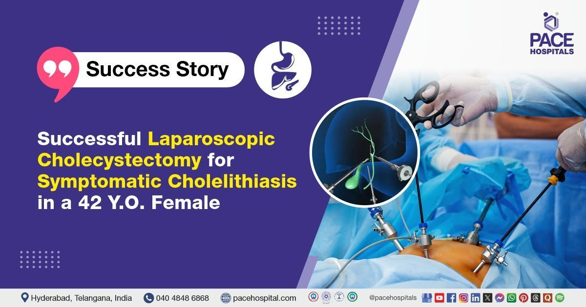
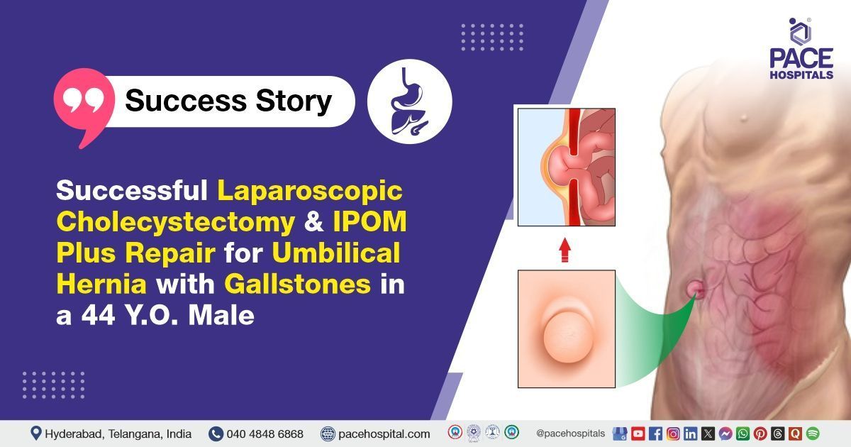
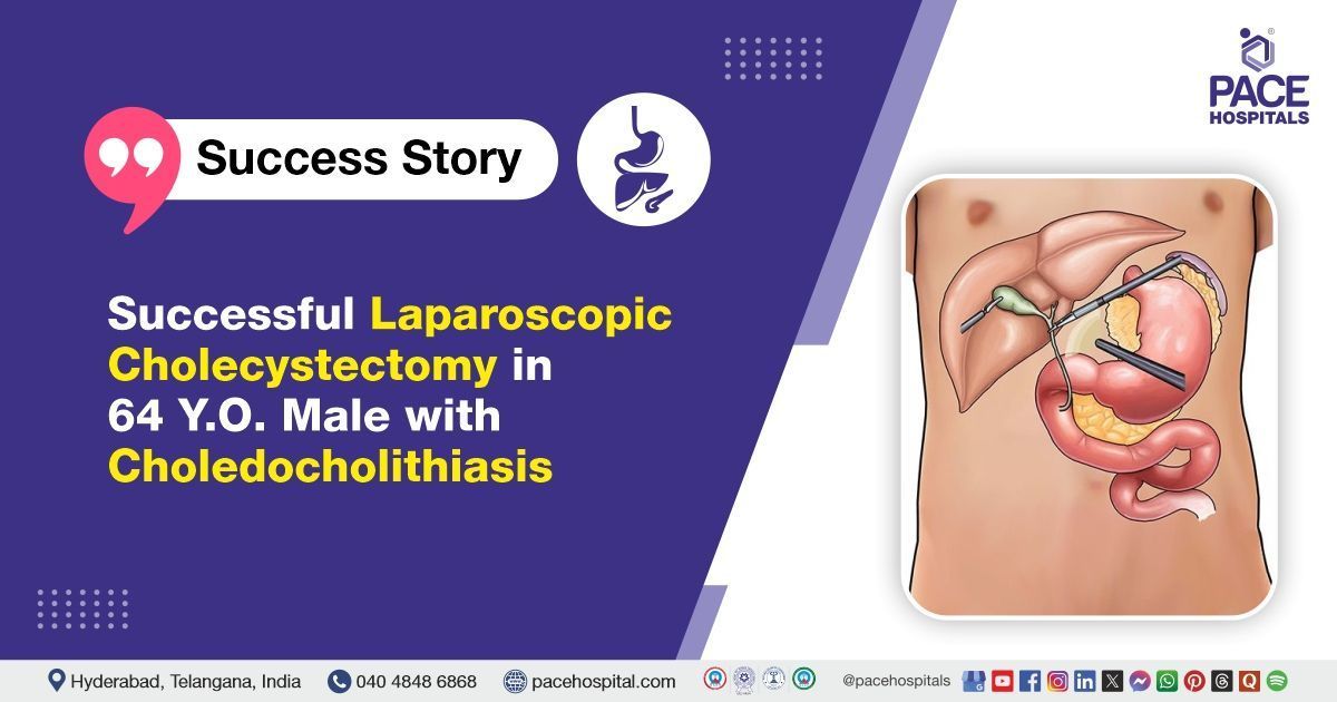
The most common surgical treatment for gallstones is laparoscopic cholecystectomy. It involves removing the gallbladder through small incisions using a camera and specialized instruments. This procedure is minimally invasive, associated with less pain, faster recovery time, shorter hospital stay, and lower complication rates compared to open surgery. It is recommended as the first-line surgical option for most patients with symptomatic gallstones or gallstone complications.
Open cholecystectomy
Open cholecystectomy involves removing the gallbladder through a larger abdominal incision. It is usually reserved for cases where laparoscopic surgery is not possible, such as patients with substantial scarring from previous procedures, severe inflammation, anatomic abnormalities, or probable gallbladder cancer. Although recovery takes longer and postoperative discomfort is greater, open cholecystectomy remains an important option in complex or high-risk situations.
Endoscopic management
Endoscopic management provides minimally invasive approaches for diagnosing and treating gallstones, particularly those in the bile ducts. Gallstone removal without surgery includes the following:
Endoscopic retrograde cholangiopancreatography (ERCP)
In ERCP, gallstones in the common bile duct are removed by first performing a sphincterotomy (cutting the bile duct opening), followed by extraction using tools like balloon catheters or retrieval baskets. A stent may be placed if necessary to maintain bile flow.
Gallstones Prognosis
The prognosis of gallstones is generally favourable. Most people with gallstones remain asymptomatic throughout life, and fewer than 50% develop symptoms or complications. Symptomatic gallstones can cause pain and digestive issues, but surgery (laparoscopic cholecystectomy) usually provides lasting relief with very low risk.
Mortality after elective laparoscopic cholecystectomy is less than 0.5%, though it rises in emergency cases. Long-term outcomes are excellent, with most patients maintaining normal life expectancy and quality of life. Rarely, complications like bile duct injury or infection may affect recovery.
Gallbladder Stone Surgery Cost in Hyderabad, India
The cost of Gallbladder Stone Treatment in Hyderabad generally ranges from ₹28,000 to ₹1,10,000 (approx. US $335 – US $1,325).
The exact treatment cost varies depending on factors such as whether the stones are single or multiple, presence of inflammation or infection (cholecystitis), whether laparoscopic or open surgery is performed, need for ERCP (if stones migrate to the bile duct), surgeon expertise, hospital stay duration, and hospital facilities — including cashless treatment options, TPA corporate tie-ups, and assistance with medical insurance wherever applicable.
Cost Breakdown According to Type of Gallbladder Stone Treatment
- Medical Management for Mild/Asymptomatic Stones – ₹5,000 – ₹12,000 (US $60 – US $145)
- Laparoscopic Cholecystectomy (Gallbladder Removal Surgery) – ₹38,000 – ₹75,000 (US $455 – US $905)
- Open Cholecystectomy – ₹45,000 – ₹85,000 (US $540 – US $1,025)
- ERCP for CBD Stone Removal (If Stones Enter the Bile Duct) – ₹25,000 – ₹55,000 (US $300 – US $660)
- Laparoscopic Cholecystectomy + ERCP (Combined Treatment) – ₹65,000 – ₹1,10,000 (US $780 – US $1,325)
Frequently Asked Questions on Gallbladder Stones Treatment
What are the common gallstones symptoms that female patients commonly present with?
Women with gallstones often feel sudden pain in the upper right part of the stomach, which may spread to the back or right shoulder. They may also experience nausea, vomiting, and occasionally fever or yellowing of the skin (jaundice). These symptoms usually happen after eating fatty foods. Women are more likely than men to get gallstones due to hormonal changes, especially during pregnancy.
Which Is the best hospital for Gallstones Treatment in Hyderabad, India?
PACE Hospitals, Hyderabad, is one of the leading centres for gallbladder stone management, offering advanced surgical and endoscopic treatment for cholelithiasis and its complications.
We have highly experienced gastroenterologists, HPB surgeons, and laparoscopic surgeons specialised in minimally invasive gallbladder removal, bile duct stone extraction, and treatment of acute gallbladder infections with high precision and safety.
Our team of experts deal with complicated cases and bring outstanding results with state-of-the-art modular operation theatres, advanced laparoscopic systems, ERCP facilities, dedicated recovery units, and comprehensive patient support, PACE Hospitals ensures safe, effective, and patient-focused gallstone treatment — supported by cashless insurance options, TPA corporate tie-ups, and assistance with medical insurance documentation.
How to prevent gallstones?
Gallstones can be prevented through healthy lifestyle choices. A balanced diet rich in fruits, vegetables, and fibre, and low in saturated fats, helps reduce the risk. Regular physical activity and maintaining a healthy weight are essential because both rapid weight loss and being overweight increase the risk of stones. Drinking enough water and avoiding long fasting periods also helps. For high-risk individuals, doctors may recommend medications to lower the chances of gallstone formation.
Is it necessary to remove the gallbladder for gallstones?
Gallbladder removal is not always necessary for gallstones. Many people have stones without symptoms and do not require surgery.Gallbladder removal, known as cholecystectomy, is advised when gallstones cause recurring pain, inflammation, or complications such as infection or blockage. Surgery is the most effective way to prevent future attack. For individuals without symptoms or those with a high surgical risk, doctors may recommend monitoring or non-surgical treatments. The decision depends on symptoms, overall health, and risks.
Do gallstones cause jaundice?
Yes, gallstones can cause jaundice if they block the bile duct, preventing bile from reaching the intestines. This blockage causes a buildup of bile pigments in the blood, which then results in the yellowing of the skin and eyes, known as jaundice. This is a warning sign that urgent treatment may be needed to unblock bile flow.
What Is the Cost of Gallbladder Stone Treatment at PACE Hospitals, Hyderabad?
At PACE Hospitals, Hyderabad, the cost of gallbladder stone treatment typically ranges from ₹26,000 to ₹95,000 and above (approx. US $310 – US $1,145), making it an affordable and reliable option for gallstone management. However, the final cost depends on:
Whether stones are symptomatic or causing complications
Type of treatment (medical management / ERCP / laparoscopic surgery)
- Whether bile duct stones are present (choledocholithiasis)
- Surgeon expertise and laparoscopic technology used
- Duration of hospital stay and anesthesia requirements
- Diagnostic tests (USG abdomen, LFT, blood tests, MRCP if required)
- Medications, consumables, and postoperative care
For uncomplicated gallstone removal via laparoscopic surgery, costs fall at the lower end; cases requiring ERCP, open surgery, or management of severe infection fall toward the higher range.
After a detailed gastroenterology evaluation and imaging review, our specialists will provide a personalised treatment plan and transparent cost estimate tailored to your medical needs.
Can gallstones affect the kidneys?
Yes, gallstones can affect the kidneys, both by increasing the risk of developing kidney stones (nephrolithiasis) and, in rare cases, through complications like acute pancreatitis, leading to fluid accumulation that stresses the kidneys. Gallstones and kidney stones have a bidirectional association, meaning having one type of stone increases the risk of forming the other. Additionally, conditions like gallstone pancreatitis can cause severe dehydration, which may lead to kidney problems.
What are gallstones, and what causes gallstones?
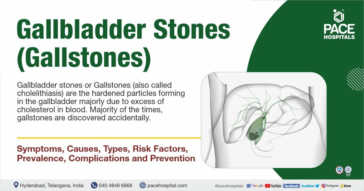
Gallstones are small, hard deposits formed in the gallbladder, a small organ that stores bile for digestion. They occur when substances in bile, primarily cholesterol and bilirubin, become imbalanced and form crystals. The primary causes of gallstones include high levels of cholesterol in bile, poor gallbladder emptying, bacterial infections, certain diseases such as diabetes and obesity, genetic factors, and alterations in gut bacteria. These factors cause bile components to clump and form stones over time.
Why is the gallbladder removed instead of removing the stones?
The gallbladder is removed because simply taking out the stones doesn’t prevent new ones from forming, and the organ itself may be inflamed or not functioning well. Removing it completely solves the problem, and the body can digest food normally without it.
Which size of gallbladder stone is dangerous?
The size of a gallstone can influence the risk of complications. Tiny stones (less than 5 mm) can slip into the bile ducts and cause blockages, leading to severe pain, jaundice, or pancreatitis. On the other hand, very large stones (over 3 cm) increase the chance of gallbladder cancer in rare cases. Stones of any size can cause pain or infection, but extremes of very small or very large are considered more dangerous.
What is the recovery time for gallstone surgery?
Recovery time varies with the type of surgery. Most people heal within 1–2 weeks after laparoscopic (keyhole) surgery, whereas open surgery may require 4–6 weeks. Patients can slowly return to normal activities, and digestion remains normal because bile moves directly from the liver to the intestine.
How is gallstone ileus diagnosed and treated in clinical practice?
Gallstone ileus is a rare condition in which a gallstone becomes lodged in the intestine, causing pain, vomiting, and constipation. Doctors diagnose it using X-rays or CT scans that show the stone and signs of blockage. Treatment usually involves surgery to remove the stone and fix the blockage. Sometimes, part of the intestine or gallbladder might need to be removed if there is severe damage.
What are the types of gallstones and their underlying causes?
The two main types of gallstones are cholesterol stones, pigment stones, and mixed stones. Cholesterol stones form when there is a large amount of cholesterol in bile, often due to a diet high in cholesterol or obesity. Pigment stones form from a substance called bilirubin, which is made when blood cells break down. Mixed stones are made of cholesterol and salts. Certain diseases, such as liver problems or blood disorders, can cause changes in pigment tone. Genetics can also play a role in the formation of stones.
What are the complications of gallstones?
Complications include blockage of bile ducts, which can cause pain, infection, and inflammation of the gallbladder (cholecystitis). Blockage of the pancreatic duct can lead to pancreatitis. Gallstones can also cause jaundice, which is characterized by yellowing of the skin and eyes, if bile flow is obstructed. In rare cases, stones may lead to an intestinal blockage known as gallstone ileus. If untreated, these issues can become serious and may require surgery.
Can gallstones be dissolved?
Certain gallstones, especially those made of cholesterol, can sometimes be dissolved using medicine containing bile acids taken over months. This works slowly and is usually for patients who cannot or prefer not to have surgery. Other options include shock wave therapy to break stones, but these treatments are less common than surgery, and stones may recur. Dissolving is not effective for all types of stones.
Can gallstones cause cancer?
Gallstones themselves do not directly cause cancer, but having long-term gallstones or inflammation in the gallbladder can increase the risk of developing gallbladder cancer over time. Chronic irritation from stones may lead to changes in the gallbladder lining. However, gallbladder cancer is rare and occurs mostly in older adults or those with a long history of gallstone disease.
Can gallstones cause gas?
Yes, gallstones can sometimes cause gas and bloating. When stones block the normal flow of bile, the digestion of fatty foods becomes less efficient. This can lead to abdominal fullness, belching, and excess gas, especially after heavy or greasy meals. While gas alone is not a specific sign of gallstones, when combined with upper abdominal pain, nausea, or discomfort after eating, it may suggest gallbladder problems that need medical evaluation.
Can gallbladder stones cause weight loss?
Gallbladder stones themselves do not directly cause weight loss. However, people with stones may avoid eating fatty foods due to pain, nausea, or indigestion, which can lead to unintentional weight loss over time. In more serious cases, complications like infection of the gallbladder, bile duct blockage, or pancreatitis can reduce appetite and contribute to weight loss. A doctor needs to continually evaluate rapid, unexplained weight loss, as it may indicate underlying complications or other health issues.
What is the survival rate for people with gallstones?
Gallstones themselves generally do not directly affect survival, as most people with gallstones lead normal lives without serious complications. However, complications like gallbladder cancer or severe infections related to gallstones can impact survival. For example, gallbladder cancer has an overall 5-year relative survival rate of about 67% when localised, but this drops significantly if the cancer spreads. Most gallstone-related surgeries, like cholecystectomy, have low mortality risks, generally under 1%. Thus, gallstones alone do not reduce survival; however, associated complications may affect outcomes if left untreated.
How to test for gallstones at home?
There are no tests to detect gallbladder stones at home. A hepatologist or gastroenterologist may be consulted in case of any potential symptoms of gallbladder stones. They will be able to advise you on the right diagnostic tests to confirm if you have gallbladder stones.
What is the Mirizzi syndrome?
Mirizzi syndrome is a common hepatic duct obstruction due to the extrinsic compression arising from a stone impacted in the cystic duct or infundibulum of the gallbladder. Patients suffering from Mirizzi syndrome have jaundice, fever, and right upper abdominal pain.
What is the difference between kidney stones and gallbladder stones?
Gallbladder stones (gallstones) are hardened deposits of digestive fluid that form in the gallbladder, while kidney stones are solid masses formed from crystals in the urine within the kidneys. Gallstones can block bile flow and cause upper abdominal pain, whereas kidney stones obstruct urine flow, leading to flank pain. Their composition, location, symptoms, and treatments differ significantly.
What are gallbladder stones made of?
Gallbladder stones are primarily composed of cholesterol, bilirubin, and calcium salts, with smaller amounts of protein and other materials. There are three main types: pure cholesterol stones (at least 90% cholesterol), pigment stones (mainly bilirubin and calcium salts), and mixed stones containing varying proportions of cholesterol, bilirubin, and calcium compounds. In Western populations, cholesterol stones are most common, while pigment stones are more frequent in certain medical conditions.
Resources
