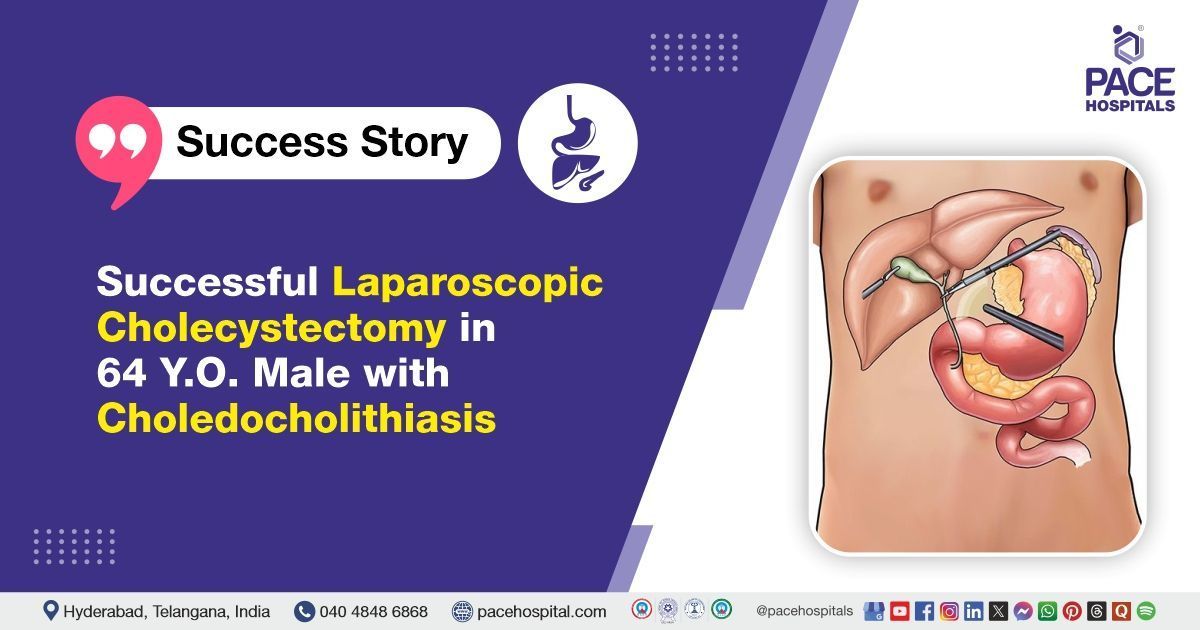
Successful Lap Cholecystectomy in 64 Y.O. Male with Choledocholithiasis
PACE Hospitals
PACE Hospitals’ expert Gastroenterology team successfully performed a laparoscopic cholecystectomy on a 64-year-old male patient with Cholelithiasis and choledocholithiasis, the procedure aimed to enhance the patient’s overall functional recovery and quality of life, ultimately relieving his symptoms and further improving his quality of life.
Chief Complaints
A 64-year-old male patient with a
Body Mass Index (BMI) of 23 presented to the gastroenterology Department at
PACE Hospitals, Hitech City, Hyderabad, for an elective laparoscopic cholecystectomy.
Past History
The patient was a known case of
hypertension and had been on regular antihypertensive medication. He also had a history of undergoing
endoscopic retrograde cholangiopancreatography (ERCP) with common bile duct (CBD) stenting in the past for
choledocholithiasis. These prior interventions were considered during the preoperative evaluation and planning to ensure optimal surgical outcomes and minimize procedural risks.
On Examination
On admission to PACE Hospitals, the patient was hemodynamically stable, with stable vital signs. General examination revealed that he was conscious, coherent, and cooperative. Cardiovascular and respiratory assessments were unremarkable, with normal heart sounds (S1, S2) and clear bilateral lung fields on auscultation, indicating no signs of cardiovascular or respiratory distress.
Diagnosis
Upon admission, the patient underwent a comprehensive clinical evaluation, along with a detailed review of his prior medical records and diagnostic reports by the Gastroenterology team.
The diagnosis of cholelithiasis (gallstones) with choledocholithiasis (stones in the bile duct) in this patient had been previously established based on a combination of clinical symptoms, laboratory investigations, and imaging studies. The patient presented with recurrent right upper quadrant abdominal pain, which raised clinical suspicion of biliary tract disease. Laboratory tests revealed elevated liver enzymes, including Alanine Transaminase (ALT), Aspartate Transaminase (AST), Alkaline phosphatase (ALP), and Gamma-glutamyl transferase (GGT), along with increased total and direct bilirubin levels, indicating possible biliary obstruction.
Abdominal ultrasonography had demonstrated the presence of gallstones and a dilated common bile duct. This was further confirmed by magnetic resonance cholangiopancreatography (MRCP), which provided detailed visualization of the biliary tree and showed stones within the common bile duct. The patient had also undergone endoscopic retrograde cholangiopancreatography (ERCP) with common bile duct stenting, during which choledocholithiasis was directly visualized and managed.
Based on the confirmed diagnosis, the patient was advised to undergo Cholelithiasis Treatment in Hyderabad, India, under the expert care of the Gastroenterology Department.
Medical Decision Making
After a thorough consultation with Dr. Govind R Verma consultant gastroenterologist and cross consultations with, Dr. Suresh Kumar S, a comprehensive evaluation was carried out to determine the most appropriate diagnostic and therapeutic approach for the patient.
Based on their expert assessment, it was concluded that a laparoscopic cholecystectomy would be the most effective procedure to address the patient’s condition.
Surgical Procedure
Following the decision, the patient was scheduled to undergo laparoscopic cholecystectomy in Hyderabad at PACE Hospitals, under the expert supervision of the gastroenterology Department.
Before undergoing the surgery, informed consent was obtained after thoroughly explaining the procedure and confirming the patient’s full understanding.
During the procedure, the following steps were carried out and findings observed:
- Preoperative Preparation: The patient was evaluated under general anesthesia, and standard preoperative protocols were followed. His previous ERCP and CBD stenting history were considered.
- Port Placement: Pneumoperitoneum was created using a Veress needle, followed by insertion of four laparoscopic ports.
- Gallbladder Exposure: The gallbladder was visualized and retracted to expose Calot’s triangle.
- Dissection: Careful dissection was performed to identify and isolate the cystic duct and cystic artery.
- Clipping and Division: The cystic duct and artery were clipped and divided safely.
- Gallbladder Removal: The gallbladder was dissected off the liver bed using electrocautery and removed through the umbilical port.
- Inspection and Irrigation: The operative field was inspected for bleeding or a bile leak, and irrigation was done as needed.
- Port Closure: All ports were removed under vision, and port sites were closed appropriately.
The surgery was successfully completed without any complications. Afterward, the patient was closely monitored to support a stable recovery and to watch for any issues at the surgical site.
Postoperative Care
The procedure was completed without complications, and the patient remained stable throughout the surgery. Postoperatively, he received intravenous fluids, antibiotics, and supportive care to promote healing and prevent infection. He showed steady improvement and was discharged in a hemodynamically stable condition with appropriate follow-up plans.
Discharge Medications
Upon discharge, the patient was prescribed oral antibiotics to prevent infection, analgesics for pain relief, proton pump inhibitors for gastric protection, and was advised to continue his regular antihypertensive medications to control blood pressure. He was advised to follow the medication schedule and attend follow-up visits.
In addition, other supportive treatments were provided to aid recovery and ensure proper healing post-surgery. These medications were tailored to the patient’s condition, addressing both the surgical recovery and the need for infection prevention and symptom management.
Emergency Care
The patient was informed to contact the Emergency ward at PACE Hospitals in case of any emergency or development of symptoms such as pain in the abdomen, fever, and vomiting.
Review and Follow-Up
The patient was advised to schedule a follow-up appointment with the Gastroenterologist in Hyderabad at PACE Hospitals, two weeks after discharge for planning CBD (common bile duct) stent removal. During this visit, the patient’s recovery would be evaluated to determine the need for any further management.
Conclusion
This case highlights the effectiveness of laparoscopic cholecystectomy in performing cholelithiasis Treatment in Hyderabad, India, enabling successful removal of the gall bladder, thereby facilitating normal function.
Significance of ultrasonography in diagnosis and management of cholelithiasis with choledocholithiasis
Ultrasonography is a vital, non-invasive imaging technique commonly used to diagnose and manage gallstones and stones in the common bile duct. It provides detailed images of the gallbladder, bile ducts, and surrounding structures, allowing detection of stones and signs of bile duct obstruction. Characteristic ultrasound features, such as the wall-echo-shadow sign and echogenic foci, help confirm the presence of gallstones. Visualization of stones within the bile duct and dilation of the ducts are strong indicators of choledocholithiasis. These findings enable gastroenterologist / gastroenterology doctor, to accurately diagnose the condition, evaluate complications, and plan appropriate treatment.
Share on
Request an appointment
Fill in the appointment form or call us instantly to book a confirmed appointment with our super specialist at 04048486868
Appointment request - health articles
Recent Articles