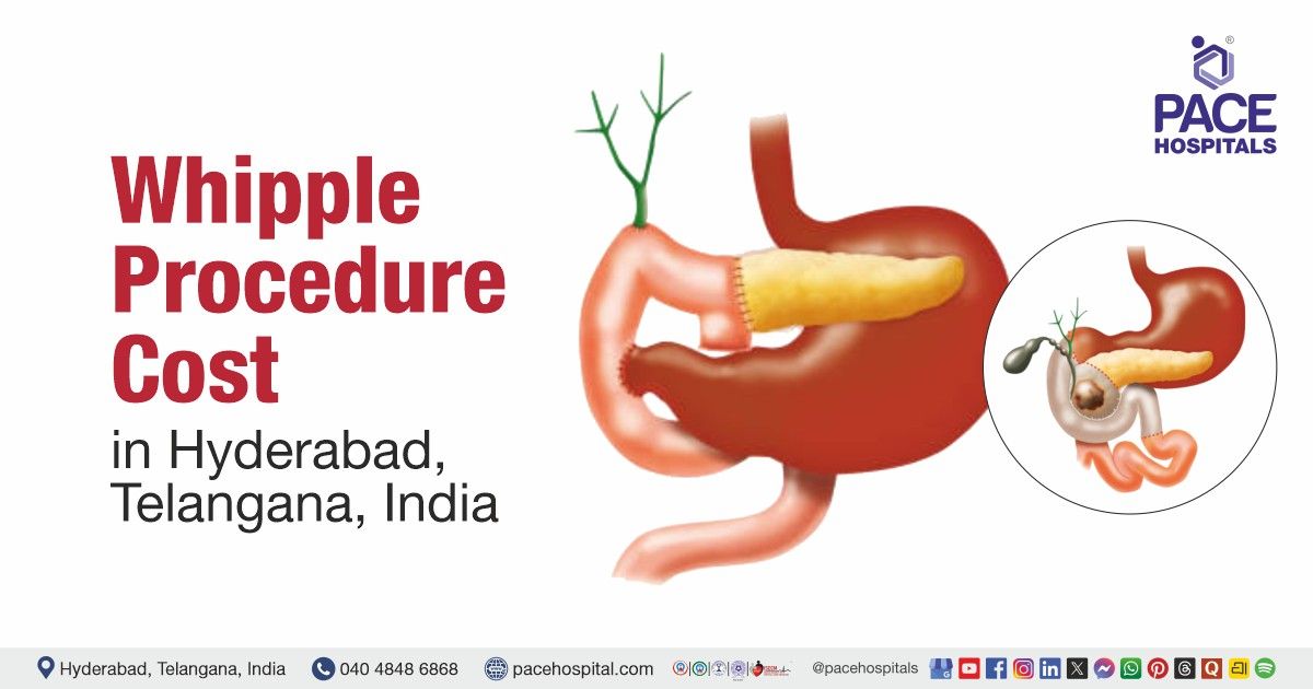
MIPPO Surgery for Left Distal Fibula Fracture in a 53-Year-Old Male
PACE Hospitals
The expert Orthopaedic team at PACE Hospitals successfully performed a Closed Reduction and Internal Fixation using Minimally Invasive Percutaneous Plate Osteosynthesis (MIPPO) with a Titanium Plate and Screws (PDL) on a 53-year-old male patient with a history of blunt trauma and persistent pain in the left ankle region. The procedure was carried out to alleviate his pain, restore mobility, and promote functional recovery.
Chief Complaints
A 53-year-old male patient with a
Body Mass Index (BMI) of 22.3 presented to the Orthopaedics Department at
PACE Hospitals, Hitech City, Hyderabad, with complaints of pain in the left ankle region. He reported a history of blunt trauma to the area prior to the onset of symptoms. Clinical examination revealed localized tenderness and mild swelling over the lateral aspect of the left ankle. The patient denied any previous similar episodes or underlying chronic joint conditions.
Past Medical History
The patient had no known allergies or significant comorbidities. There was no history of chronic illnesses such as
diabetes,
hypertension, or
cardiovascular disease, and he was not on any long-term medications. His overall health status was stable, with no underlying conditions that were likely to affect his treatment or recovery.
On Examination
Upon admission to PACE Hospitals, the patient's vital signs were stable. Examination of the left ankle revealed a visible deformity, palpable crepitus, and tenderness over the lateral malleolar region. Abnormal mobility was noted, and the range of motion at the ankle joint was significantly restricted and painful. No distal neurovascular deficits were identified, indicating preserved circulation and nerve function.
Systemic examination revealed normal findings. The respiratory system showed bilateral air entry, the cardiovascular system had audible S1 and S2 heart sounds, the abdomen was soft and non-tender, and the central nervous system examination indicated that the patient was alert, conscious, and oriented to time, place, and person.
Diagnosis
As the patient arrived, the Orthopaedic team conducted a comprehensive assessment, which included a thorough review of the patient’s medical history and a detailed clinical evaluation. Based on the presenting symptoms and physical findings, a provisional diagnosis of post-traumatic spiral fracture of the left distal fibula was made.
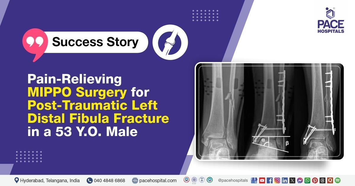
To confirm the diagnosis, imaging studies were performed. Plain radiographs (X-rays) of the left ankle in anteroposterior (AP), lateral, and oblique views revealed a post-traumatic spiral fracture of the left distal fibula. The fracture pattern was consistent with the reported mechanism of injury and accounted for the patient’s symptoms of localized pain, swelling, deformity, and restricted ankle movement. These radiographic findings supported clinical suspicion and provided a clear understanding of the extent of the injury, guiding further management and treatment planning.
Based on the confirmed diagnosis, he was advised to undergo Post Traumatic - Left Distal Fibula Spiral Fracture Treatment in Hyderabad, India, under the care of the Orthopaedic Department, ensuring comprehensive management of the fracture.
Medical Decision Making (MDM)
After a thorough evaluation by Dr. Anand V Agroya, Orthopaedic Surgeon, a comprehensive assessment of the patient’s clinical condition and imaging findings was performed. Considering the patient’s symptoms, functional limitations in daily activities, and the confirmed diagnosis of a post-traumatic left distal fibula spiral fracture, surgical intervention was deemed necessary.
The patient and his family were thoroughly counselled about the nature of the injury, the planned surgical procedure, potential risks, and the necessity for surgery. The procedure involved the use of a titanium plate and screws with Pre-Contoured Distal Lateral (PDL) plating for the distal fibula fracture.
Surgical Procedure
Following the decision, the patient was scheduled to undergo MIPPO Surgery in Hyderabad at PACE Hospitals, under the expert supervision of the Orthopaedic Department. The following steps were carried out during the procedure:
- Preparation and Anesthesia: The patient was positioned supine on the operating table with support under the affected hip. The left leg was cleaned and draped in a sterile fashion. Preoperative imaging was reviewed, and the surgical site was marked. Spinal anesthesia was administered.
- Closed Reduction: Using manual traction and manipulation, the fractured fibula was realigned without making a large incision. Fluoroscopy (real-time X-ray) was used to confirm that the bone fragments were properly aligned.
- Minimally Invasive Approach (MIPPO): A small incision was made laterally on the lower leg, away from the fracture site. A subcutaneous tunnel was created to pass the pre-contoured titanium plate along the lateral aspect of the fibula without disturbing the fracture.
- Plate Placement and Fixation: The Pre-Contoured Distal Lateral (PDL) titanium plate was inserted through the tunnel and positioned on the fibula. Multiple screws—both cortical and locking—were inserted percutaneously through small stab incisions to secure the plate firmly to the bone. Fluoroscopy was used throughout to ensure accurate placement.
- Closure: The incisions were closed in layers with sutures and a sterile dressing applied.
Postoperative Care
The patient was transferred to the recovery room immediately after surgery, where his vital signs were closely monitored to ensure stability. Continuous assessment of the neurovascular status of the operated limb was carried out to detect any early signs of complications such as impaired circulation or nerve injury. Intravenous antibiotics, analgesics, antiemetics, antipyretics, and other supportive treatments were administered. The surgical wound remained healthy, with no signs of infection observed.
The patient showed symptomatic improvement and was prepared for discharge with detailed post-discharge care instructions and follow-up recommendations.
Discharge Medication
Upon discharge, the patient was prescribed a course of antibiotics to prevent postoperative infection, analgesics for effective pain control, and a proton pump inhibitor (PPI) to protect the gastric lining from potential irritation caused by pain medications. He was advised to adhere strictly to the medication schedule to support recovery and prevent complications.
The patient was advised not to bear weight on the left lower limb and to use a walker for mobility with strict non-weight-bearing precautions. He was instructed to keep the left lower limb elevated at all times to reduce swelling and promote healing. The surgical dressing was to be kept clean and dry, and the patient was specifically advised not to wet the dressing to prevent infection or wound complications.
Emergency Care
The patient was informed to contact the Emergency ward at PACE Hospitals in case of any emergency or development of symptoms such as fever, pain in the abdomen and vomiting’s.
Review and Follow-Up
The patient was advised to return for a follow-up visit with the Orthopaedic Department at PACE Hospitals after 5 days for a dressing change and further evaluation.
Conclusion
This case demonstrates the effectiveness of Closed Reduction and Internal Fixation (MIPPO) with Titanium Plate and Screws (PDL) in the treatment of a distal fibula fracture, providing significant symptom relief, faster recovery, and a reduced risk of complications.
Significance of X-rays in interpreting and managing spiral fractures
X-rays play a vital role in the interpretation and management of spiral fractures, as they provide clear visual confirmation of the fracture, allowing for accurate assessment of its severity and characteristics. They help identify the exact location of the bone break, the pattern of the fracture, and any displacement, which is essential in determining whether the fracture is stable or unstable. X-rays also guide the orthopedic doctor/orthopedic surgeon in selecting the most appropriate treatment approach—whether conservative management or surgical intervention is needed. Additionally, they are used in follow-up assessments to monitor the progress of fracture healing and evaluate overall bone health throughout the recovery period.
Share on
Request an appointment
Fill in the appointment form or call us instantly to book a confirmed appointment with our super specialist at 04048486868
Appointment request - health articles
Recent Articles