Pancreatic Cyst Treatment in Hyderabad, India
PACE Hospitals is regarded as one of the best hospitals for pancreatic cyst treatment in Hyderabad, Telangana, India. Our expert team of gastroenterologists, endocrinologists, surgeons, and dietitians offers a multidisciplinary approach to diagnosing and managing various types of pancreatic cysts, including pseudocysts, mucinous cystic neoplasms (MCNs), and intraductal papillary mucinous neoplasms (IPMNs). We use advanced imaging techniques such as MRI, CT, and endoscopic ultrasound (EUS) to assess cyst characteristics and tailored treatment plans that may include medications, enzyme therapy, drainage procedures, or surgery—focused on symptom relief, preventing complications, and improving quality of life.
Book an appointment for Pancreatic Cyst Treatment
Pancreatic Cyst Treatment Online Appointment
Thank you for contacting us. We will get back to you as soon as possible. Kindly save these contact details in your contacts to receive calls and messages:-
Appointment Desk: 04048486868
WhatsApp: 8977889778
Regards,
PACE Hospitals
HITEC City and Madeenaguda
Hyderabad, Telangana, India.
Oops, there was an error sending your message. Please try again later. Kindly save these contact details in your contacts to receive calls and messages:-
Appointment Desk: 04048486868
WhatsApp: 8977889778
Regards,
PACE Hospitals
HITEC City and Madeenaguda
Hyderabad, Telangana, India.
Why Choose PACE Hospitals for Pancreatic Cyst Treatment?

Advanced Diagnostic Facilities: CT scan, MRI with MRCP, EUS, Pancreatic Function Tests (PFTs)
Top Pancreatic and Hepatobiliary (HPB) Specialists in Hyderabad
Minimally Invasive and Surgical Treatment Options
Affordable & Transparent Pancreatic Cyst Treatment with Insurance & Cashless Options
Diagnosis of Pancreatic Cyst
Before recommending diagnostic investigations, a gastroenterologist conducts a comprehensive clinical assessment to evaluate the likelihood of a pancreatic cyst and determine whether immediate imaging or further follow-up is warranted.
Accurate diagnosis and timely management are essential to prevent complications, including progression to pancreatic cancer. This overview outlines the key aspects of initial clinical assessment, available diagnostic tools, treatment approaches, and differential diagnoses associated with pancreatic cysts.
Initial Assessment Before Diagnosis
- Presented Signs and Symptoms
- Age, Family History, and General Health of the Patient
- Medical and Medication History
- Results of Previous Medical Tests
- Physical Examination
Presented Signs and Symptoms
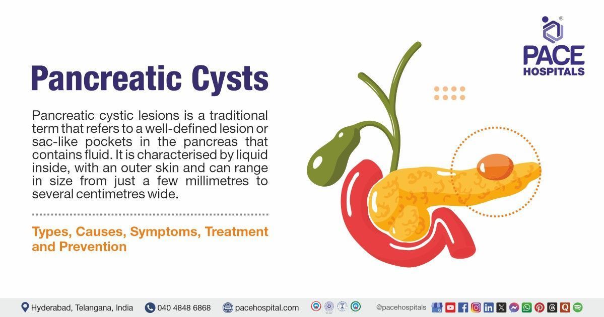
Pancreatic cysts are often asymptomatic and incidentally discovered during imaging for unrelated issues. When symptoms manifest, they may include abdominal pain, nausea, vomiting, bloating, and unexplained weight loss. Jaundice can occur if the cyst obstructs the bile duct. These nonspecific symptoms necessitate further evaluation to determine the cyst's nature.
Age, Family History, and General Health of the Patient
The prevalence of pancreatic cysts increases with age, particularly in individuals over 60. A family history of pancreatic diseases or genetic syndromes like von Hippel-Lindau disease elevates the risk. Assessing the patient's overall health is crucial, as comorbidities can influence both the presentation and management strategies.
Medical and Medication History
A thorough medical history can reveal risk factors such as chronic pancreatitis or previous abdominal trauma, which are associated with pseudocyst formation. Medication history is also pertinent, as certain drugs can induce pancreatitis, leading to cyst development.
Results of Previous Medical Tests
Reviewing prior imaging studies, laboratory results, and endoscopic findings provides insight into the cyst's progression and assists in differentiating benign from malignant lesions. Trends in tumor markers or changes in cyst characteristics over time are particularly informative.
Physical Examination
While often unremarkable, a physical exam may reveal abdominal tenderness or a palpable mass in cases of large cysts. Signs of
jaundice or weight loss can also be observed, prompting further diagnostic workup.
Diagnostic Tests
Based on the above evaluation, doctors select the most appropriate diagnostic tests to confirm the presence of a pseudocyst and guide treatment:
Blood tests
- Complete blood count (CBC)
- Liver function tests (LFTs)
- Kidney function test
Imaging studies
- CT scan
- MRCP
- MRI
- Trans Abdominal Ultrasound
- Endoscopic ultrasonography (EUS)
- Endoscopic retrograde cholangiopancreatography (ERCP)
Biopsies
- EUS-guided FNA
Cytology, smears
- Cyst fluid analysis
Blood tests
- Complete blood count: A CBC can detect signs of infection or inflammation, such as elevated white blood cell counts, which may suggest an infected cyst or pancreatitis. Anaemia might indicate chronic disease or bleeding within the cyst.
- Liver Function Tests (LFTs): Elevated liver enzymes and bilirubin levels can indicate biliary obstruction due to cyst compression, necessitating prompt intervention to prevent liver damage.
- Kidney Function Tests: Assessing renal function is essential, especially before administering contrast agents for imaging studies. Impaired kidney function may limit diagnostic and therapeutic options.
Imaging Studies
- Computed Tomography (CT): CT scans provide detailed cross-sectional images, aiding in assessing cyst size, location, and potential involvement of adjacent structures. They are instrumental in detecting calcifications or septations within the cyst.
- Magnetic Resonance Cholangiopancreatography (MRCP): MRCP offers non-invasive visualization of the pancreatic and biliary ducts, helping to identify communication between the cyst and ductal systems, which is crucial for classification and management.
- Magnetic Resonance Imaging (MRI): MRI provides high-contrast images, allowing for better characterization of cyst contents and wall features. It is particularly useful in differentiating mucinous from non-mucinous cysts.
- Transabdominal Ultrasound: This initial imaging modality can detect cysts and assess their size and location. However, its sensitivity is limited by patient body habitus and overlying bowel gas.
- Endoscopic Ultrasonography (EUS): EUS offers high-resolution images and enables fine-needle aspiration (FNA) for cyst fluid analysis. It is invaluable in evaluating cyst wall characteristics and vascular involvement.
- Endoscopic Retrograde Cholangiopancreatography (ERCP): ERCP allows direct visualization and intervention within the pancreatic and biliary ducts. It is primarily used when therapeutic procedures, such as stenting are indicated.
Biopsies
- EUS-Guided Fine-Needle Aspiration (FNA): EUS-guided FNA enables the collection of cyst fluid for cytological and biochemical analysis. This procedure aids in distinguishing benign from malignant cysts and guides management decisions.
Cytology and Smears
- Cyst Fluid Analysis: Analysing cyst fluid for carcinoembryonic antigen (CEA) levels helps differentiate mucinous from non-mucinous cysts, with elevated CEA suggesting a mucinous origin. Amylase levels can indicate communication with the pancreatic duct, as seen in pseudocysts. Viscosity, glucose levels, and molecular markers further refine the diagnostic process.
Differential Diagnosis of Pancreatic Cysts
When evaluating pancreatic cysts, it's important to consider several conditions that can appear similar but require different management. Some of the most common and potentially confusing conditions include:
- Chronic pancreatitis vs. intraductal papillary mucinous neoplasms (IPMNs): These can have overlapping features, but IPMNs carry a higher risk of malignancy.
- Pseudocysts after pancreatitis vs. serous or mucinous cystic neoplasms: Pseudocysts are usually non-cancerous and result from inflammation, while mucinous cysts may become cancerous.
- Serous cystic neoplasms vs. branch-duct IPMNs vs. acinar cell cystadenomas: These all-form fluid-filled cysts, but have different structures and risks.
- Solid variants of serous cystic neoplasms vs. neuroendocrine tumors vs. solid pseudopapillary tumors: These can look similar on imaging but vary widely in behaviour and treatment needs.
- Cystic appearances of typically solid tumors: Some solid tumors can develop cyst-like areas, which can complicate diagnosis.
- Rare cystic lesions in or near the pancreas (e.g., epithelial cysts): These are uncommon but should be considered, especially if the diagnosis is unclear.
Understanding these possibilities is essential for choosing the right diagnostic tests and guiding appropriate treatment.
Treatment Considerations
Before selecting the appropriate treatment for a pancreatic pseudocyst, healthcare professionals carefully evaluate several clinical factors to determine the most effective and safe approach for each patient. Key considerations include:
- History: Healthcare professional considers the patient complete history including medical, medication, social, family history and also physical evaluation.
- Risk stratification: It is vital for determining the need for surgery or close surveillance. Criteria include:
- High-risk stigmata: enhancing solid components, main pancreatic duct ≥10 mm, obstructive jaundice.
- Worrisome features: cyst ≥3 cm, thickened/enhancing walls, MPD size 5–9 mm, abrupt duct cut-off, lymphadenopathy.
- Cysts with high-risk stigmata often warrant surgical resection, while those with worrisome features may need further evaluation via EUS-FNA.
- Role of Tumor Markers in Cyst Fluid
Tumor markers and biochemical tests from cyst fluid obtained via EUS-FNA are essential in differentiating pseudocysts from other cystic pancreatic lesions.
- Several biomarkers enhance diagnostic accuracy:
- Carcinoembryonic Antigen (CEA): High levels (>192 ng/mL) suggest mucinous cysts.
- Amylase: Elevated in pseudocysts due to pancreatic enzyme content; low in mucinous cysts
- Glucose: Low glucose (<50 mg/dL) in mucinous cysts.
- DNA Analysis: KRAS and GNAS mutations support a diagnosis of mucinous cysts/IPMNs.
- Molecular markers may soon guide targeted therapies as research progresses.
Treatment strategies depend on the cyst's type, size, symptomatology, and malignant potential. Options range from conservative management to surgical intervention, including:
- Conservative treatments
- Medications
- Antibiotics
- Enzyme replacement therapy
- Monitoring & Observation
- Regular Imaging
- Observation
- Drainage Procedures
- Endoscopic Drainage
- Percutaneous Drainage
- Surgical Treatments
- Cyst Resection
- Pancreaticoduodenectomy (Whipple Procedure)
- Other Procedures
- Sclerotherapy
- Ethanol Injection
Conservative treatments
Conservative management of pancreatic cysts is aimed at relieving symptoms, promoting spontaneous resolution, and preventing complications in stable patients. Here are some of the non-surgical treatment approaches that may be considered for the conservative management of a pancreatic cyst:
Medications
Symptomatic relief can be achieved with analgesics and pancreatic enzyme supplements, especially in cases of exocrine insufficiency. These measures do not alter the cyst's course but improve the quality of life.
- Antibiotics: Infected cysts or those at risk of infection may require antibiotic therapy. Empirical treatment is initiated based on clinical suspicion and adjusted according to culture results.
- Enzyme Replacement Therapy: For patients with pancreatic exocrine insufficiency, enzyme supplementation aids digestion and nutrient absorption, mitigating symptoms like steatorrhea and weight loss.
Monitoring & Observation
Asymptomatic cysts without high-risk features can be monitored with periodic imaging to detect changes in size or characteristics that might necessitate intervention. This approach minimizes unnecessary procedures.
- Regular Imaging: Scheduled imaging studies, such as MRI or EUS, track cyst evolution over time, ensuring timely detection of concerning changes. The frequency of surveillance depends on initial cyst features.
- Observation: A watchful waiting approach is suitable for low-risk cysts, especially in elderly or comorbid patients for whom surgical risks outweigh potential benefits. Regular follow-up ensures prompt action if the cyst evolves.
Drainage Procedures
These are recommended when a pancreatic cyst becomes symptomatic, infected, or causes complications such as obstruction or pain. These procedures help relieve symptoms, reduce the risk of rupture or infection, and are often used when the cyst is not suitable for immediate surgical removal:
- Endoscopic Drainage: Endoscopic techniques allow internal drainage of pseudocysts into the gastrointestinal tract, providing symptom relief and resolving the cyst without external incisions. This minimally invasive approach reduces recovery time.
- Percutaneous Drainage: Under imaging guidance, a catheter is inserted through the skin to drain the cyst externally. This method is typically reserved for infected or symptomatic pseudocysts not amenable to endoscopic drainage.
Surgical Treatments
These are recommended for pancreatic cysts that show high-risk features for malignancy, are increasing in size, or are symptomatic and cannot be managed with drainage alone. Surgery allows for the complete removal of the cyst, reducing the risk of cancer development and recurrence.
- Cyst Resection: Surgical removal of the cyst is indicated for symptomatic, large, or potentially malignant lesions. The extent of resection depends on the cyst's location and involvement of surrounding structures.
- Pancreaticoduodenectomy (Whipple Procedure): This extensive surgery involves removing the pancreatic head, duodenum, gallbladder, and part of the bile duct. It is reserved for cysts with high malignant potential or confirmed cancer in the pancreatic head.
Other Procedures
Other procedures like sclerotherapy and ethanol injection are considered for pancreatic cysts that are not suitable for surgery, especially in patients with high surgical risk or small, well-defined cysts. These minimally invasive options aim to reduce cyst size or eliminate it while avoiding major surgery.
- Sclerotherapy: Sclerotherapy involves injecting a sclerosing agent into the cyst to induce fibrosis and obliteration. It is considered for select cysts, particularly when surgery is contraindicated.
- Ethanol Injection: Ethanol ablation entails injecting alcohol into the cyst cavity to destroy the epithelial lining, leading to cyst resolution. This minimally invasive technique is an option for certain mucinous cysts.
Follow-up Guidelines
Based on Cyst Type and Size, Surveillance recommendations vary by international guidelines (e.g., AGA, Fukuoka):
- <1.5 cm cysts without worrisome features: Imaging every 2–3 years
- 1.5–2.5 cm cysts: Annual imaging
- 3 cm or with features: Shorter intervals, consider EUS
- No further follow-up after 5–10 years of stability in low-risk cysts, depending on age and comorbidities
Pancreatic Cyst Treatment Cost in Hyderabad, India
The cost of Pancreatic Cyst Treatment in Hyderabad generally ranges from ₹1,000 to ₹1,15,000 and above (approx. US $12 – US $1,380).
The exact cost varies depending on the type of cyst (pseudocyst, IPMN, MCN, serous cystadenoma), size and complications, diagnostic evaluation (CT, MRI, EUS, cyst fluid analysis), the need for drainage or surgery, hospitalization, and hospital facilities — including cashless insurance, TPA corporate tie-ups, and complete support with medical insurance wherever applicable.
Cost Breakdown According to Type of Pancreatic Cyst Treatment
- Gastroenterologist / HPB Specialist Consultation – ₹1,000 – ₹2,000 (US $12 – US $24)
- Blood Tests (CBC, LFT, Amylase, Lipase) – ₹900 – ₹2,500 (US $11 – US $30)
- Ultrasound Abdomen – ₹1,000 – ₹2,500 (US $12 – US $30)
- CT Abdomen (Contrast) – ₹4,500 – ₹9,000 (US $54 – US $108)
- MRI Abdomen / MRCP – ₹4,500 – ₹10,000 (US $54 – US $120)
- Endoscopic Ultrasound (EUS) – ₹8,500 – ₹18,000 (US $100 – US $215)
- EUS-Guided Cyst Fluid Aspiration / FNAC – ₹10,000 – ₹22,000 (US $120 – US $265)
- Cyst Fluid Analysis (CEA, Cytology, Amylase) – ₹3,000 – ₹8,000 (US $36 – US $96)
- Medical Management (Pain, Infection, Enzyme Therapy) – ₹1,000 – ₹4,000 (US $12 – US $48)
- Endoscopic Drainage Procedure – ₹20,000 – ₹45,000 (US $240 – US $540)
- Surgical Management (If Required) – ₹65,000 – ₹1,15,000 (US $780 – US $1,380)
- Hospitalization (1–5 Days) – ₹8,000 – ₹25,000 (US $95 – US $300)
- Follow-up Ultrasound / CT – ₹1,000 – ₹2,500 (US $12 – US $30)
Frequently Asked Questions (FAQs) on Pancreatic Cysts
What are the treatment options for pancreatic cysts?
Treatment may include monitoring with imaging, endoscopic drainage, or surgical removal, depending on the cyst’s type, size, and risk of malignancy.
Which Is the best hospital for Pancreatic Cyst Treatment in Hyderabad, India?
PACE Hospitals, Hyderabad, is one of the most trusted centres for diagnosing and treating pancreatic cysts, offering advanced imaging, endoscopic, and surgical expertise.
We have team of highly experienced gastroenterologists, HPB surgeons, interventional radiologists, and endoscopy experts who manage all types of pancreatic cysts, including:
- Pancreatic pseudocysts
- IPMN (Intraductal Papillary Mucinous Neoplasm)
- MCN (Mucinous Cystic Neoplasm)
- Serous cystadenoma
- Solid–cystic lesions
- Cysts associated with pancreatitis
- Cysts causing pain, jaundice, or digestive symptoms
With CT, MRI/MRCP, EUS-guided aspiration, advanced endoscopy, minimally invasive drainage, and HPB surgery, PACE Hospitals ensures accurate diagnosis, safe treatment, and effective long-term outcomes — supported by cashless insurance, TPA tie-ups, and complete documentation support.
What are the common signs and symptoms of a pancreatic cyst?
Common signs and symptoms of a pancreatic cyst include abdominal pain, nausea, vomiting, and a feeling of fullness or bloating. Some cysts may also cause jaundice or back pain, while others remain asymptomatic and are found incidentally during imaging.
Can pancreatic cysts lead to cancer?
Yes, some types like IPMNs and MCNs can progress to pancreatic cancer if not monitored or treated appropriately.
Are there lifestyle changes that can reduce the risk of developing pancreatic cysts?
Yes, avoiding alcohol, quitting smoking, maintaining a healthy weight, and managing chronic pancreatitis can help lower the risk.
How much does pancreatic cyst surgery cost in Hyderabad at PACE Hospitals?
At PACE Hospitals, Hyderabad, the cost of pancreatic cyst Surgery typically ranges from ₹1,000 to ₹1,05,000 and above (approx. US $12 – US $1,260), offering a comprehensive and competitively priced solution for pancreatic cyst management. However, the final cost depends on:
- Type of pancreatic cyst (pseudocyst, IPMN, MCN, etc.)
- Size and location of the cyst
- Need for MRI, CT, EUS, or cyst fluid analysis
- Requirement for endoscopic drainage or surgical removal
- Presence of complications (infection, obstruction, pancreatitis)
- Duration of hospitalization and supportive care
- Follow-up imaging and long-term monitoring
Small, uncomplicated cysts treated with medications and observation fall at the lower range, while cysts requiring EUS-guided aspiration, drainage, or surgical management fall toward the higher range.
After clinical evaluation, imaging, and specialist consultation, our gastroenterology and HPB surgery team will provide a personalized treatment plan and a transparent cost estimate based on the cyst type and severity.
Can pancreatic cysts cause long-term complications?
Yes, untreated or complex cysts can cause recurrent pancreatitis, infections, ductal obstruction, or even progress to pancreatic cancer over time.
Which types of pancreatic cysts are most likely to become cancerous?
Mucinous cystic neoplasms (MCNs) and intraductal papillary mucinous neoplasms (IPMNs) have the highest potential for malignancy among pancreatic cysts.
What causes pancreatic cysts to form?
Pancreatic cysts can form due to pancreatitis, trauma, genetic mutations, or as a result of abnormal ductal growths in the pancreas.
Are there risk factors for developing pancreatic cysts?
Yes, risk factors include chronic pancreatitis, family history of pancreatic diseases, smoking, heavy alcohol use, and certain genetic syndromes.
What is a precancerous pancreatic cyst?
A precancerous pancreatic cyst is a fluid-filled sac in the pancreas that has the potential to develop into pancreatic cancer over time. Common types include intraductal papillary mucinous neoplasms (IPMNs) and mucinous cystic neoplasms (MCNs), which require careful monitoring or surgical removal based on size, symptoms, and risk of malignancy.
What symptoms might indicate the presence of a pancreatic cyst?
Symptoms may include abdominal pain, bloating, nausea, jaundice, or unintended weight loss, depending on the cyst’s size and location.
Can pancreatic cysts be asymptomatic?
Yes, many pancreatic cysts are asymptomatic and are discovered incidentally during imaging for unrelated health issues.
When is surgery recommended for a pancreatic cyst?
Surgery is considered when the cyst shows suspicious features, causes symptoms, grows over time, or has malignant potential.
What size is considered a large pancreatic cyst?
A pancreatic cyst is generally considered large if it measures 3 cm (30 mm) or more in diameter. Larger cysts, especially those over 3 cm, may carry a higher risk of malignancy and often require closer monitoring or further evaluation.
What is the long-term outlook for someone with a pancreatic cyst?
The outlook depends on the type and behaviour of the cyst; benign cysts often require monitoring, while precancerous or cancerous ones need intervention.