PACE Hospitals is one of the Best Orthopedic Hospital in Hyderabad, India, providing holistic and patient centric orthopedic treatment. The team of experienced and skilled orthopedic doctor, orthopedic surgeon and sports medicine doctor have vast expertise in managing all kind of deformities of bone, joints, muscles, ligament and tendons, including:
Thank you for contacting us. We will get back to you as soon as possible. Kindly save these contact details in your contacts to receive calls and messages:-
Appointment Desk: 04048486868
Whatsapp: 8977889778
Regards,
PACE Hospitals
Hitech City and Madinaguda
Hyderabad, Telangana, India.
Oops, there was an error sending your message. Please try again later. Kindly save these contact details in your contacts to receive calls and messages:-
Appointment Desk: 04048486868
Whatsapp: 8977889778
Regards,
PACE Hospitals
Hitech City and Madinaguda
Hyderabad, Telangana, India.
Providing treatments to wide range of musculoskeletal disorder including deformities in the bone, joint, muscles, ligament, tendons and sports injuries.
Equipped with advanced and latest diagnostic equipment, robotic and minimally invasive surgical facilities for musculoskeletal disorders treatment.
Team of experienced orthopaedic doctor, orthopaedic physician, sports medicine doctor, orthopaedic surgeon with vast experience in minimally invasive procedures.
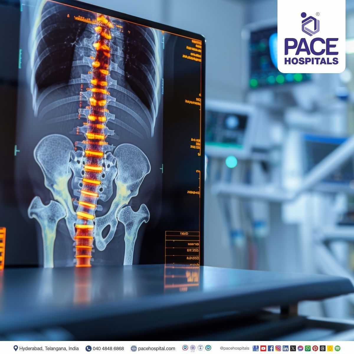
The Orthopaedic Department at PACE Hospitals is one of the Best Hospitals in Hyderabad for orthopaedic treatment. The Department encompasses a team of skilled and experienced orthopaedic doctors, rheumatologists, sports injury doctors, and orthopaedic surgeons who provide comprehensive expert orthopaedic care for a range of disorders related to bone, joints, muscles, ligaments and tendon injuries. They have vast expertise in diagnosing and managing critical conditions related to the musculoskeletal systems and are well-versed with the latest treatment modalities catering to holistic and precise orthopaedic treatment with a high success rate.
The Department of Orthopaedics is equipped with state-of-the-art and cutting-edge facilities that offer a wide range of comprehensive bone and joint care, including advanced and high-resolution imaging systems, digital X-rays, MRI, and the latest treatment modalities, including robotic surgeries, minimally invasive procedures for full joint and knee replacements, and advanced rehabilitation facilities, ensuring quicker recovery and lasting results.
A team of the best Orthopaedic Doctor in Hyderabad, India, having extensive expertise in diagnosing and managing a wide range of musculoskeletal diseases and disorders, including arthritis, fractures, osteoporosis, scoliosis, joint pain, ligament injuries, and sports-related injuries are committed to delivering patient-centric, evidence-based and compassionate musculoskeletal disorders treatment to the pediatric, adult and geriatric patients. The skilled and experienced team of orthopaedic specialists and sports injury doctor thoroughly analyse the diagnosis report to understand the root cause of discomforts to cater to complete orthopaedic care tailored to individual needs. Orthopaedic surgeons are highly skilled in using advanced procedures like joint & knee replacements and minimally invasive techniques to treat complex and critical musculoskeletal disorders with minimal complications and high success rates.
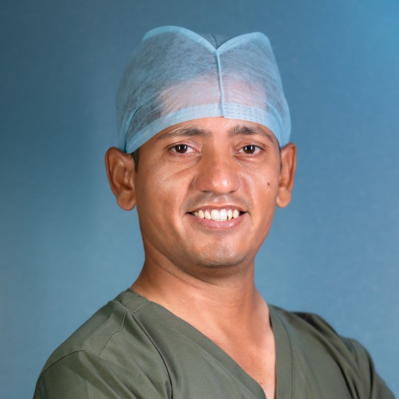
MBBS, D.Ortho, Fellow in Joint Replacement, Arthroscopy & Sports Medicine
Experience : 10+ years
Senior Orthopaedic Consultant, Trauma Surgeon & Sports Medicine Specialist | Expert in Knee & Joint Replacement & Arthroscopy Surgery
He has expertise in advanced orthopaedic care and comprehensive sports injury management, focusing on knee and joint replacement, arthroscopy surgery, ligament reconstruction, fracture management, cartilage preservation, and joint restoration techniques. He supports patients in regaining strength, alleviating pain, and enhancing their quality of life.
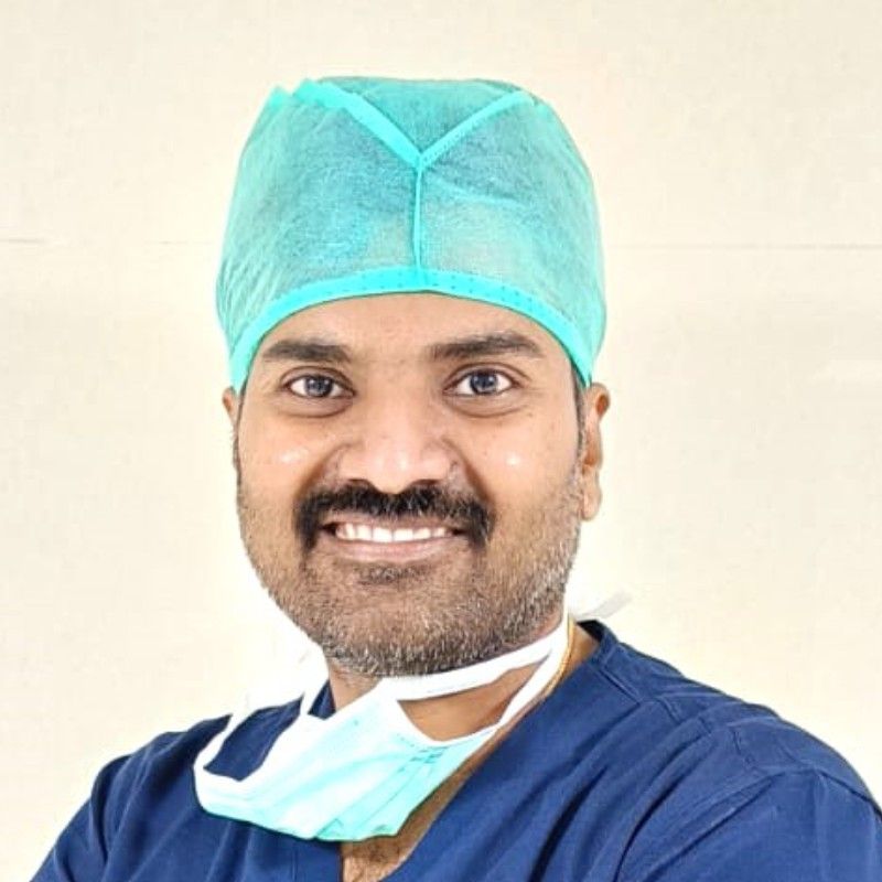
MBBS, DNB ORTHO, Fellowship in Joint Replacement and Arthroscopy, Fellowship in Shoulder and Upper limb, Sports medicine and Replacement
Experience : 10+ years
Orthopaedic Consultant, Trauma, Shoulder and Knee Arthroscopic Surgeon, Hip and Knee Joint Replacement Specialist
He is a specialist in diagnosing, managing, treating problems and performing procedures related to Complex and Failed Traumas, Non-Union and Malunion Fractures, Joint Replacements, Revision Surgeries, Geriatric Trauma Surgeries, Spine Problems, Foot and Ankle Problems, Knee and Shoulder Pain, Sports Injuries, Arthritis etc.
Struggling with chronic pain and swelling in your joints, dealing with a broken bone or injury, facing back pain, numbness or tingling in your hands, severe sports injuries or seeking treatment for rheumatoid arthritis, osteoarthritis, rotator cuff injuries, meniscus tear, torn ligaments (ACL & MCL), herniated disc problems, scoliosis, bone tumours, bone infections, tendonitis, bursitis, tennis elbow, hip dysplasia, compound fractures, multiple fractures, we provide evidence-based treatment tailored to your needs. Our team of skilled and experienced orthopaedic doctors, sports medicine physician, orthopaedic surgeon provides comprehensive and compassionate care for geriatric, adult and pediatric patients.
Orthopaedics is the medical branch that deals with diseases and injuries of the human musculoskeletal system, which includes joints, bones, tendons, ligaments, and muscles.
Orthopaedic departments at any multi-speciality hospital specialize in diagnosing, managing and treating an extensive range of common and critical orthopaedic conditions impairing the musculoskeletal system. Some common orthopaedic diseases and disorders include:
The most common musculoskeletal conditions for which they might need ortho medical care are arthritis (osteo, rheumatoid, elbow), fibromyalgia, sprains or strains, fractures, back and neck pain, hand and knee pain, bursitis, hip fracture, foot pain, shoulder pain, osteoporosis, kyphosis, Paget's disease, and soft tissue injuries.
The person with a fracture may have heard or felt a grinding or snap noise during the injury. In addition to these, bruising, swelling, pain and tenderness are seen at the site of the injury. In some severe cases, the injured part looks deformed, and the affected person may feel dizzy or faint as a result of shock due to bone fracture.
You should see an orthopedic doctor if you are experiencing any of the following symptoms:
The most common orthopedic procedures done by orthopedic surgeons are:
Some of the common diagnostic tests and examinations performed to assess musculoskeletal system disabilities are as follows:
Orthopedists are specialists in the musculoskeletal system, which consists of bones, joints, muscles, ligaments, and nerves. These experts are trained in diagnosing and treating bone and joint diseases and injuries, which may involve orthopaedic surgery.
Rheumatologists are specialists in the management of inflammatory autoimmune diseases. In diseases like lupus or rheumatoid arthritis, the immune system of the patient attacks healthy body tissue rather than invading foreign pathogens.
Orthopedists are specialized doctors and surgeons with expertise in neuromusculoskeletal systems. Chiropractors are holistic healthcare practitioners who are skilled at maintaining the healthy position of the spine.
An orthopedist is a doctor who specializes in the prevention or correction of deformities, disorders, or injuries of a patient's skeleton. A physical therapist (PT) is a healthcare expert who helps people improve their movement and function.
Anyone seeking treatment for diseases and disorders of bone, joints, muscles, ligaments, and tendons or looking for an appointment with the best orthopedic doctor, sports medicine doctor, or orthopedic surgeon in the locations near Hitech City, Madhapur, Kondapur, Gachibowli, KPHB, or Kukatpally can visit the PACE Hospital's Orthopedic Department webpage and fill out the Appointment Form. They can also directly visit the hospital located near the Hi-Tech City metro station or call 04048486868 to book a hassle-free appointment.
We specialize in treating and managing a wide range of orthopaedic diseases and disorders affecting the musculoskeletal systems, including bone, joints, muscles, ligaments and tendons. From common sports injuries and orthopaedic conditions like osteoporosis, osteoarthritis, osteomyelitis (bone infections), fractures, tendonitis, bursitis, frozen shoulder, shoulder dislocation, ankle sprains, clubfoot, tennis elbow, runner's knee, meniscus tear, compound fractures to complex, critical and chronic conditions like rheumatoid arthritis, bone tumours, herniated or slipped discs, kyphosis, degenerative disc disease, sciatica, torn ligaments (ACL, MCL tears), hip dysplasia, developmental dysplasia of the hip, multiple fractures, pelvic fractures, traumatic amputations, our team of orthopaedic physician, orthopaedic surgeon and sports medicine doctor is committed to cater compassionate, patient-centric comprehensive solutions for your bone and joint health.
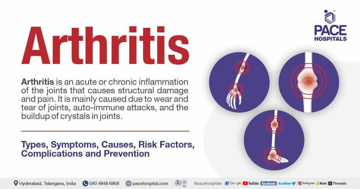
The term "arthritis" is often called inflammation of the joints. The joint is the part of the body where two or more bones meet to allow movement, such as elbows and knees. There are different types of arthritis conditions with different causes and specific treatments. The symptoms of arthritis include redness, pain, swelling, and joint heat. In some arthritis cases, it also affects the heart, eyes, and skin.
Arthritis is mainly caused by joint wear and tear, autoimmune attacks, and the buildup of crystals in joints. In addition, gender, age, weight, hereditary factors, injuries, and infections also lead to arthritis.
Inflammation of the bursa is often termed bursitis. The bursa is a fluid-filled sac that acts as a cushion to decrease the friction between the tissues. Bursae are in the hips, shoulders, knees, and elbows next to the tendons. Bursitis is temporary and does not cause any deformities; it limits motion or movement. It can occur at any location in the body. Based on location, bursitis is divided into many types.
Bursitis symptoms include swelling and redness at the affected site, limited motion or movement, pain, and tenderness at the injury site. In chronic conditions, these symptoms occur repetitively. The causes of bursitis are injury, overuse, and some infections. In addition to this, bursitis is associated with conditions such as gout, tendonitis, arthritis, diabetes, and thyroid disease.
Bunions are also known as 'Hallux valgus' (HV). HV is the common forefoot condition with a deviation of the proximal phalanx to lateral and first metatarsal head to medial due to adduction of the first meta tarsus, often called metatarsus primus varus. The symptoms of bunions are medial dorsal cutaneous nerve neuritis, ulcerations, blisters, interdigital keratosis, and irritated skin. These symptoms may also cause morbidity and limit physical activity.
The cause of HV is not known, but theories have stated that it is caused by genetic factors, dorsiflexed or short first metatarsal, abnormal foot mechanics, and hypermobility. It is also commonly seen in people with connective tissue disorders, muscle imbalances in the foot, and wearing heels or tight shoes.
A fracture to the humerus (ball) or glenoid (socket) of the shoulder joint due to trauma is often called a shoulder fracture. Most of these injuries are treated without surgery. Few cases are treated with surgery because they may have a high risk of arthritis if left untreated. Fractures are of two types, either displaced or non-displaced. Nearly 80 per cent of all shoulder fracture cases are non-displaced.
Symptoms of shoulder fractures are swelling, shoulder pain, deformities, tenderness, discolouration and inability to move the shoulder. The causes include direct collisions, trauma or injury, and falls.
Carpal tunnel syndrome (CTS) is a neurological disorder caused when the palm's median nerve gets squeezed or pressed at the wrist. It may cause weakness, numbness, and pain in the hand and wrist; the fingers may become swollen and useless. This condition makes you wake up and feel like shaking your hand or wrist. The symptoms of CTS are numbness and tingling sensation in fingers, mild to severe pain which worsens at night, weakness in hands, inability to grasp objects and can't perform tasks.
The causes of CTS are injury or trauma to the wrist, ageing, imbalance in either the thyroid or pituitary gland, application of vibrating tools, bent wrist, metabolic disorders, rheumatoid arthritis, etc.
Cubital tunnel syndrome is a nerve problem that causes numbness, tingling, swelling, and pain in the ulnar nerve due to compression or irritation of the nerve at the elbow. Other symptoms include hand pain, tingling and numbness in the small finger or hand, muscle weakness, and ache pain at the elbow.
The common causes of cubital tunnel syndrome are stretching (elbow bent, repetitive use), injuries (dislocations, fractures, effusions and swelling), and pressure (injury, trauma).
Knee pain is common among adults and is associated with general tear and wear from daily activities such as standing, walking, bending, and lifting. The knee consists of the tibia, femur, and patella. It bears excessive stress every day. Common knee problems are torn cartilage, sprained or strained ligaments, tendonitis, and arthritis.
Symptoms of knee pain include pain and inflammation. The causes of knee pain are injury, ageing, repeated stress on the knee, obesity, infections, degenerative disorders, and autoimmune conditions.
Damage or injuries to the spinal discs due to wear and tear result in degenerative disc disease. Spinal discs act like shock absorbers between bones and spinal cord vertebrae and provide flexibility for bending and twisting.
The symptoms of degenerative disc disease are pain in the buttocks and thigh region, inability to bear the pain when walking and moving, and feeling worse when performing activities such as bending, twisting, and lifting. The causes of degenerative disc disease are dryness in discs due to water loss due to ageing and cracking due to stress from daily activities.
The shoulder joint is one of the mobile joints in the body. It can turn in many directions, but it is easy to dislocate from its position. A partial dislocation (subluxation) is a condition where the head of the humerus comes partially outside of the socket (glenoid). A complete dislocation is a condition where the socket is completely out. Both partial and absolute dislocations cause pain and instability in the shoulder.
Symptoms of this condition include weakness, bruising, deformity, pain and swelling. Causes include falls, injuries or trauma, and sporting activities.
Elbow arthritis is a condition in which the cartilage in the elbow becomes worn or damaged. This condition may be due to overuse, repetitive activities, or an injury (fractures and dislocations). It is extremely painful and can interfere with daily tasks such as arm bending.
Symptoms of elbow arthritis are pain, stiffness, swelling, unable to move the elbow, and tingling or numbness in elbows. The causes of elbow arthritis are injuries or trauma, rheumatoid arthritis, osteoarthritis, gout, and juvenile arthritis.
A fracture is a break or cracks in the bone. A condition where the broken bone punctures the skin is called an open or compound fracture. Depending on the location of the injury, there are several types of fractures. Symptoms of fractures include deformities of the limbs, intense pain, bruising and swelling of the bones, tenderness at the site of injury, inability to move the limbs, tingling, and numbness.
The fractures are usually caused due to sports injuries, accidents, osteoporosis, low bone densities, and overuse of the limbs.
Fibromyalgia is a chronic or long-lasting disorder that causes pain, tenderness all over the body, trouble sleeping, and fatigue. Currently, there is no cure for this condition, but doctors manage the symptoms through movement and psychological and medical therapies.
The symptoms of fibromyalgia are chronic pain all over the body, fatigue, disturbed sleep, joint or muscle stiffness, tenderness, altered memory or concentrations, bloating, constipation and sensation to light or noise.
Fibromyalgia is idiopathic (the cause is unknown), but researchers found it might be happening due to genetic factors. Fibromyalgia is most likely in conditions such as osteoarthritis, rheumatoid arthritis, systemic lupus erythematosus, depression, and inflammatory bowel disease.
Fractures result from injury or break to the bones of the foot and ankles. The foot and ankles contain many bones that intricately work together. Extreme pain and impairment of mobility occur when a fracture occurs.
The symptoms of foot and ankle fractures are bruising, throbbing pain, pain during activity, difficulty walking and bearing weight, and deformities in the foot or anklefractures include weakness of ligaments and tendons, overstretching or overextending, extreme force on ankles or feet, hard falls, and injuries.
The humerus, radius and ulna together form an elbow joint. These bones are well organised together by ligaments, tendons and muscles. Fractured elbow, often called an olecranon fracture. In this condition, breakage of "pointy bone" takes place; it occurs due to a direct blow to the elbow.
Symptoms of an elbow fracture include tenderness at the site of injury, pain when moving, swelling, bruising, and inability to move the elbow. This condition is mainly caused by an injury or trauma to the elbow (direct blow or impact on the elbow joint).
FAI, also called hip impingement, is a clinical syndrome caused by the anatomic abnormalities of the femoral head. It results in abnormal contact between the two hip motions, especially hip flexion and rotation, which eventually leads to cartilage, labral damage, and hip pain.
Symptoms include pain or stiffness in the thigh and groin region, and pain worsens during activities. Causes of hip impingement are sporting activities, athletics, and slipped capital femoral epiphysis.
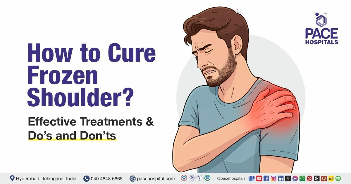
Frozen shoulder is also called Adhesive capsulitis (AC), which is a painful shoulder condition that has been present for more than three months due to inflammation. This inflammatory condition causes fibrosis of the glenohumeral joint capsule, accompanied by progressive stiffness and significant restriction. The symptoms of a frozen shoulder are restricted or limited motions, neck pain, stiffness, pain in activities, and inflammation.
The cause of the frozen shoulder is not clearly determined. However, some possible causes of frozen shoulder are injuries or trauma to the shoulders, diabetes mellitus, thyroid disorder, Dupuytren disease, Parkinson's disease, cancer and complex regional pain syndrome.
Ganglion cysts are synovial cysts filled up with gelatinous mucoid material, and the condition is commonly seen in orthopaedic practice. Studies have stated that they arise due to repetitive trauma, resulting in the degeneration of connective tissue. These cysts are soft tissue masses, especially found in the hand and wrist.
Most of the cysts are asymptomatic. The patient may experience tenderness, pain, and weakness in wrist motion. Patients with volar ganglion cysts may present with carpal tunnel syndrome secondary to compression in the median nerve and intrusion of flexor tendon sheath as well as ulnar nerve neuropraxia.
No exact cause has been reported for this condition until now. Theories in 1746 and 1926 stated that ganglion cysts are caused by herniation of synovial tissues from joints and mucinous degeneration of connective tissue, respectively.
Golfer's elbow, also known as baseball elbow, is often called medial epicondylitis in medical terms. It is a condition in which injury to the tendons that curve the wrist towards the palm occurs. Tendons are tough cords of tissue connecting muscles and bones. This condition is characterised by pain at the elbow.
The symptoms of this condition are pain when bending the wrist, inability to move the elbow, swelling, and bruising. This condition is mainly caused by excessive force when bending the wrist or elbow. Other possible causes include weak shoulder or muscles, javelin throw, carrying heavy objects, frequent use of hand tools, and repetitive use of the elbow or wrist.
A groin pull or strain results from too much stress on the groin and thigh muscles. When pressure is applied, these muscles become overstretched or torn. It is commonly seen in people who play sports that require running and jumping.
A groin pull is caused by jumping or suddenly changing direction. This condition is also seen in people who play soccer and football, making up about 10% of all injuries in professional hockey players.
Symptoms of groin pulls include pain and tenderness in the groin region, pain when raising the knee and bringing the legs together, and a popping or snapping sensation during injury.
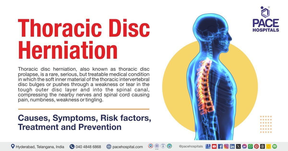
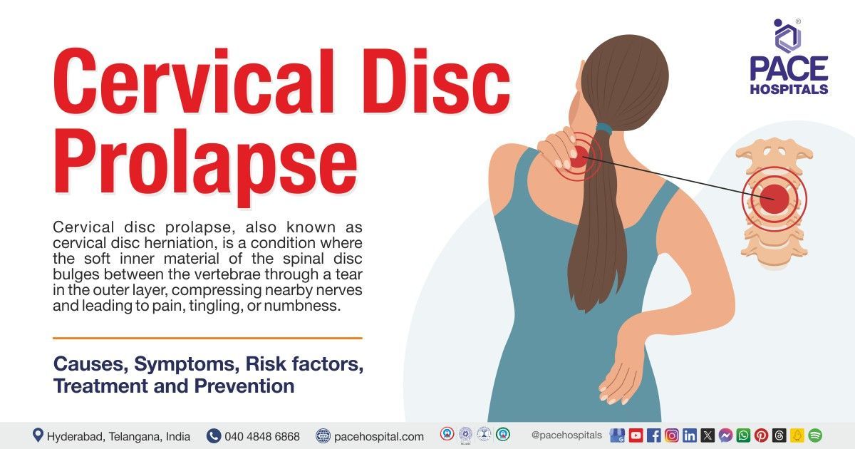
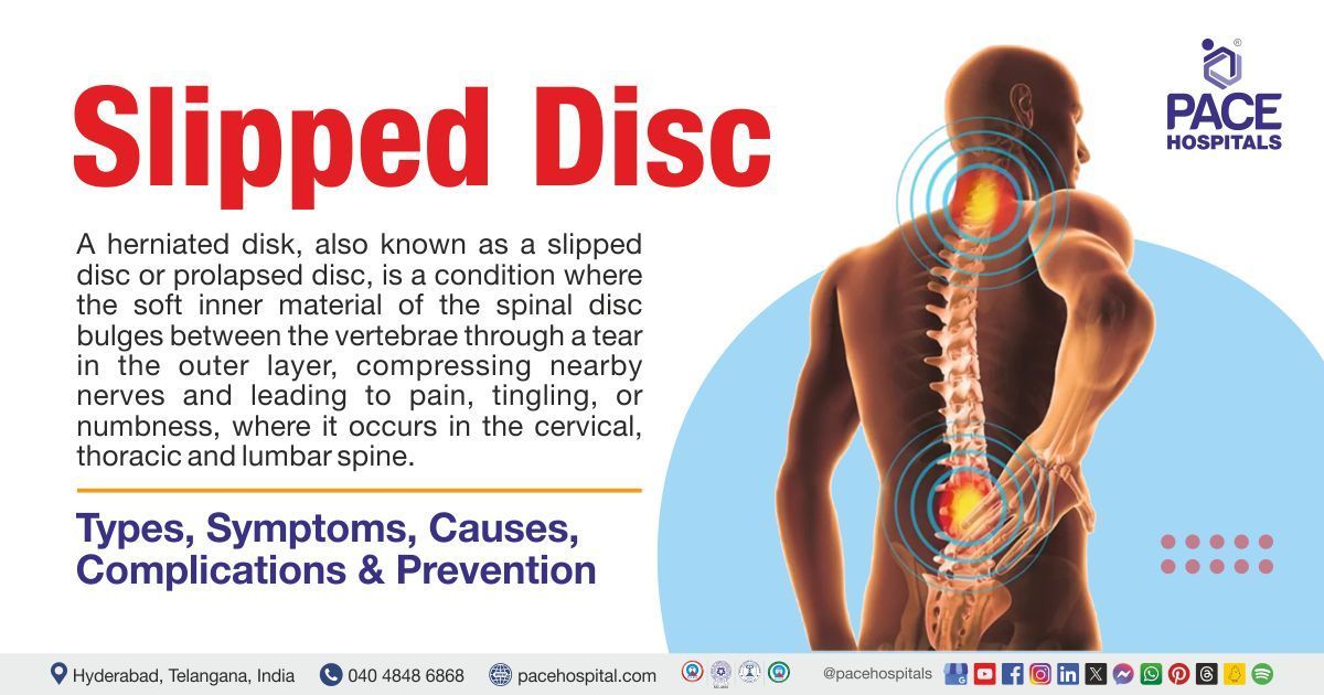
Herniated discs are often called slipped, bulged, or ruptured discs. Disk herniation occurs when the nucleus pulposus (located in the centre of the disc) is displaced from the intervertebral space.
The symptoms of herniated discs include low back pain, sciatica or radiculopathy, which induces burning, numbness, and tingling sensations, and cervical radiculopathy, which induces sharp pain.
The causes of disc herniation are degeneration due to ageing, trauma or injury, connective tissue disorders, and congenital disorders. In addition to these, smoking, obesity, lifting heavy things, repetitive twisting or bending, standing or sitting for many hours in the same place, and an inactive lifestyle are the factors that may cause herniated discs.
A hand fracture, also called a broken hand, is a condition in which the breakage of hand bones occurs; the breakage may be in phalanges (small bones of fingers) or metacarpals (long bones of the palm). Most cases heal with non-surgical treatments, and few require splints, straps, and surgeries to realign the broken bone.
The symptoms of hand fracture are swelling, tenderness, unable to move the finger, bruising, deformity, shortened finger and sunken knuckle (boxer’s fracture). The common causes of hand fractures are falls, twisting, crush injuries and direct contact in sports.
Hammer toe is a common forefoot deformity resulting in an imbalance between the weak intrinsic muscles and the stronger extrinsic muscles surrounding the lesser digits' metatarsophalangeal joints (MTPJ).
It is a condition with a deformity that involves flexion at the interphalangeal joints (IPJ) and is categorised as a classic hammer toe, mallet toe, or claw toe. Initially, the patient gets conservative management; if it fails and the condition worsens, the patient may advise a surgical approach.
Hammer toe symptoms include the inability to move the toe, tenderness on wearing shoes or walking, callus formation on the foot, and bent toe. The causes of hammer toe are congenital factors, biomechanical dysfunction, diabetes, neuromuscular functions, hallux valgus, pes planus, long metatarsals, inflammatory arthropathies, wearing of inappropriate shoes, and some neuromuscular conditions.
Hamstring pulls, commonly called hamstring muscle injuries, are frequently seen in athletes. This condition occurs if there is one or more injuries to the back thigh muscles. Hamstring muscles run downwards at the back thigh; these are categorised into semitendinosus, semimembranosus, and biceps femoris. They start from the bottom of the pelvis, cross the knee joint, and end at the lower leg. It helps to extend the leg straight and bend the knee.
Symptoms of this condition include swelling, pain, bruising, weakness, and discolouration at the back of the leg below the knee. Causes of hamstring pulls include muscle overload, tightness, weak muscles, fatigue, imbalance, and sporting or athletic activities.
Hip pain is a condition involving any pain in and around the hip joint. The person may not feel pain from the hip directly but experiences pain in the groin, thigh, or knee regions.
Symptoms include hip pain, bruising, swelling, leg deformities, bleeding, and inability to move the hip. Some serious signs include fever and rash with pain. The causes of hip pain are fractures, labral tears, infections, osteonecrosis, arthritis, injuries or trauma, and strains.
Joint dislocation is a condition in which the two bones are separated or misplaced from their range. It is more painful and leads to temporary deformity. Joint dislocations commonly occur in the shoulders and fingers; they may also occur in elbows, hips, and knees.
The symptoms of joint dislocations are intense pain, discolouration at the site of injury, joint deformities, numbness or tingling sensation at the joint, swelling at the site of injury, and limited movements.
The cause of joint dislocations is fall or blows, trauma or injuries, and sporting activities.
Kyphosis is also called “hunch back”. It is a condition with the spine curvature measuring 50 degrees or greater on the X-ray; a normal spine appears straight. However, a spine affected by kyphosis shows a curvature of the vertebrae in the upper region of the back, resulting in an abnormally rounded or "humpback" appearance. It is more common in females. Different types of kyphosis are postural, Scheuermann’s and congenital. The signs and symptoms of kyphosis are different in shoulder and shoulder blade height, height of upper back, tight back thigh muscles, and back pain.
Neuromuscular conditions, metabolic problems, osteoporosis, low bone density, and hereditary factors cause kyphosis.
Injury to the tissue (labrum) which holds the ball and socket of the hip together is often called labral tears. The hip joint is also called the ball and socket joint, composed of the ball at the top of the pelvis femur. Usually, it is seen due to progressive wear on the labrum.
The symptoms of labral tears are hip stiffness, clicking or locking sensation at the hip joint, and pain in the groin, hip, or buttocks. Some tears may cause no symptoms and are not identified for a few years. The causes of hip labral tears include hip injuries or trauma, hip overuse or repetitive motions, wear and tear of the hip, hip deformities, and osteoarthritis.
Lordosis, commonly called sway back, is a condition characterized by exaggerated inward curvature of the spine. It typically occurs in the lower back (lumbar lordosis) and neck (cervical lordosis). This condition doesn't cause symptoms except in severe cases.
The symptoms of lordosis are neck or back pain radiating to the legs and feet and a numbness or tingling sensation. In rare conditions, it may cause severe leg pain, weakness, and loss of control of bowel and bladder habits. The exact cause of this condition is usually unknown, but factors such as osteoporosis, obesity, osteosarcoma, spondylolisthesis, muscular dystrophy, and static encephalopathy may lead to it.
This condition is often referred to as torn meniscus. The menisci are located between the tibia (lower leg bone) and femur (thigh bone), which protects the lower part of the leg; the medial meniscus is inside the knee, whereas the lateral meniscus is outside the knee. The meniscus tear or torn meniscus is commonly seen in athletes due to turns or twists of the upper leg when the foot is placed and the knee is bent.
The symptoms of meniscal injuries are swelling and pain at the knee joint, especially in the lateral or medial position, locking or catching (a sign of cartilage degeneration), inability to move or bend the knee joint, and limping (difficulty walking). Meniscal injuries are commonly caused by rotational or shearing forces, lifting or carrying heavy weights, activities with rapid acceleration or deceleration, jumping, and trauma.
Muscle contusion, also called a muscle bruise, in this condition bruise occurs in the muscle. It is the second leading cause of sports injuries. In most cases, they heal quickly, but in severe cases, they may cause tissue damage and lead to complications.
The symptoms of contusions are pain and swelling at the site of injury, bluish discolouration on torn blood vessels, and muscle becoming weak, stiff, and lumpy. In some conditions, they may lead to dislocation of joints, sprains, and other injuries. Muscle contusions are caused by direct blows (injury or trauma).
Muscular dystrophy is caused by more than 30 genetic conditions, resulting in muscle weakness and muscle-related symptoms that worsen over time. This condition may present at birth or develop in childhood or adulthood.
Muscle atrophy, irregular walking (toe walking), tightening of muscles, stiff or loose joints, muscle pain, and spasticity are symptoms of muscular dystrophy. Genetic mutations, such as recessive, X-linked, and dominant inheritance, cause muscular dystrophy.
Arthritis in the neck and back is often referred to as spinal arthritis. In this condition, inflammation is seen in facet joints of the spine or sacroiliac, especially between the spine and pelvis. Spinal arthritis is related to autoimmune disorders, infections or wear and tear conditions. Inflammation might affect the sites where the ligaments and tendons attach to the bones. Spinal arthritis can be painful, sometimes leading to chronic inflammation. Symptoms include headaches, popping noise or grinding sensation of the neck on movements, loss of balance, generalised weakness in hands and legs, muscle spasms at neck or shoulders, and rarely numbness.
The causes of neck and back arthritis are autoimmune triggers, genetic components, age factors, obesity, and conditions such as Lyme disease, irritable bowel syndrome, tuberculosis, diabetes, gout, and psoriasis.
Due to injury, sprain of ligaments and strain of muscles in the neck and back regions tend to these conditions. It occurs mainly due to abnormal rotation or twisting of the neck and back region. The pain might be mild or severe and appears later after injury. Symptoms of this condition include back pain or neck pain on movements, pain and muscle spasms in the shoulder, headache on the backside of the head, difficulty in sleeping and concentrating, weakness and numbness in the arms, irritability, etc.
Hard falls, vehicle collisions, and injuries to the neck and back are common causes of these sprains and strains.
A tear or tagged opening (ruptured tendon) on the wrist, forearm, fingers and palms results in this condition. Tendons are cord-like structures that connect muscles to bones. The nervous system is made up of sensory and motor nerves in the body; these nerve controls the sensation, motor and moment co ordinations in the body. A cut or a tear due to injuries to the nerves results in nerve lacerations. Symptoms of tendon laceration include inability to bend the joints in fingers, numbness in fingertips, pain on bending, and tenderness. The common causes of tendon injuries are cuts, injuries in sporting and wrestling activities, and rheumatoid arthritis.
Whereas symptoms of nerve injury are burning sensation, severe pain, loss of sensation, and tingling. Causes include bruising, cuts or tears, gunshot wounds, and electrical or injection injuries.
Injury to the neck and back leads to pain, which can range from mild to severe, such as dull or annoying aches. Back pain restricts mobility and quality of life. The pain is acute in some cases, whereas chronic in others; it may eventually be continuous or intermittent.
Symptoms of neck and back pain include dull or sharp pain, tingling or numbness above the knee, sharp pain from the lower back to the buttock, and consistent aches after sitting or standing. Causes include strenuous activities, trauma, injury, tumours, infection, degeneration of vertebrae, poor muscle tone, obesity, spasms, strain or sprain, arthritis, ligament or muscle tears, osteoporosis, and congenital abnormalities.
Infection of the deep neck tissues leads to neck abscesses, which are complicated to diagnose, access, and manage. The affected tissues are deep and unable to be seen externally. Inflammation is seen in nearby structures and leads to bony, neurovascular, and airway issues.
The symptoms of neck abscess may include severe pain, upper respiratory infections, neck or mouth trauma, respiratory distress, dysphagia or dysphonia, spiking fevers, and shortness of breath. The causes of neck abscess are tonsillitis, pharyngitis, odontogenic (in adults), a history of surgical procedures, infections, trauma, malignant lymph nodes, cleft anomalies, and a suppressed immune system.
The bones comprise living cells that require a blood supply to stay healthy. In osteonecrosis, blood flow to part of a bone is disrupted. This results in the death of bone tissue, and thereby, the bone can eventually break down and cause the joint to collapse. Osteonecrosis is also called Aseptic necrosis, avascular necrosis, or ischemic necrosis of bone.
Initially, the symptoms are not experienced, but after the gradual increase of the condition, the patient may experience pain in the thigh bone and groin regions, joint stiffness, limited motion, and osteoarthritis over time. Causes of osteonecrosis are injury, use of corticosteroids, excessive tobacco and alcohol use, and certain medical conditions such as gout, cancer, blood disorders, rheumatoid arthritis, etc.
Osteoporosis is a type of bone disease that is developed due to a decrease in bone mass and mineral density or due to the structural changes of bone. Osteoporosis leads to a decrease in bone strength, increasing the risk of fractures. Osteoporosis is a "silent" disease because it is asymptomatic, and individuals may not know it is undeveloped due to decreased bone mass and mineral density or structural changes in bone. Ittil they experience bone breakage. Osteoporosis is the major source of fractures in postmenopausal women and older men.
Symptoms of osteoporosis include severe back pain, spine malformations, loss of height, and kyphosis (abnormal postural deformities). The causes of osteoporosis are deterioration in ageing, medications such as glucocorticoids and anti-epileptics, anorexia, hyperparathyroidism, and renal failure. In addition, obesity, age, family history, female gender, smoking and alcohol intake, and history of fractures may also lead to this condition.
Osteoarthritis (OA) is the most common type of arthritis that causes joint inflammation. It can be classified into primary and secondary osteoarthritis. OA is a monoarticular or polyarticular condition characterized by joint pain and loss of function; however, the disease is clinically variable and can present merely as asymptomatic. This condition affects 3.3-3.6% of the population globally.
The symptoms of osteoarthritis are stiffness, pain, locomotor restriction, joint deformities, bony swelling, instability, and joint swelling. The causes of osteoarthritis include ageing, anatomical factors, female gender, muscle weakness, occupational or sporting activities, trauma, congenital disorders, necrosis, infections, etc.
Infections to the bones are called osteomyelitis. Osteomyelitis is an acute or long-lasting infection of the bone. It is a severe infection involving the inflammatory process in the bone and its structures due to pyogenic organisms that spread through fractures, the bloodstream, or surgery. The pyogenic organisms are bacteria, fungi and mycobacteria.
The symptoms of acute osteomyelitis include swelling, erythema, warmth, and fever or chills. In sub-acute cases, the symptoms include mild pain and malaise with fever. At the same time, the symptoms of chronic osteomyelitis occur for a longer duration of time. The main cause of osteomyelitis is bacterial, fundal or germ infections due to trauma or fractures. The factors that may increase the risk of osteomyelitis are diabetes, injuries, poor blood supply, bone surgeries, haemodialysis and a weak immune system.
Paget's disease of bone is a long-lasting disorder in which bones grow larger and weaken. This disease commonly affects bones such as the spine, skull, pelvis, femur, and tibia. Usually, specific symptoms are not seen in this condition, but bone changes may cause pain, fractures, and inflammation. In this condition, new bones form abnormally, so the bones are misshapen and weaker.
The exact cause of Paget’s disease is unknown, but factors such as age, genetics, family history, and people of Anglo-Saxon descent increase the risk of Paget's disease of bone.
This condition is often referred to as patellofemoral pain syndrome. In this condition, the pain is seen around the kneecap due to overuse and exposure to frequent / too much strain. This condition is commonly seen in mountaineering, cycling, jogging and sports activities. The symptoms of this condition are dull or aching pain at the back or next to the kneecap (patellofemoral pain) and feel of crunching or crackling on movement (noisy knees).
Runner's knee is caused due to overuse of the knee, foot deformities, anatomical abnormalities such as having one leg longer than the other, weak thigh and hip muscles, short muscles and having knock-kneed or bow-legged.
An injury to the rotator cuff is termed a rotator cuff tear. The rotator cuff is a part of the shoulder that holds the muscles and tendons in place and allows the arm to be lifted. It is the common cause of pain and disability in adults.
Symptoms of a rotator cuff tear are recurrent pain, limited arm movement, muscle weakness, and grating or crackling sounds. The causes of the rotator cuff are injury (tear) and degeneration with age.
Rheumatoid arthritis (RA) is an autoimmune disease in which the immune system mistakenly attacks its own healthy cells, leading to inflammation of affected parts of the body. RA affects the joints, commonly in the hands, knees, and wrists. In this condition, the tissue lining is damaged due to inflammation, causing deformities, chronic pain, and lack of balance.
The symptoms of RA include pain in joints, joint stiffness, fever, tenderness, swelling, weight loss, weakness and fatigue. The specific cause of RA is unknown, but factors such as sex, age, early life exposures, genetic traits, history of live births, and obesity may be associated and increase the risk for this condition.
Sports injuries are injuries caused by sporting activities and exercises. Usually, these sports injuries affect the musculoskeletal system. The muscles, tendons, bones, tissues, and ligaments collectively form the musculoskeletal system, providing stability and allowing movement. Sports injuries are classified into acute and chronic sports injuries. Acute sports injuries occur due to sudden falls, twists, or blows to a joint. "E.g.," dislocations and sprains, whereas chronic sports injuries result from overuse. "E.g.," stress fractures, shin splints.
Symptoms include sudden severe pain, bruising and swelling of joints or bones, weakness, unable to move the limbs, and dull ache. The common causes of sports injuries include overuse of limbs, trauma or history of sudden falls, and ankle twists. In addition, overtraining, running or jumping on hard surfaces, and not using proper equipment for activities may cause sports injuries.
Shin splints are a condition in which pain and inflammation are seen behind the tibial bone (the large lower leg bone). The common symptoms of shin splints are pain on the front and outside of the shin, tenderness at the site of the shin, and pain above the ankle when standing or rolling inwards. Shin splints are commonly seen in gymnasts, runners, military recruits, and dancers.
Shin splints are caused by heavy or hard exercises, sporting activities, and repetitive activities; these factors cause inflammation and pain in the tendons, muscles, and tissues covering the shin.
Spinal deformity is when the alignment or spinal curvature deviates from its range. These deformities affect the coronal, axial and sagittal planes. The signs of spinal deformities include kyphosis (abnormally rounded upper back), scoliosis (S or C-shaped back), and lordosis (lower back with inward curve). The symptoms of this condition are dyspnoea (shortness of breath), ataxia (coordination problems), and severe back pain.
The causes of spinal deformities are degeneration due to ageing, neuromuscular diseases, congenital abnormalities, trauma, imbalance of vertebral muscles and poor postures.
Sciatica is a condition which weakens the person. The patient with sciatica experiences pain, numbness and paraesthesia in the sciatic nerve or lumbar sacral nerve. It is mistakenly referred to as leg or lower back pain. Sciatica is the symptom of a medical condition; it is not the medical condition itself. Sciatica symptoms include a dull ache, tingling and burning sensation, and unilateral pain in the leg, hip, calf, or sole of the foot. The condition worsens during bowel movements, breathing, standing, sitting, coughing, sneezing, walking and bending.
The causes of sciatica are compression of the sciatic nerve due to various conditions, bulging or lumbar herniated disc, lumbar spine stenosis in elderly patients, spondylolisthesis, or misalignment of vertebra, pelvic or lumbar muscle spasms, inflammation, malignancy, and epidural hematoma or abscess.
A spinal tumour is a condition where the abnormal mass of cells or tissues surrounding the spinal column or spinal cord. The tumours may be malignant (cancerous) or benign (non-cancerous). Primary tumours originate from the spinal cord, whereas secondary or metastatic tumours originate from other sites to the spine, spreading cancer.
Symptoms of spinal tumours include back pain in the middle or lower back and spreading to arms, feet, hips, and legs. Additional symptoms of spinal tumours include a stiff neck, neurological symptoms, difficulty in walking, loss of bladder function, paralysis, and scoliosis. The exact cause of spinal tumours is unknown. but some factors contribute to this condition, such as exposure to cancer-producing agents, lymphoma, and genetic conditions such as neurofibromatosis-2 and Von Hippel-Lindau diseases.
Spinal stenosis is the condition where the spine becomes narrow and exerts pressure on the spinal cord and nerve roots. The narrowing is seen in one or more areas, commonly in the cervical and lumbar spine. It might occur in any age or any gender.
Symptoms of spinal stenosis are based on the location where the spine gets narrowed. Symptoms of the lower back include lower back pain, burning pain radiating to buttocks, weakness in feet and legs, numbness, etc. Upper back symptoms are generalised weakness in arms or hands, neck pain and numbness.
Severe stenosis may interfere with sexual function, bladder, and bowel function. The causes of spinal stenosis are ageing, trauma or injury, genetic factors, and other diseases.
Spondylolisthesis is the condition where the slippage of one vertebral body with the adjacent vertebral body takes place, thus causing mechanical symptoms or pain. This condition may be caused due to congenital, acquired, or idiopathic factors. Spondylolisthesis is classified and graded based on the degree of slippage.
The symptoms of spondylolisthesis are pain like muscle strain, back stiffness, difficulty in walking or standing, tingling, numbness, and weakness in the legs. The major causes of spondylolisthesis are degenerative (disc degeneration), isthmic (injuries related to sporting activities), trauma, dysplastic (congenital abnormalities), and pathologic (connective tissue and bone disorders).
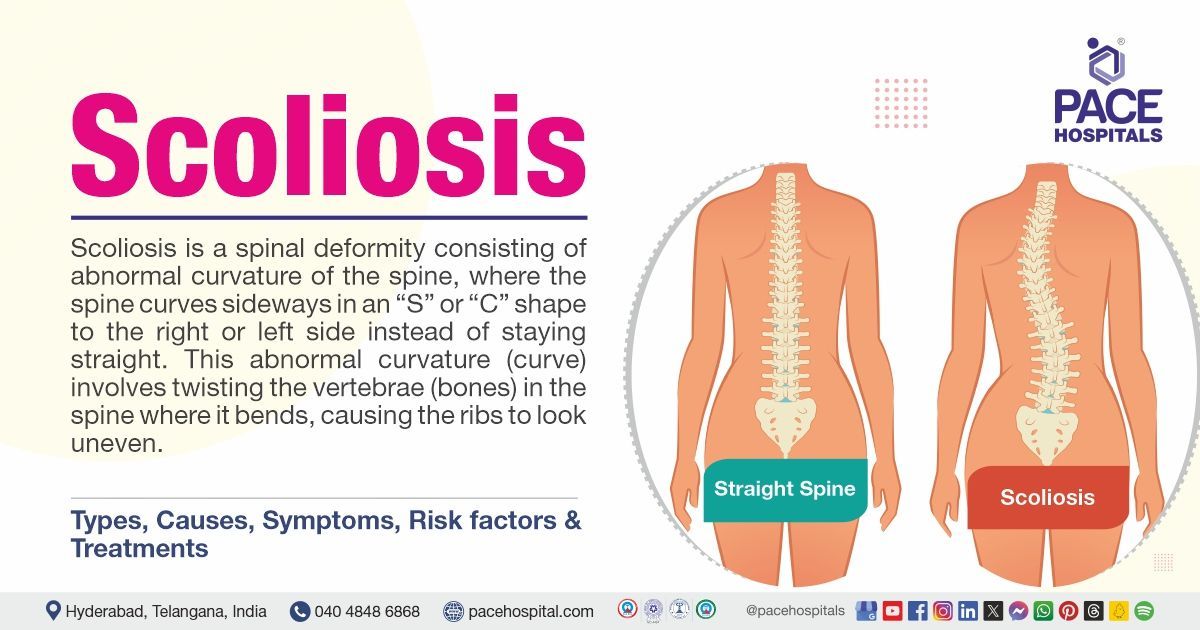
Scoliosis is the sideway spine curvature in which the curve may occur on the left or right side. If the angle of curvature is diagnosed with more than 10° on the X-ray, then the condition is considered scoliosis. Usually, doctors describe this condition as 'C' or 'S'. There are three types of scoliosis: congenital, neuromuscular, and degenerative.
Symptoms of scoliosis include curves on the back, uneven shoulders or hips, lower back pain, stiffness, ribs postured on the sides of the body, dyspnoea (breathing difficulties), fatigue, etc. The causes of scoliosis are ageing, genetic factors, neuromuscular conditions, uneven leg length, and osteoporosis. In addition to these factors, poor posture, spinal curvatures, connective tissue disorders, and some injuries may also cause scoliosis.
Soft tissue masses are also called soft tissue sarcomas (STC). STCs are a group of more than 60 neoplasms commonly seen in individuals of extreme age. They can originate from any location all over the body and are evaluated by imaging and biopsy studies. Benign lipomas and metastatic angiosarcomas of skeletal muscle, connective tissues, and adipose tissues are clinically seen.
The symptoms include painless lumps, swelling under the skin and near the lungs, and swelling in the abdomen. These symptoms, in turn, cause constipation, abdominal pain, breathlessness and cough. The causes of soft tissue masses are germline mutations (Neurofibromatosis Type 1 (NF1) Von Recklinghausen Disease, Li-Fraumeni Syndrome, Familial Adenomatous Polyposis (FAP), carcinogens, lymphedemas and radiation.
Ankle sprains are conditions in which ligaments tear and stretch beyond their limits. Ligaments are fibrous tissues that help connect bones. Sprains are common in people of all ages. In most cases, sprains heal by conservative management, but severe fractures and sprains need surgical reconstruction.
The symptoms of ankle sprains include pain in rest / weight-bearing, tenderness to touch, bruising, swelling, and instability. Causes include exercises or walking on uneven surfaces, tripping, falling down, and sporting and athletic activities.
Shoulder sprains occur if the shoulder's ligament tears or is stretched beyond its limitation. The ligament is the connective tissue which connects joints, bones, and organs. The clavicle, scapula and sternum are the three bones responsible for the shoulder function. The symptoms of shoulder sprains are pain, bruising, unable to move the shoulder, swelling, and pop sound during injury.
Causes of shoulder sprains include trauma and injuries due to falling, twisting, hitting and overuse (sporting activities and occupation).
A sprain is an injury or trauma to the ligaments. The ligament is the connective tissue which connects joints, bones, and organs together and holds them in their places. Sprains occur if the shoulder's ligament tears or is stretched beyond its limitation.
Symptoms of sprains include swelling, joint or muscle pain, bruising, and discolouration. Causes of sprains include trauma and injuries due to falling, twisting, hitting and overuse (sporting activities and occupation).
Shoulder bursitis is a condition that results due to inflammation of the bursa. The bursae are fluid-filled sacs of the skeletal system. They cushion the space between connective tissues and bones, allowing tendons, muscles, and bones to move together. Bursae make the bones and tendons glide without friction. These are acute, chronic and infectious.
The symptoms of shoulder bursitis are pain, stiffness, and swelling, especially at night when lying. The causes of shoulder bursitis are overuse of repetitive movement and conditions such as arthritis, uraemia, diabetes, rheumatoid arthritis, and thyroid disease.
Shoulder arthritis is a condition in which damage to the cartilage inside the shoulder joint is seen. The shoulder has two joints, the ball-and-socket joint and the glenohumeral joint; the cartilage covers these two joints. In shoulder arthritis conditions, the breakdown of this cartilage occurs on the surface and deeper layers.
Symptoms of shoulder arthritis include pain at the shoulder joint, a clicking, grinding, or cracking sensation, joint stiffness, and a limited range of motion. Causes include trauma or injury, rotator cuff tear arthropathy, avascular necrosis, rheumatoid arthritis, and osteoarthritis.
Fractures to the vertebrae of the spine often lead to spine compression fractures. Vertebrae are the spine's bones; spine compression fractures are commonly called compression fractures or vertebral compression fractures.
The symptoms of this condition are pain in the middle or lower spine, intermittent sharp 'knife-like' pain, loss of height, over posture, numbness or tingling sensation, loss of control of bowel or bladder, difficulty in walking, and weakness. The causes of spine fractures are trauma, tumours that start in the bone or spread to the bone from other sites, and multiple myeloma (tumours in the spine).
Septic arthritis is a condition characterized by joint inflammation caused by joint infections by bacteria and occasionally by fungal, mycobacterial, viral, or other uncommon pathogens. It is usually a monoarticular infection involving one large joint, such as the knee or hip; however, polyarticular septic arthritis is also seen in some cases. In some conditions, septic arthritis leads to severe infection and may lead to morbidity or mortality.
The symptoms of septic arthritis include joint pain, swelling, fever, and refusal to move the joints; in some cases, severe sepsis with associated conditions is seen. Only local symptoms are seen in children affected with this condition.
The local symptoms are joint swelling, fever, and erythema. In comparison, systemic symptoms such as tachycardia, fever, irritability and loss of appetite are seen. Dermatological symptoms such as dermatitis and tenosynovitis are also seen. The causes of septic arthritis are bacterial, viral and fungal infections due to trauma or injuries (open wounds), surgeries, and abdominal infections. Individuals with leukaemia and rheumatoid arthritis conditions develop septic arthritis.
The ligament is a part of the knee that holds bones together and enables motion. The two ligaments that intersect inside the knee are termed the anterior cruciate ligament (ACL) and posterior cruciate ligament (PCL). The ACL is commonly injured due to sudden foot rotation during sporting activities like soccer, football, and basketball. The injury is sometimes heard with a pop or snap, resulting in severe pain.
The symptoms of an ACL tear are swelling of the knee, instability of the knee, inability to bear weight on the leg, and a pop or snap at the time of the injury. The causes of an ACL tear are accidents, work injuries, rapid change of direction and sudden stoppage during sporting activities, direct collision or attacks during a football tackle, and landing incorrectly while jumping.
MCL (medial collateral ligament) is the ligament that holds bones and cartilage in the knee joint. It is the connective link that connects the lower part of the thigh bone to the top of the shin bone. The injury or tear to the MCL is due to too much stretching. The tear of MCL may be partial or complete.
The symptoms of MCL are pain and 'pop' on a knee injury, swelling, and instability of the knee, which may lead to a fall or stumble. The common causes of MCL are changing direction, twisting the knee during running, and knee twisting on sudden jumps and landing. In addition to this, the players may experience the MCL sprain during sporting activities such as skating, skiing, football, soccer, tennis, and basketball.
Tendonitis, often called an inflamed or irritated tendon, is the tissue band that connects muscles to bones. It may occur anywhere in the body, but it is commonly seen in ankles, wrists, shoulders, and elbows.
Tendonitis may be acute (sudden onset) or chronic (long-term inflammation, where the tendon degenerates progressively). The symptoms of tendonitis are swelling, grating sensation on tendon movement, lump on the tendon, pain, tenderness at the site of the affected tendon, and decreased motion or movement of the tendon.
The exact cause of tendonitis is not known. However, it might be caused by injury, overuse, excessive straining, or exercise. Tendonitis is also related to rheumatoid arthritis, diabetes, or infections.
Trigger function contributes to functional impairment. In this condition, inflammation and swelling occur in the tendons and wrist due to repetitive use. Narrowing of the flexor pulley with hypertrophy leads to trigger finger or stenosing tenosynovitis. This condition is mostly seen in the thumb and ring finger. Symptoms of this condition include locking digits on extension or flexion, problematic extensions, pain, odd aches during movement, and snapping sounds on extension.
Usually, the trigger finger is caused by multiple factors. The trigger finger is associated with conditions such as trauma, rheumatoid arthritis, osteoarthritis, amyloidosis, gout, diabetes, and carpel tunnel syndrome. In children, most cases are idiopathic but are associated with inflammatory and congenital conditions.
Thumb arthritis is a state in which the cartilage wears out from the thumb. The sites that are commonly affected are basal joints (carpals and metacarpals). The symptoms of thumb arthritis are pain while gripping, swelling, tenderness, loss of strength, limited motion, aching, and enlarged out-of-joint.
The causes of thumb arthritis are osteoarthritis (main cause), wear and tear on ageing, injuries (fractures and dislocations), and infections. In addition to these, several factors such as sex, weight, genetics, age, hormones and occupation may also contribute to this condition.
The tennis elbow is often called lateral epicondylitis. It is a condition characterized by swelling of tendons that bend the wrist backwards away from the palm. The tendon is a tough cord of tissue connecting muscles and bones. Extensor carpi radialis brevis is the tendon likely involved in tennis elbow. Tennis elbow is usually seen in men and women between the ages of 30 and 50.
Symptoms include pain, burning sensation, and ache at the elbow or forearm. Tennis elbow, as the name itself says, is caused by hitting the ball to the backhand while playing tennis. The factors that cause tennis elbow are weak shoulders or muscles, improper backhand stroke, and using a tight or short tennis racket. In addition to painting with the brush, frequent use of hand tools and repeated hand motions may also lead to this condition.
Tarsal tunnel syndrome is often called tibial nerve dysfunction or posterior tibial nerve neuralgia. It is a neuropathic condition associated with the compression of tarsal tunnel structures. This condition is similar to carpal tunnel syndrome. The tarsal tunnel is a small fibro-osseous space that moves behind and inferior to the medial malleolus. The symptoms of tarsal tunnel syndrome are numbness on the plantar surface, sharp shooting pain in the foot, paraesthesia, burning or tingling sensation, and pain in extremities.
The causes of tarsal tunnel syndrome are categorised into two types: extrinsic and intrinsic. Extrinsic causes include trauma, anatomical and biochemical abnormalities, oedema, poor shoe fitting, post-surgical scarring, systemic diseases, and arthropathies. Intrinsic causes include tendinopathy, tenosynovitis, lipoma, neoplasm, and nerve ischemia.
Wrist fractures are often called distal radius fractures. In this condition, the neck portion of the distal radius is involved. Likewise, distal ulna, radioulnar, joint, and radiocarpal joints may or may not involved in this condition. It is associated with high energy mechanisms in younger ones, whereas low energy mechanisms in lower ones.
Wrist fractures are caused by falls, compressions, high-energy injuries, falls from height, injuries during sporting activities and athletic participation, simple falls from standing, and motor vehicle accidents. Symptoms include wrist pain, swelling, bruising, wrist deformity, and tenderness.
A Patient from Somalia with Osteoarthritis in both knee with 90-degree Flexon Deformity, treated successfully with
Total Knee Replacement.
We provide a comprehensive and precise orthopaedic diagnosis to carry out evidence-based treatment for any kind of impairments in the musculoskeletal system. Our advanced and latest screening approach examines all sorts of abnormalities and impairments in bone, joints, muscles, ligaments, and tendons, which results in the early diagnosis and accurate evaluation, enabling our orthopedist, sports injury doctor, and orthopaedic surgeon to make an informed decision to proceed with the appropriate treatment modalities and surgical procedures.
1.Electromyography (EMG): Electrodiagnostic studies are used to detect neuromuscular abnormalities, measuring the electrical activity in muscles and nerves to assess nerve function and evaluate injuries. These studies include electromyography (EMG) and nerve conduction studies (NCS), which are commonly ordered during the assessment of patients with suspected peripheral nerve compression. These tests are often conducted together, and the combined evaluation is commonly referred to as two and abbreviated as an EMG/NCS or commonly called an EMG test. During the test, thin needles are placed in specific muscles, and then a healthcare professional asks the patient to mobilize the muscle to look at the electrical signals.
2. Bone Scan: A bone scan is also called a skeletal scintigraphy, a specialized radiology procedure used to examine the abnormal areas or damage in various bones of the skeleton, which is performed to detect areas of chemical and physical changes in bone. During this procedure, a tiny amount of radioactive material is injected into a person's vein, travels through the blood, collects in the bones and is detected by a special camera that takes images of the inside of the body called a scanner. This scan is advised to detect bone non-cancerous tumours or cancers that have spread to the bone and to diagnose other bone conditions, including bone infections, fractures, etc.
3. Arthroscopy: Arthroscopy is a keyhole surgery (minimally invasive surgical procedure) used to examine and treat problems inside the joint. During this test, a surgeon inserts a thin tube attached to a camera (arthroscope) into the joint through small incisions to view the joint on a monitor and address issues such as cartilage damage, tears, or loose fragments.
4. Dual-energy X-ray Absorptiometry (DEXA): Dual-energy X-ray Absorptiometry (DEXA) is a medical imaging technique primarily used to assess bone mineral density (BMD) and body composition. It employs two different X-ray energy levels to distinguish between bone and soft tissue, providing precise measurements of bone density, which is crucial for diagnosing conditions like osteoporosis, monitoring the effectiveness of osteoporosis treatments and predicting the likelihood of bone fractures can also include measuring muscle and fat composition in specific body areas.
5. Nerve Conduction Studies: A nerve conduction velocity (NCV) test is performed to measure how fast (velocity) an movement of electrical impulse moves through the person's nerve. During this examination, two electrodes are placed on the skin over a muscle or over a nerve where one electrode stimulates the nerve with a very mild electrical impulse, and others record it, which is repeated for each nerve being tested. Healthcare professionals measure the distance between electrodes and the time taken for electrical impulses to travel between them to calculate the speed of electrical impulses.
6. Muscle Biopsy: Muscle biopsy is a common diagnostic procedure used by clinicians to evaluate patients with weakness that may be caused by muscle disease. It involves removing a small tissue sample for testing in a laboratory to diagnose neuromuscular disorders, infections that affect muscle, and other abnormalities in muscle tissue, including muscular dystrophy (MD), myasthenia gravis (MG), polymyositis (chronic disease involving skeletal muscles) etc.
1. Arthroplasty: Arthroplasty is a surgical procedure that is done to restore the functionality of a joint by resurfacing the bones. An artificial joint (often called a prosthesis) is used in this procedure. This surgical procedure is indicated when other medical treatments are no longer effective in relieving joint pain and disability.
2. Achilles Tendon Repair: Achilles tendon repair is a type of surgery that is done to fix the damaged Achilles tendon. During this procedure, an incision is made in the backside of the calf. The surgeon will stitch the tendon if it ruptures. In some occasional scenarios, the Achilles tendon repair surgery will be minimally invasive with several small incisions. A special scope with a tiny camera and a light is used in this procedure to help or guide during the surgery.
3. ACL (Anterior Cruciate Ligament) Reconstruction: ACL reconstruction surgery is indicated to reconstruct the ligament in the centre of the knee. This procedure is usually done with the help of knee arthroscopy by inserting a tiny camera into the knee through a small surgical cut. The surgeon will make necessary small cuts around the knee and insert medical instruments to fix or correct the abnormality. After the insertion, the surgeon will stitch with sutures and covers the area with a dressing.
4. Bracing and casting: Hip and knee braces are commonly used to support post-surgery patients to prevent or reduce the severity of injuries. The braces are meant for stabilization, mechanical correction, immobilization, and rehabilitation. The casting material is a Plaster of Paris or synthetic used to immobilize the fractures and complex ligament injuries of the lower leg and maintain the foot position during conservative management. Casting is a temporary measure for definitive fracture fixation.
5. Bunionectomy: Bunion surgery, or bunionectomy, is a treatment that is used to correct bunions. A bunion (hallux valgus), is a bony bump-like structure present aside from the big toe joint. There are a few types of bunion surgery that are involved in repositioning the big toe to get relief from the pain and to improve its function. This surgery uses different techniques such as Exostectomy, Osteotomy, and Arthrodesis.
6. Carpal Tunnel Release: Carpal tunnel release is a kind of surgery indicated to treat and heal painful carpal tunnel syndrome. During this procedure, the surgeon cuts through the ligament, pressing down on the carpal tunnel, making more space for the tendons and median nerve to pass through the tunnel, thereby reducing the pain and improving the function. This outpatient procedure is usually done in traditional or open carpal tunnel release and endoscopic carpal tunnel release.
7. Decompression Surgery: Decompression surgery is indicated to treat chronic lower back pain. This surgery involves either the partial or complete removal of anatomical structures from the lumbar spine, which causes neural impingement. The types of decompression surgery are laminectomy and discectomy, whereas the procedures include open conventional and microscopic. The indications of decompression surgery are disc herniations, neurological dysfunction and spinal stenosis.
8. Electrical Stimulation: Electrical stimulation is a therapy developed in recent years for healing bone abnormalities or fractures related to bones. Bone stimulators are the devices that are often used in this procedure to treat fractures which are failed to heal on their own. These types of fractures are called "non-unions."
9. Extracorporeal shock wave therapy (ESWT): Extracorporeal shockwave therapy (ESWT) is a non-invasive treatment used to treat several conditions, such as chronic plantar fasciitis, tendonitis, and Achilles tendonitis. Shockwave therapy helps to induce the body's normal healing process. Extracorporeal shockwave therapy works by using either focused shockwaves or radial pressure waves. These are outpatient procedures that last for 5–15 minutes. On turning on the ESTW machine, the instrument or shockwave gun is pressed against the affected site to deliver rapid impulses to the patient.
10. Fracture Repair: Bone fracture repair is a type of surgical procedure that fixes a broken bone using pins, plates, metal screws, or rods to hold the bone in its place. This procedure is also called open reduction and internal fixation (ORIF) surgery.
11. Hammertoe correction: Hammer toe surgery is one of the outpatient procedure which requires anaesthesia or numbing medications. The type of procedure is suggested based on the severity of the hammer toe. A flexible toe requires tendon transfer, whereas joint resection or joint fusion is suggested for stiff or fixed toes.
12. Hip Replacement: Hip replacement, often called hip arthroplasty, is a surgical procedure used to treat hip pain. This surgery replaces parts of the hip joint with an artificial implant. Hip replacement surgery may require replacement of one or both parts. The motto of hip replacement is to allow the individual to resume daily activities and exercises.
13. Joint Aspiration: Joint aspiration is a procedure that removes the fluid from the space surrounding a joint using a needle and syringe. This procedure requires local or regional anaesthesia to relieve swelling or obtain fluid for analysis to diagnose a patient with a joint disorder or problem. Joint aspiration is mostly done on the knee. However, this procedure can also be used to remove the fluid from other joints, such as the hip, shoulder, elbow, ankle, or wrist.
14. Joint Debridement: Debridement is referred to as the surgical removal of damaged tissue. This procedure is usually done with an arthroscopy technique and requires general or local anaesthesia; Arthroscopic debridement is an accessible treatment modality that involves chondroplasty, synovectomy, toilet lavage, and meniscectomy which aims to reduce the symptoms by removal of inflammatory factors and cartilaginous debris. It is well-indicated for mild to moderate osteoarthritis, especially in the knee and joints. Irrigation, often known as "lavage," is essential to rinse out the fluid from the knee.
15. Joint Distension: Joint distension, commonly known as arthrographic or hydrodialtion, is performed under local anaesthesia. This procedure initially injects a dye into the joint to visualize it clearly on an imaging device. This procedure involves injecting sterile water into the joint to expand the area and make the adhesions loose and pull away. This frees up the space near the joints and improves the range of movement. It is a less invasive and the best alternative to shoulder injury procedures.
16. Joint Manipulation: Joint manipulation is a procedure that involves the synovial joint's opposing articular surfaces being split apart (gapped) and thereby causing cavitation in the joint's synovial fluid. Joint manipulation is a brief manual therapy technique that only affects the target joint and may produce a slight pop. Joint manipulations can offer short-term pain relief and mobility improvement in a dysfunctional location.
17. Knee Replacement: Knee replacement, commonly called total knee replacement, or knee athroplasty, is a surgical procedure to resurface a knee damaged due to arthritis. Metal and plastic parts are incorporated to seal the bones' ends that form a knee joint alongside the kneecap. This surgery is considered for individuals with severe arthritis or a severe knee injury.
18. Laminectomy: Laminectomy is a type of procedure involving the removal of a part or total of the vertebral bone (lamina). This procedure reduces pressure on the spinal cord or the nerve roots that are caused by injury, stenosis, herniated disc, or tumours. The laminectomy is suggested if the medical therapy is failed. This procedure is usually done for neck or back pain that continues on medical treatment or is done when the pain is due to nerve damage, such as numbness / weakness in the arms or legs.
19. Meniscus Repair: Meniscus repair will be done based on the grade of the condition (meniscus tear). The meniscus tear of grade-3 is suggested with surgeries such as either arthroscopic repair or arthroscopic partial meniscectomy, or total meniscectomy. The arthroscopic procedure is done by inserting the arthroscope to look at the tear and placing small devices like darts to stitch it. Arthroscopic partial meniscectomy involves the removal of a piece of the torn meniscus to make the knee functional, whereas total meniscectomy involves the complete removal of the meniscus.
20. Osteotomy: An osteotomy is a type of surgical procedure used to cut and reshape the bones. This procedure is commonly used to repair the joint and to shorten or lengthen the deformed bones. During this osteotomy procedure, general or regional anaesthesia is given prior to the procedure, and then the surgeon will make a small cut in the skin to guide a special wire that measures the bone. The section is taken out by using a special surgical saw, followed by filling the open space. Tiny screws and metal plates are commonly used in this procedure to hold the bones. The types of osteotomy are knee, hip, spine, toe, chin, and jaw.
21. Rotator Cuff Repair: Rotator cuff (tendons and muscles that form a cuff on the shoulder) repair is a surgery used to correct a torn tendon in the shoulder. The procedure can be done with an open incision or with arthroscopy. Prior to this procedure, general anaesthesia is given. Three common techniques that are used to repair the rotator cuff tear are open repair (used for large or complex tears), shoulder arthroscopy, and mini-open repair surgery.
22. Spinal Fusion: Spinal fusion is a type of surgical procedure indicated to correct the problems that are found in the bones of vertebrae (spine). Spine fusion is a welding process done to eliminate pain and restore the spine's stability. This surgery is usually recommended when the ortho surgeon confirms the source of the pain. This procedure uses an imaging test, such as X-rays, computed tomography scans (CT-scan), and magnetic resonance imaging (MRI) scans.
23. Synovectomy: Synovectomy is a surgical procedure involving destroying or removing the synovial membrane. A synovial joint is the largest articulation, is frequently affected by chronic inflammation, and often undergoes synovectomy. Open surgical, radiation, chemical, and arthroscopic synovectomies are the surgical types indicated to remove potentially damaging synovium from the knee. The common indications for synovectomy are chronic inflammatory arthritis conditions, benign neoplastic disorders and recurrent hemarthrosis.
24. Tendon Repair: Tendon repair is a surgical procedure indicated to treat a damaged or torn tendon. Tendon repair surgery is commonly suggested to people with tendon injuries (lacerations and sports injuries) that are painful and make it difficult to move. The aim of this surgery is to bring back the normal movement. This procedure requires a small incision followed by sewing the torn ends of tendons; thereby, the incision is closed with stitches and dressings. Prior to the procedure, regional or local anaesthesia is given.
25. Total Joint Replacement: Total joint replacement is a surgical technique in which arthritic or diseased joint pieces are taken out and replaced with a metal, plastic, or ceramic prosthesis. The prosthesis can replace a natural, healthy joint's motion. Joint replacement surgery can be done on various joints, such as the elbow, shoulder, ankle, and wrist. Hip and knee replacements are the most often performed joint replacements.
26. Visco-supplementation: Visco supplementation is a procedure where the ortho surgeon injects hyaluronic acid into the joints. This fluid reduces the pain and swelling in the joints. Before this procedure, numbing medicine is injected into the space around your knee joint, followed by draining of fluid that causes swelling and pain. This method is commonly indicated to treat arthritis conditions.
27. Prolotherapy: Prolotherapy is a type of medical therapy indicated to treat joint and muscle pain. Prolotherapy is commonly called proliferation therapy or regenerative injection therapy. This procedure involves an injection of saline or sugar substance into the muscle or sore joints, this substance acts as an irritant, and thereby the body sends the immune cells and other chemicals to the injected area, which in turn starts the natural healing process. This process is used to repair the damaged soft tissue in muscle or joint areas such as muscle tissues, nerves and blood vessels.
By clicking on Subscribe Now, you accept to receive communications from PACE Hospitals on email, SMS and Whatsapp.
Thank you for subscribing. Stay updated with the latest health information.
Oops, there was an error. Please try again submitting your details.
Payment in advance for treatment (Pay in Indian Rupees)
For Bank Transfer:-
Bank Name: HDFC
Company Name: Pace Hospitals
A/c No.50200028705218
IFSC Code: HDFC0000545
Bank Name: STATE BANK OF INDIA
Company Name: Pace Hospitals
A/c No.62206858997
IFSC Code: SBIN0020299
Scan QR Code by Any Payment App (GPay, Paytm, Phonepe, BHIM, Bank Apps, Amazon, Airtel, Truecaller, Idea, Whatsapp etc)
Disclaimer
General information on healthcare issues is made available by PACE Hospitals through this website (www.pacehospital.com), as well as its other websites and branded social media pages. The text, videos, illustrations, photographs, quoted information, and other materials found on these websites (here by collectively referred to as "Content") are offered for informational purposes only and is neither exhaustive nor complete. Prior to forming a decision in regard to your health, consult your doctor or any another healthcare professional. PACE Hospitals does not have an obligation to update or modify the "Content" or to explain or resolve any inconsistencies therein.
The "Content" from the website of PACE Hospitals or from its branded social media pages might include any adult explicit "Content" which is deemed exclusively medical or health-related and not otherwise. Publishing material or making references to specific sources, such as to any particular therapies, goods, drugs, practises, doctors, nurses, other healthcare professionals, diagnoses or procedures is done purely for informational purposes and does not reflect any endorsement by PACE Hospitals as such.