Kidney Stones Treatment in Hyderabad, India
PACE Hospitals is recognized as the Best Hospital for Kidney Stone Treatment in Hyderabad, providing advanced care for patients with kidney stones. Our expert team of doctors and specialists offers comprehensive treatment options, including lifestyle modifications, medications, and minimally invasive kidney stone surgery.
With state-of-the-art diagnostics and highly experienced kidney stone specialists, we ensure safe and effective kidney stone surgery, helping patients relieve pain, prevent recurrence, and restore urinary health. We also provide transparent guidance on operation cost, ensuring quality care with affordability.
Book an appointment for
Kidney Stone Treatment
Kidney Stones Treatment Appointment
Thank you for contacting us. We will get back to you as soon as possible. Kindly save these contact details in your contacts to receive calls and messages:-
Appointment Desk: 04048486868
WhatsApp: 8977889778
Regards,
PACE Hospitals
HITEC City and Madeenaguda
Hyderabad, Telangana, India.
Oops, there was an error sending your message. Please try again later. Kindly save these contact details in your contacts to receive calls and messages:-
Appointment Desk: 04048486868
WhatsApp: 8977889778
Regards,
PACE Hospitals
HITEC City and Madeenaguda
Hyderabad, Telangana, India.
Why Choose PACE Hospitals for Kidney Stone Operation?

Advanced Diagnostic Facilities: Ultrasound KUB, X-Ray, CT KUB, IVP and Urine/Blood Tests for Accurate Kidney Stone Evaluation
Specialized Kidney Stone Treatment by Leading Urologists in Hyderabad
Comprehensive Minimally Invasive Options: Ureteroscopy (URS), Retrograde Intrarenal Surgery (RIRS), Percutaneous Nephrolithotomy (PCNL) for Safe and Effective Stone Removal
Affordable & Transparent Kidney Stone Treatment with Insurance & Cashless Options
Kidney Stone Diagnosis
The diagnosis of kidney stones involves a careful assessment by the urologist, who relies on medical history and physical findings to determine the diagnosis. Since kidney stones can cause pain and complications, the choice of diagnostic tests depends on the patient’s symptoms and overall health.
The urologist considers the following before selecting the appropriate tests to diagnose a kidney stone:
- Medical history
- Physical examination
Medical history
- Urologists begin by obtaining a detailed medical history and inquiring about the symptoms the patient is experiencing. Common kidney stone symptoms include severe pain in the side or back, haematuria (blood in the urine), pain during urination, and frequent urination.
- Information is collected regarding the time of onset, severity, and location of the pain.
- The absence of these features makes kidney stones less likely and directs attention to other potential causes, such as urinary tract infections (UTI) or gastrointestinal problems.
- Past medical history, including prior episodes of stones, current medications, metabolic disorders, dehydration, or family history of urolithiasis, is also explored.
- If no risk factors are present and the symptom profile does not align with typical renal colic, the doctor can reasonably rule out the possibility of kidney stones.
Physical examination
- A physical examination may be performed to assess for signs of tenderness or pain in the abdomen or back, as it allows the doctor to determine whether the patient's symptoms match the common presentation of stone-related pain.
- During the examination, the doctor palpates the flank and abdomen to identify areas of tenderness. Kidney stone pain is usually sharp, radiates to the groin, and worsens with pressure over the costovertebral angle. If such tenderness is absent and the pain pattern suggests other causes, such as muscular strain, appendicitis, or gastrointestinal discomfort, then kidney stones are considered less likely.
- Additionally, the doctor evaluates vital signs, such as fever or low blood pressure, which may indicate an infection or another systemic disease rather than stones. Careful examination, therefore, helps distinguish kidney stones from other abdominal or urinary conditions, guiding the need for further testing.
Kidney Stone Tests
Based on the above information, a urologist may recommend tests to confirm the presence of renal stones, assess the extent of obstruction, and rule out potential complications, such as infection.
The following are the diagnostic tests that may be suggested for the diagnosis of kidney stones:
- Laboratory test
- Blood tests
- Urinalysis
- Imaging studies
- X-rays (KUB)
- Ultrasound
- Computed tomography (CT) scan
- Intravenous pyelogram (IVP)
- Plain renal tomography
- Nuclear renal scanning
- Magnetic resonance imaging (MRI)
Laboratory tests
Laboratory testing is critical in the initial diagnosis and further evaluation of kidney stones. These diagnostic tests help recognise the presence of a kidney stone, identify underlying metabolic-related abnormalities, assess kidney function, and detect any related infections.
Complete blood cell count (CBC)
A CBC is useful for checking for signs of infection or inflammation, which may occur when a kidney stone causes a blockage and leads to a secondary urinary tract infection. An elevated white blood cell count (leukocytosis) can indicate an infection, while anaemia may suggest bleeding complications associated with the stones. Thus, CBC supports the evaluation of kidney stone complications.
- C-reactive protein (CRP): CRP is an inflammatory marker of the body. If levels are high, it suggests an active infection or inflammatory response, which may accompany obstructing kidney stones, particularly in severe conditions like pyelonephritis or urosepsis. Elevated CRP helps doctors determine the severity of the condition and the need for urgent treatment.
- Coagulation testing: These tests (such as PT, APTT, INR) evaluate the blood’s ability to clot. They are not specific to kidney stones, but are important before surgical or interventional procedures (like lithotripsy or stone removal). Abnormal results may increase the risk of bleeding during treatment, so they guide safe management in stone-related cases.
- Parathyroid hormone (PTH) test: The parathyroid hormone test is essential when hyperparathyroidism is suspected as a cause of recurrent calcium stones to identify metabolic or hormonal contributors.
- Serum creatinine level: Creatinine levels reflect kidney function. Elevated creatinine may indicate impaired kidney function due to obstruction caused by a stone. Monitoring creatinine levels helps evaluate the degree of obstruction and determine the urgency of intervention. Prolonged blockage can harm the kidneys.
- Blood urea nitrogen (BUN): BUN measures the levels of urea nitrogen in the blood, a waste product formed from the breakdown of protein. Normally, the kidneys filter out urea from the blood into the urine. If a kidney stone obstructs the urinary tract, it can impair this filtering process and cause elevated BUN levels. An increased BUN, along with serum creatinine, indicates reduced kidney function and possible obstruction due to stones.
- Serum electrolytes: Serum electrolytes (such as sodium, potassium, calcium, chloride, and bicarbonate) are important in evaluating kidney function and metabolic disturbances caused by kidney stones. For example, abnormal calcium levels may indicate a risk of calcium kidney stones, which are the most common type. Potassium and bicarbonate levels help detect acid-base imbalances. These imbalances can occur in conditions such as renal tubular acidosis, which can increase a person's likelihood of developing stones. In situations where stones cause prolonged obstruction, changes in electrolyte levels indicate declining kidney function.
- Serum uric acid: Elevated uric acid levels in the blood may indicate uric acid stone formation and guide specific management approaches, such as drugs to lower uric acid and dietary modifications. This test not only helps confirm the stone type but also aids in preventing kidney stones from recurring by managing the underlying metabolic cause. Hence, serum uric acid testing is valuable for both diagnosis and long-term care planning in patients with kidney stones.
Urinalysis
Urinalysis provides important clues for diagnosing kidney stones. The presence of blood in urine (haematuria) is a common finding when stones irritate the urinary tract. Detecting crystals can indicate the sort of stone, such as calcium oxalate or uric acid. Elevated white blood cells or nitrites may indicate a urinary tract infection linked to stones. Protein in the urine could indicate renal involvement.
- 24-hour urine profile: This test evaluates risk factors for stone formation by measuring urine volume and metabolic constituents, guiding personalised prevention plans, especially for individuals with a history of recurrent stone formation.
- Urine culture: Urine Culture is performed when there is suspicion of a urinary tract infection, particularly in conjunction with kidney stones. It involves growing bacteria from a urine sample to identify the specific infecting organisms and determine their sensitivity to antibiotics. Certain types of kidney stones, like struvite stones, are closely linked to infections. Detecting and treating infections is crucial to prevent complications, such as kidney damage or sepsis.
- Urine pH: Measuring urine pH helps determine the type of stone a patient is likely to form. Acidic urine (low pH) favours uric acid or cystine stones, while alkaline urine (high pH) is associated with struvite or calcium phosphate stones. Monitoring pH of the urine is also helpful for evaluating treatment efficiency, such as urine alkalinization in uric acid stones.
Imaging studies
Imaging studies are important for confirming the presence, size, and location of kidney stones. These studies help identify urinary tract obstruction, assess complications, and guide appropriate treatment decisions. Imaging is frequently the most direct and reliable way to visualize stones in the kidneys or ureters.
- X-rays (KUB): Plain X-rays of the abdomen and/or kidneys (known as a X-ray KUB) are a standard imaging test used to diagnose renal stones. It primarily detects radiopaque calcium-containing stones; it often misses radiolucent stones. KUB X-rays help determine the size, shape, number, and kidney stone location, particularly those that contain calcium. These stones are visible because they are denser than the nearby tissues. This information is important to plan treatment, such as deciding if a stone is suitable for shock wave lithotripsy or surgical removal.
- Ultrasound: This test uses sound waves to create real-time images of the kidneys, ureters, and bladder. This is useful for detecting hydronephrosis and most stones, though less sensitive for small ureteric stones compared to CT. Ultrasound is a radiation-free option, especially useful for children, pregnant women, and detecting stones that are not visible on X-rays.
- Computed tomography (CT) scan: A Non-contrast CT scan is recognised as the gold standard for diagnosing kidney stones. A CT scan produces detailed cross-sectional images that can detect very small stones anywhere in the urinary tract. It not only confirms the presence or absence of stones but also precisely locates them, assesses their size, and detects any related problems like blockage or inflammation. CT scans are quick, widely available, and can also help evaluate other causes of abdominal or flank pain.
- Intravenous pyelogram (IVP): IVP is an older imaging test that assesses the urinary tract using X-rays and contrast dye. Due to their greater accuracy and lower risk, CT scans have largely replaced this method, which was once used to detect stones and urinary obstruction. When CT or other imaging is not available or is not recommended, IVP may still be utilised in certain circumstances for a thorough anatomical evaluation.
- Plain renal tomography: This imaging method involves taking multiple X-ray images of the kidneys at different angles. It can help detect stones that may not be clearly visible on a standard abdominal X-ray. By providing more defined layers of the kidney, it improves visualisation of calcifications and abnormalities. However, with the advancement of CT scans, plain renal tomography is now rarely used, but it can still help rule out kidney stones when more advanced imaging is unavailable.
- Nuclear renal scanning: Nuclear renal scanning is primarily used to assess kidney function, perfusion, and drainage, rather than to directly detect stones. It can help evaluate if a stone is obstructing by showing impairment of kidney drainage, but it does not visualise the stones themselves.
- Magnetic resonance imaging (MRI): MRI can identify secondary changes, such as swelling of the kidney (hydronephrosis), that indicate obstruction. Advanced techniques like MR urography improve visualisation of the urinary tract and can indirectly rule out kidney stones by showing whether any obstruction or abnormality is present.
Once a kidney specialist has diagnosed a kidney stone, they will develop a treatment plan based on the size, location, and type of stone.
Stages of Kidney Stones
Kidney stones usually do not form suddenly. Instead, they progress through different stages, beginning with chemical changes in the urine and ending with the passage or removal of the stone. Understanding these stages helps urologists identify kidney stones early, provide timely treatment, and prevent complications. Stages of kidney stones include:
Stage 1: Kidney stone formation (crystal nucleation)
Kidney stones begin as microscopic crystals formed from supersaturated urine containing stone-forming substances like calcium, oxalate, phosphate or uric acid. These crystals act as the nidus (starting point) for further stone growth, driven by physicochemical factors and crystal aggregation in the kidney tubules or collecting system.
Stage 2: Crystal growth
Once crystals form, they may stick together and gradually enlarge. Over time, the crystal clusters develop into small stones. If the stone remains in the kidney, it can either stay small and unnoticed or continue to grow. This stage may still be painless and without obvious symptoms.
Stage 3: Movement into the ureter
Once stones reach a size that can obstruct urinary flow, they may move into and travel through the ureter, causing intense episodes of cramp-like pain known as renal colic. This stage is marked as the symptomatic stage and generally the time of initial clinical presentation.
Stage 4: Obstruction and complications
A stone lodged in the ureter obstructs urine flow, causing hydronephrosis (swelling of the kidney) and severe pain. This increases the risk of urinary tract infection and kidney damage. The presence of fever or signs makes it a medical emergency.
Stage 5: Passing and intervention
The final stage, depending on the size and location of the kidney stone, it may pass on its own through the urinary tract. Smaller than 5mm kidney stones often pass naturally with increased fluid intake and pain-management drugs, or they can be relaxed with ureteral dilation. Larger stones or those causing persistent symptoms usually require medical treatment such as lithotripsy, ureteroscopy, or surgery.
Kidney Stone Differential Diagnosis
The signs of kidney stones usually include haematuria (blood in urine), acute flank pain, nausea, vomiting, and sometimes urinary symptoms such as urgency or frequency. However, these symptoms are not unique to kidney stones and may resemble those of other medical conditions affecting the gastrointestinal system, urinary tract, or musculoskeletal structures. Therefore, a careful differential diagnosis is essential to distinguish nephrolithiasis (kidney stones) from other potential causes of similar symptoms.
Below are some of the conditions that are included in the differential diagnosis of a kidney stone:
- Appendicitis: This condition can resemble kidney stone pain because both cause severe abdominal discomfort. While appendicitis generally produces pain in the right lower abdomen with fever and nausea, kidney stones cause colicky pain radiating to the flank or groin, often with blood in the urine.
- Benign familial haematuria: This inherited disorder causes blood in the urine without any underlying structural kidney disease. Since haematuria is also a common feature of kidney stones, confusion can occur. The lack of pain or obstruction helps distinguish this condition from stone disease.
- Cholecystitis: This inflammation of the gallbladder causes right upper abdominal pain, nausea, and vomiting. These symptoms can mimic those of kidney stones, but cholecystitis pain is usually confined to the upper abdomen and not linked to urinary issues.
- Costochondritis: This inflammation of the chest wall cartilage produces sharp, localized pain. It may resemble the severe discomfort of kidney stones, but the pain is worsened by chest movement or pressure and is not associated with urinary abnormalities.
- Diverticulitis: This condition involves inflammation of small pouches in the colon. It causes lower abdominal pain, fever, and nausea, which can resemble symptoms of kidney stones. Changes in bowel habits help differentiate diverticulitis from kidney stones.
- Focal nephronia: This localized kidney infection presents with flank pain, fever, and malaise. It may resemble stone symptoms, but the presence of infection markers, rather than mechanical obstruction, helps distinguish the two.
- Glomerulonephritis: This inflammation of the kidney’s filtering units results in blood and protein in the urine. It may be confused with kidney stone disease, but it is usually associated with immune or systemic causes rather than obstruction.
- Hernia: This condition can cause groin or lower abdominal pain, similar to the pain associated with kidney stones. Unlike renal colic, hernia pain worsens with activity and improves with rest, and urinary findings are absent.
- Pyelonephritis: This kidney infection generally causes flank pain, fever, and chills. It overlaps with kidney stone symptoms, but the presence of pus in the urine, a high fever, and systemic illness indicates an infection rather than obstruction alone.
Considerations of a Urologist / Nephrologist before treating kidney stones
Before planning treatment for kidney stones, a urologist or nephrologist analyses the factors listed below:
- The location and size of the stone within the urinary tract.
- Whether the stone is causing pain, obstruction, or infection.
- Whether the stone is likely to pass on its own, based on its size and position.
- The severity of symptoms such as haematuria, nausea, vomiting, or urinary difficulties.
- Whether there is any fever or signs of systemic infection, which may indicate a medical emergency.
- Patient’s overall health, medical history, and any co-existing conditions (e.g., single kidney, chronic kidney disease).
- History of previous kidney stones and treatments used in the past.
- Whether the patient is pregnant, as specific imaging and treatment methods may be contraindicated.
- Patient preferences regarding treatment options, especially for elective or preventative procedures.
Kidney Stones Treatment Goals
- The main treatment goals for kidney stones are to:
- Relieve symptoms and pain associated with the stones.
- Facilitate the passage or removal of stones, either naturally or through medical/surgical intervention.
- Prevent complications such as urinary tract obstruction, infection, or kidney damage.
- Prevent the recurrence of stones by addressing the underlying causes, including lifestyle, diet, and metabolic abnormalities.
Kidney Stones Treatment
Some kidney stones / renal stones can pass out on their own, particularly if they are small (<5 mm). If a stone becomes lodged in the urinary tract, causes obstruction, or is associated with a urinary infection or complications, it requires active intervention.
Management and kidney stone treatment depend on the size, exact location, and consistency of the stones. Based on the patient's condition, initially, drug therapy, including diet and lifestyle modifications, is advised. If the stone has not yet passed, kidney stone removal surgery is performed to prevent deterioration of kidney function and further complications. In cases of complications such as urinary infection or kidney damage, open surgery is performed.
- Non-pharmacological therapy
- Dietary changes
- Observation (watchful waiting)
- Pharmacological therapy
- For acute management
- Analgesics
- Medical Expulsive Therapy (MET) (Alpha blockers)
- Antiemetics
- For prevention (based on stone type)
- Thiazide diuretics
- Antibiotics
- Xanthine oxidase inhibitors
- Urinary alkalizer
- Endoscopic and percutaneous techniques
- Shock wave lithotripsy (SWL)
- Retrograde intrarenal surgery (RIRS)
- Ureterorenoscopic lithotripsy (URSL) with Holmium Laser
- Percutaneous nephrolithotomy (PCNL)
- Surgical treatment
- Open surgery
- Laparoscopic Surgery
Non-pharmacological therapy
Non-pharmacological therapies are generally used for small kidney stones and preventive care.
Dietary changes
Dietary changes are important for treating and preventing kidney stones by modifying the substances in urine that contribute to stone formation.
- Increasing fluid intake, especially water, dilutes urine and reduces crystal formation, making stone development less likely.
- Consuming foods rich in calcium with meals helps bind oxalate in the digestive tract, reducing its absorption and urinary excretion.
- Limiting high-oxalate foods (such as spinach, nuts, and chocolate), reducing sodium intake, and moderating animal protein consumption also lowers stone risk by decreasing urinary calcium and oxalate levels and improving urinary citrate, a natural inhibitor of stone formation.
- Additionally, citrus fruits like lemons and oranges can help prevent stone formation. These dietary adaptations aim to correct metabolic imbalances that lead to stone formation and prevent recurrence.
Observation (watchful waiting)
It is a management option for small kidney stones that cause few symptoms and are likely to clear naturally without intervention. During observation, patients are checked for pain, stone passage, and symptoms of complications such as infection or blockage.
This approach avoids unnecessary invasive or surgical treatments and focuses on symptom control with pain management and hydration. However, careful follow-up with imaging is required to address any worsening symptoms or failure of stone passage promptly.
Pharmacological therapy
Pharmacological therapy plays a crucial role in both the acute management and long-term prevention of kidney stones. These kidney stone medications include the following:
For acute management
- Analgesics: These medications help relieve the intense pain caused by renal stones moving through the urinary tract. Commonly used analgesics are Nonsteroidal anti-inflammatory drugs (NSAIDs), as they reduce both pain and inflammation by lowering pressure in the urinary system. Pain management is important not only for patient comfort but also to allow other treatments, such as natural stone passage or medical expulsive therapy, to be effective.
- Medical expulsive therapy (MET) : These drugs, such as alpha blockers, relax the smooth muscles of the ureter, making it easier for the stone to move toward the bladder. Alpha blockers improve the probability of natural stone transit by reducing ureteral spasms and lowering resistance, particularly for stones in the lower ureter. This therapy lowers the time to stone expulsion and reduces the need for surgery.
- Antiemetics: These medications are used to control nausea and vomiting, which are common symptoms during acute kidney stone attacks. By reducing gastric distress, antiemetics improve hydration and the patient's comfort.
For prevention (based on stone type)
- Thiazide diuretics: These medications reduce calcium levels in the urine by increasing calcium reabsorption in the kidneys. Since high urinary calcium is a major risk factor for calcium-based kidney stones, thiazides lower the chance of recurrence. They are especially useful for patients with recurrent calcium oxalate or calcium phosphate stones linked to hypercalciuria.
- Antibiotics: Antibiotics are used when kidney stones are associated with urinary tract infections, especially with struvite stones that form in response to infection by urease-producing bacteria. Antibiotics help eradicate the infection, preventing further stone growth and complications. Antibiotics are not used to treat stones, but play a critical role in managing infected stones or preventing infection during stone treatment.
- Xanthine oxidase inhibitors : These drugs help in treating uric acid stones. They work by reducing the formation of uric acid in the body, lowering the uric acid concentration in the urine. This prevents the formation of uric acid crystals and minimises the chance of recurring stone formation in persons with gout or hyperuricemia.
- Urinary alkalizers: These medications increase the pH of urine, making it less acidic. This helps pass or manage uric acid stones and prevents the formation of new ones. Alkalinization also reduces the risk of cystine stone formation by improving cystine solubility in urine. This therapy is commonly used for patients prone to uric acid or cystine stones.
Endoscopic and percutaneous techniques
Minimally invasive surgical procedures are commonly employed in the management of kidney stones that are too large to pass spontaneously, are causing obstruction or infection, or have failed to respond to conservative treatment.
- Extracorporeal shock wave lithotripsy (SWL): In this non-invasive procedure, non-electrical shockwaves are transmitted through the body to break the kidney stones into smaller pieces, allowing them to pass through the urine.
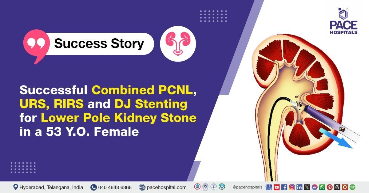
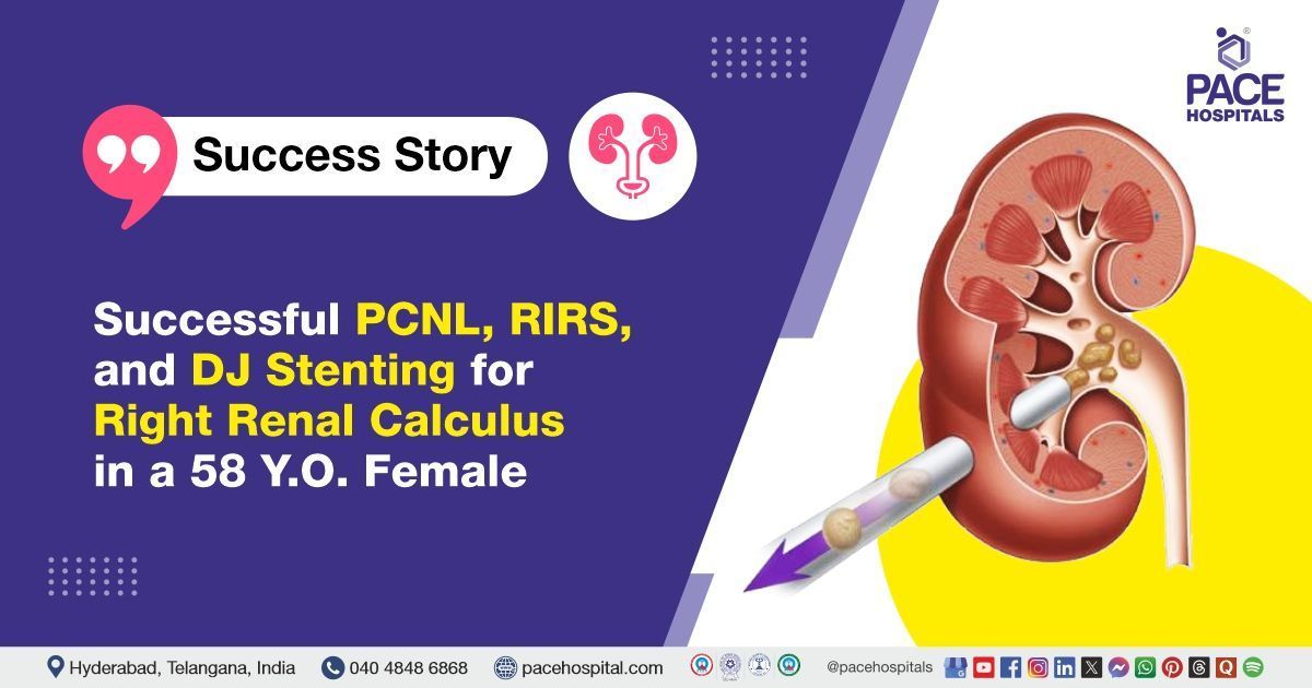
- Retrograde intrarenal surgery (RIRS): Ureterorenoscopy using a flexible ureteroscope is called Retrograde Intrarenal Surgery (RIRS). This surgery is used for intrarenal stones.
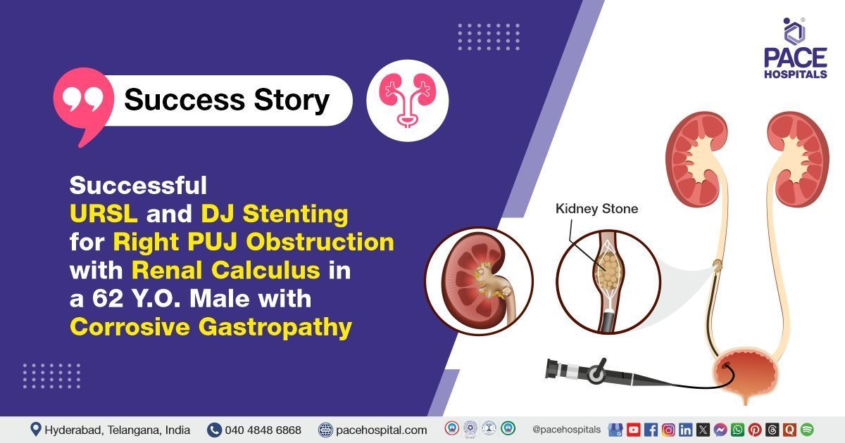
- Ureterorenoscopic lithotripsy (URSL) with Holmium Laser: A surgeon uses a thin, flexible device called a Ureteroscope to reach the kidney through the bladder and ureter when a stone is stuck within the ureter or bladder. The laser fibre is used to transmit the Holmium energy that breaks up the kidney stone, and the surgeon removes the pieces. Smaller pieces pass through the urine.
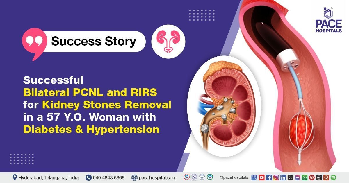
- Percutaneous nephrolithotomy (PCNL): The surgeon creates a tiny incision in the side or back and inserts a nephroscope to locate and remove the kidney stones. In cases of larger stones, shock waves or lasers are used to break them into smaller pieces.
Surgical treatment / kidney stone surgery
Surgical intervention is considered a last-line treatment for kidney stones and is usually reserved for cases where minimally invasive methods have failed or are not feasible. It may be indicated for patients with very large kidney stones, complex or recurrent stones, anatomical abnormalities, or in situations where minimally invasive procedures are contraindicated. The choice of surgical procedure depends on the size of the stones, their location, the patient's anatomy, and their overall health condition.
- Open surgery: This traditional method involves making a large incision in the flank or abdomen to access the kidney or ureter and remove the stone directly. It is rarely used today due to the availability of less invasive techniques, but it may still be required for very large stones, complex anatomy, or when other treatments fail. Open surgery is effective but carries higher risks of complications, longer recovery time, and more postoperative pain compared to modern approaches.
- Laparoscopic Surgery: This technique uses small incisions and specialised instruments to remove kidney or ureteral stones. A laparoscope (a flexible tube with a camera) allows surgeons to visualise the urinary tract while removing or fragmenting the stone. Compared to open surgery, laparoscopic surgery causes less pain, reduces blood loss, shortens hospital stay, and speeds recovery. It is often chosen for moderately large or difficult-to-reach stones when endoscopic methods are not suitable.
Kidney Stones Prognosis
The prognosis of kidney stones is generally good, especially when diagnosed early and treated appropriately. Most small stones (≤5 mm) pass spontaneously in 70–90% of cases without long-term complications, though they may cause significant acute pain. Larger stones may require medical or surgical intervention, but modern minimally invasive techniques such as ureteroscopy and percutaneous nephrolithotomy have excellent success rates with low complication risks.
This condition, however, is known for its high recurrence rate, with up to 50% of patients developing another stone within 10 years if preventive measures are not taken. Recurrence risk depends on factors such as stone composition, metabolic abnormalities, dehydration, diet, and underlying conditions like hyperparathyroidism or gout. By implementing long-term preventive measures into practice, such as dietary and medical management, recurrence rates are decreased, and results are improved.
Kidney Stone Operation Cost in Hyderabad, India
The Kidney Stone Operation cost in Hyderabad, India, typically ranges from ₹24,500 to ₹1,79,500 (US$49 to US$359). However, the final cost may vary depending on multiple factors, including the size, location, and number of stones, as well as the treatment modality chosen and hospital infrastructure.
Factors Affecting Kidney Stone Treatment Cost
- Type of Procedure: Shock wave lithotripsy (ESWL), laser treatment (RIRS or URSL), PCNL (Percutaneous Nephrolithotomy), or open surgery.
- Stone Characteristics: Size, location (kidney, ureter, bladder), number, hardness, and recurrence can significantly influence the treatment approach and cost.
- Hospital Infrastructure & Room Type: General ward, semi-private room, private room, or deluxe room options.
- Surgeon’s Expertise: Urologist's experience and hospital reputation can affect pricing.
- Anesthesia Type: Regional or general anesthesia requirements may impact cost.
- Insurance Coverage: Whether cashless treatment or reimbursement is available via insurance, corporate tie-ups, or government health schemes.
Breakdown of Kidney Stone Surgery Cost in Hyderabad, Telangana, India
Kidney Stone Laser Treatment Cost (RIRS/URSL): ₹69,500 – ₹1,79,500 (US$139 – US$359)
(RIRS - Retrograde Intrarenal Surgery / URSL - Ureteroscopic Lithotripsy) Minimally invasive laser procedures are ideal for stones in the kidney or ureter. Uses flexible scopes and Holmium laser to fragment stones. Quick recovery, no incision.
PCNL (Percutaneous Nephrolithotomy) Cost: ₹74,500 – ₹1,49,500 (US$149 – US$299)
(Standard treatment for large or complex kidney stones. A small incision is made in the back to remove stones directly from the kidney. May involve mini-PCNL or tubeless PCNL variations.)
ESWL (Extracorporeal Shock Wave Lithotripsy) Cost: ₹24,500 – ₹59,500 (US$49 – US$119)
(Non-invasive treatment using high-energy shock waves to break stones into smaller fragments. Suitable for small to medium-sized stones. May require multiple sessions.)
Open Kidney Stone Surgery Cost: ₹59,500 – ₹1,19,500 (US$119 – US$239)
Rarely performed nowadays, but used for very large or multiple stones where endoscopic procedures fail. Involves an abdominal incision and longer hospital stay.
DJ Stent Insertion or Removal Cost: ₹9,500 – ₹24,500 (US$19 – US$49)
(Sometimes required before or after laser or PCNL procedures to ensure smooth urine flow and prevent blockage.)
Follow-up Imaging and Investigations: ₹1,500 – ₹9,500 (US$3 – US$19)
(Includes ultrasound, X-ray KUB, NCCT abdomen, urine tests, and blood investigations which are essential before and after the procedure.)
Kidney Stone Laser Treatment Cost in Hyderabad, using RIRS or URSL, is slightly costlier than traditional procedures like ESWL, but it is highly effective with minimal discomfort, a reduced hospital stay, and a faster return to normal life. It is ideal for stones that are difficult to access or in patients who prefer non-invasive techniques.
At PACE Hospitals, Hyderabad, we offer comprehensive and advanced kidney stone treatment in Telangana, India. Our team of experienced urologists and endourologists use cutting-edge technology including Holmium:YAG lasers, flexible ureteroscopes, and advanced imaging, ensuring safe, precise, and cost-effective outcomes.
Frequently Asked Questions (FAQs) on Kidney Stones Treatment
Which treatment is best for kidney stones?
There is no single “best” treatment for kidney stones. The choice of therapy depends on the stone size, location, composition, associated symptoms, and the patient’s overall health.
Small stones (≤5 mm): Usually pass spontaneously with conservative management (hydration, analgesics, medical expulsive therapy).
Stones 5–20 mm: Minimally invasive procedures such as ureteroscopy (URS/URSL with laser), shock wave lithotripsy (SWL), or retrograde intrarenal surgery (RIRS) are commonly used, depending on location.
Large stones (>20 mm or >2 cm): Percutaneous Nephrolithotomy (PCNL) is considered the gold standard.
Very large or complex staghorn stones, anatomical abnormalities, or failed minimally invasive methods: Open or laparoscopic surgery may be done, though it is rarely used today.
The “best” treatment is therefore individualised, balancing effectiveness, safety, and patient needs.
Whom to consult for kidney stones treatment?
If a person is diagnosed with kidney stones (renal stones), then they should consult a urologist. Based on the size, location of the kidney stone, symptoms and medical conditions, a urologist might prescribe medication to break down the kidney stone into smaller pieces so that they can pass out through urine. In the case of a bigger kidney stone that is not coming out with medication, a urologist will perform kidney stone removal surgery to prevent infection and further complications in the future.
How are kidney stones formed?
Kidney stones form when substances like calcium, oxalate, or uric acid in urine become too concentrated, leading to crystallisation. Usually, urine contains chemicals that prevent crystals from sticking together, but when the balance is disrupted, stones can grow. Dehydration, excess salt, and more protein intake make urine concentrated, increasing stone risk. Over time, these crystals join into larger deposits, which may lodge in the kidney or travel down the urinary tract, causing pain.
How to prevent kidney stones?
Prevention mainly involves lifestyle and dietary changes:
- Drinking plenty of water throughout the day helps keep urine diluted, reducing stone formation.
- Limiting salt and animal protein lowers risk, while a diet rich in fruits, vegetables, and sufficient calcium is protective.
- Avoiding excess oxalate-rich foods like spinach, beets, and nuts may help those prone to stones.
- Maintaining a healthy BMI (body mass index) and managing conditions like diabetes or gout are also important for long-term prevention.
How to stop kidney stone pain immediately?
Kidney stone pain, known as renal colic, is often severe and requires medical care. Immediate relief usually involves pain-relieving medicines such as non-steroidal anti-inflammatory drugs (NSAIDs) or stronger prescription painkillers. Staying hydrated may help small stones pass, but sudden, severe pain often needs emergency treatment. In hospitals, patients may also receive medications that relax the urinary tract, helping the stone move more easily. Home remedies alone are rarely effective in stopping acute pain quickly.
What are kidney stones, and what causes kidney stones?
Kidney stones are small, hard deposits that form inside the kidneys when certain minerals and salts build up in urine. They can vary in size, ranging from small grains to larger stones. The primary causes include dehydration, more salt intake, obesity, high protein intake, certain medical conditions, and a family history of these conditions. When urine becomes too concentrated, crystals form and join together to make stones, which can block urine flow and cause severe pain.
What are the most common kidney stones?
The most common type of kidney stone is a calcium oxalate stone, which accounts for the majority of cases. These stones form when calcium combines with oxalate, a substance found in foods like spinach, nuts, and tea. Less common types include uric acid stones, struvite stones (associated with infections), and cystine stones (due to a rare genetic disorder). Among them, calcium oxalate stones remain the most frequent, influenced by diet, fluid intake, and genetics.
Is a banana good for kidney stone patients?
Bananas are beneficial for kidney stone patients because they are high in vitamin B6, potassium, and magnesium while being low in oxalates. Potassium helps balance calcium in the body, and magnesium prevents the growth of calcium oxalate crystals. Vitamin B6 helps remove harmful substances from the kidneys. Thus, bananas may reduce the risk of kidney stone formation and support kidney health.
How to dissolve kidney stones?
Kidney stones cannot be completely dissolved by medicines. No current medication can make a kidney stone disappear entirely. Treatment emphasises symptom management and allowing stones to pass naturally. Small stones frequently pass, and drinking plenty of water helps flush them out. For larger stones, shock wave therapy uses sound waves to break stones into tiny pieces that can be passed more easily in urine. Medications may also relax urinary muscles to aid stone passage.
What size of kidney stone requires surgery?
Kidney stones that are 20 mm or larger are generally recommended to be removed surgically, as they are unlikely to pass naturally and often cause serious obstruction or complications. For stones of sizes 6 mm and 20 mm, treatment may range from medication and less invasive procedures, depending on symptoms, stone location, and risk of complications. Stones smaller than 5 mm often pass on their own; larger stones may need intervention.
Can kidney stones cause kidney failure?
Yes, kidney stones can cause kidney failure if they block urine flow for an extended period of time or result in recurring infections. Blockage gradually damage kidney tissue, reducing its function. If both kidneys are affected, if the obstruction is not addressed promptly, permanent damage may occur. However, most cases are diagnosed and managed early, preventing such severe outcomes. Kidney failure from stones is uncommon but possible in untreated or complicated situations.
Can kidney stones cause UTI?
Kidney stones may increase the risk of urinary tract infections. Stones may block normal urine flow or act as a place where bacteria can grow, making infections more common and harder to treat. Removing stones often helps resolve frequent UTIs, highlighting the close connection between these two problems.
Is a 6mm kidney stone dangerous?
A 6 mm kidney stone is considered moderately large and has a 50% chance of spontaneous passage. It can cause pain, block urine flow, and increase the risk of kidney damage or infection if left untreated. In some cases, a urologist may recommend medical procedures, especially if symptoms are severe or complications develop.
How does a kidney stone look?
A kidney stone is a hard lump made of minerals and salts from urine. Its look can vary depending on its type. Most stones are yellow, brown, or black, while some are whitish. They may be round and smooth, but many have jagged or spiky edges, which is why they cause pain when moving. Stones can be as small as a grain of sand or as big as a marble, and rarely even larger.
Can kidney stones be seen in the toilet?
Yes, sometimes kidney stones can pass naturally and be seen in the toilet. Small stones may look like tiny grains of sand, pebbles, or crystals. They can be yellow, brown, or dark in colour. Because some stones are very small, they may be hard to notice unless urine is strained through a medical strainer. Larger stones are more visible and may pass with pain, often leaving blood in the urine.
What is the survival rate for a kidney stone?
The survival rate for kidney stones is effectively very high because kidney stones themselves are not life-threatening. Most kidney stones pass spontaneously without complications. About 80-85% of stones pass on their own, especially smaller stones under 5 mm, often within one to two weeks. Larger stones or those causing obstruction may require medical intervention or surgery.
Resources