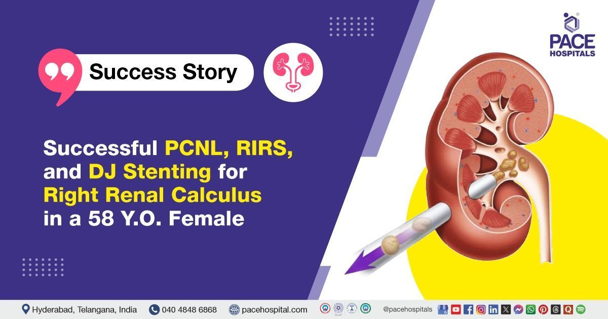
Successful PCNL, RIRS, and DJ Stenting for Right Renal Calculus in a 58-Y.O Female
PACE Hospitals
The PACE Hospital's expert Urology team successfully performed Right Percutaneous Nephrolithotomy (PCNL) with Retrograde Intrarenal Surgery (RIRS) and DJ stenting on 58-years -old female patient diagnosed with right renal calculus (Kidney stone). The procedure was carried out to remove renal stones, relieve obstruction, and restore normal urinary function.
Chief Complaints
A 58-year-old female patient with a
body mass index (BMI) of 21 presented to the Urology Department at
PACE Hospitals, Hitech City, Hyderabad, with complaints of right flank pain. A comprehensive evaluation was undertaken to identify the underlying cause of her symptoms.
Past Medical History
The patient had a known history of type 2 diabetes mellitus and hypertension, for which she was receiving regular treatment. No known drug allergies were reported.
On Examination
Upon admission, the patient was conscious, coherent, and oriented. She was afebrile, and her vital signs were stable. Physical examination revealed mild tenderness in the right flank region on deep palpation, consistent with her presenting symptoms. There was no abdominal distension, guarding, rebound tenderness, or palpable masses. Cardiovascular and respiratory system examinations were within normal limits. No peripheral edema or signs of fluid overload were observed.
Diagnosis
Upon admission to PACE Hospitals, the patient was thoroughly evaluated by the Urology team, including a detailed review of her medical history and a comprehensive clinical examination. Presenting with right flank pain, there was a strong clinical suspicion of obstructive uropathy secondary to renal calculi.
Imaging studies, including a non-contrast CT scan of the kidneys, ureters, and bladder (CT KUB), revealed multiple calculi bilaterally, with a predominant obstructive calculus measuring approximately 10 x 11 mm in the lower calyx of the right kidney indenting the renal pelvis. The left kidney showed smaller non-obstructing calculi along with faint medullary nephrocalcinosis. No significant obstruction was noted along the ureters. Additional findings included a small umbilical hernia with omental content.
Laboratory investigations, including urine examination and blood counts, were unremarkable aside from mild anemia and neutrophilic predominance, with no active infection detected.
Based on the confirmed findings, the patient was advised to undergo
kidney Stone treatment in Hyderabad, India, under the expert care of the Urology Department.
Medical Decision Making
After a detailed consultation with Dr. Abhik Debnath, Consultant Laparoscopic Urologist, a comprehensive evaluation was conducted to determine the optimal diagnostic and therapeutic approach for the patient presenting with right flank pain and confirmed right renal calculus. Based on detailed clinical evaluation and imaging studies, including non-contrast CT KUB, surgical intervention was deemed necessary.
It was determined that the patient had a right renal calculus responsible for her right flank pain. Right PCNL combined with Retrograde Intrarenal Surgery (RIRS) and DJ stenting was identified as the most effective intervention to achieve complete stone clearance, relieve obstruction, and improve her overall condition.
The patient and her family were informed about her condition, the procedure, its associated risks, and its potential to alleviate symptoms and enhance her quality of life.
Surgical Procedure
Following the decision, the patient was scheduled for Right Percutaneous Nephrolithotomy (PCNL) with Retrograde Intrarenal Surgery (RIRS) and DJ stenting in Hyderabad at PACE Hospitals, under the expert care of the urology department.
The procedure involved the following steps:
- Preoperative Preparation and Anesthesia: The patient was taken to the operating room and placed under general anesthesia to ensure complete comfort and immobility throughout the procedure. Standard aseptic precautions were followed. The patient was positioned appropriately to allow optimal access to the right kidney and ureter.
- Access and Initial Ureteroscopy (RIRS): A flexible ureteroscope was introduced through the urethra and bladder into the right ureter. Due to the tortuous and narrow ureter, careful navigation was required to reach the renal pelvis. A retrograde pyelogram was performed to delineate the anatomy and confirm the location of the calculus.
- Percutaneous Access and Tract Dilation: Under fluoroscopic guidance, a posterior inferior calyceal puncture (subcostal approach) was made to access the right kidney’s collecting system directly. The access tract was then dilated up to 24 French (Fr) to allow passage of instruments necessary for stone removal.
- Stone Fragmentation and Clearance: The obstructive right renal pelvic calculus, measuring approximately 13 mm, was located. Pneumatic lithotripsy energy was used to fragment the hard calculus into smaller pieces. Complete stone clearance was achieved by removing the fragments through the percutaneous tract.
- Placement of Double J (DJ) Stent and Closure: After stone clearance, a Double J (DJ) stent was placed to ensure proper drainage of urine and to prevent ureteric obstruction during healing. The stone fragments were sent for FTIR stone analysis. The procedure was completed without any intraoperative or postoperative complications, and the patient was monitored for recovery.
Postoperative Care
After surgery, the patient was closely monitored to ensure stability and an uncomplicated recovery. Her intra and post operative course was uneventful. During her hospital stay, she received intravenous antibiotics to prevent infection, analgesics for pain control, and proton pump inhibitors (PPIs) to reduce gastric acid secretion and protect the stomach lining. The patient was discharged in stable condition with appropriate follow-up instructions.
Discharge Medications
The patient was prescribed a broad-spectrum antibiotic to prevent postoperative infections commonly associated with urological interventions. Analgesic and antipyretic were included to manage post-surgical pain and reduce inflammation. A proton pump inhibitor was advised to minimize gastric acid secretion and protect the stomach lining, especially in the context of multiple concurrent medications. A urinary alkalinizer was included to help maintain optimal urine pH, thereby preventing stone recurrence and easing urinary discomfort. An alpha-blocker was prescribed to promote relaxation of the urinary tract muscles, aiding in the smooth passage of any residual stone fragments and improving patient comfort with the DJ stent.
To ensure regular bowel movements and avoid straining, a stool softener/laxative was recommended. An antioxidant and nutritional supplement was included to support renal recovery and counter oxidative stress. For chronic disease management, a combination antihypertensive was continued to control blood pressure, and a statin was maintained to manage lipid levels. The patient was also advised to continue her regular antidiabetic medications to maintain glycemic control in the postoperative period.
Advice on Discharge
At the time of discharge, the patient was advised to maintain an adequate fluid intake of 3 to 4 liters per day to promote urinary flow and support stone clearance. Citrus fruits and juices were recommended to help prevent stone recurrence. The patient was instructed to avoid lifting heavy weights, forward bending, and strenuous physical activities. Straining during bowel movements should be avoided, and prolonged travel by bus, auto, or two-wheeler is not advised during the recovery period. It was explained that the passage of small stone fragments in the urine is common after the procedure. Mild flank or suprapubic discomfort, occasional burning sensation during urination, cloudy urine, or small amounts of blood in the urine may occur and are expected to resolve within 7 to 10 days.
Dietary Advice
The patient was advised to follow a low-salt, diabetic-friendly diet and to avoid spinach.
Emergency Care
The patient was informed to contact the emergency ward at PACE Hospitals in case of any emergency or development of symptoms like fever, abdominal pain, bleeding, or vomiting.
Review and Follow-up Notes
The patient was advised to return for a follow-up visit with the Urologist in Hyderabad at PACE Hospitals after 2 months for removal of the DJ stent and to review the Fourier Transform Infrared (FTIR) stone analysis report. Additionally, ongoing follow-up with the physician for diabetes management was recommended.
Conclusion
This case highlights the effective management of a right renal calculus using combined minimally invasive procedures. The patient had an uneventful recovery with complete stone clearance and stable postoperative condition. Ongoing follow-up for stent removal and general health management is essential for long-term well-being.
Advancements in Minimally Invasive Techniques for Renal Calculus Management
Minimally invasive procedures like percutaneous nephrolithotomy (PCNL) combined with retrograde intrarenal surgery (RIRS) have revolutionized the treatment of complex kidney stones by enabling complete stone clearance with reduced morbidity. These techniques are particularly valuable in patients with multiple or large renal calculi, offering a safer alternative to open surgery. Careful perioperative planning, including the use of DJ stents, optimizes outcomes and facilitates recovery.
Multidisciplinary coordination among the
urologist / urology doctor, anesthesiologists, and radiologists is essential for procedural success. In this case, the combination approach resulted in effective stone removal with no complications, highlighting the importance of advanced endourological methods in modern urology practice.
Share on
Request an appointment
Fill in the appointment form or call us instantly to book a confirmed appointment with our super specialist at 04048486868
Appointment request - health articles
Recent Articles