Successful PCNL & Ureterolithotomy for Complex Kidney Stone Disease in a 66-Year-Old Male
PACE Hospitals
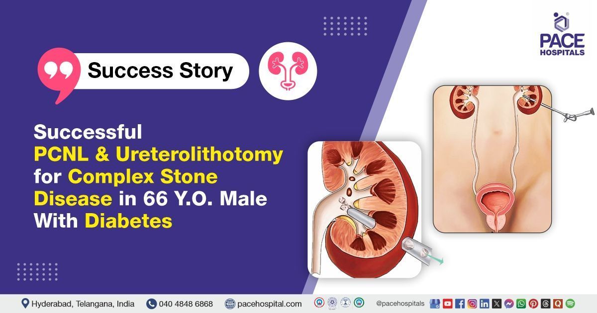
The PACE Hospital's expert Urology team successfully performed a left Percutaneous Nephrolithotomy (PCNL), right Laparoscopic Ureterolithotomy, and bilateral DJ stenting on a 66-year-old male patient who had presented with left flank pain. These procedures were performed to remove kidney and ureteric stones, effectively relieve the patient’s symptoms, and restore normal kidney function.
Chief Complaints
A 66-year-old male patient with a
Body Mass Index (BMI) of 23 presented to the Urology Department at
PACE Hospitals, Hitech City, Hyderabad, with left flank pain. A detailed evaluation was carried out to determine the underlying cause of his symptoms.
Past Medical History
The patient had a known history of
diabetes mellitus and was on regular antidiabetic medication. He also reported a documented allergy to sulfur-containing medications, which had previously caused adverse reactions. This information was considered during treatment planning to avoid potential complications and ensure safe medication use.
On Examination
On examination, the patient was conscious, coherent, and oriented, and appeared to be in a stable condition. Vital signs were within normal limits. Abdominal examination revealed mild tenderness in the left flank region, with no palpable masses or signs of peritonitis. Left renal angle tenderness was noted, which was suggestive of kidney pathology, such as kidney stones or infection. Cardiovascular, respiratory, and neurological examinations were within normal limits.
Diagnosis
The patient initially underwent a comprehensive clinical assessment at PACE Hospitals, including a detailed history and physical examination by the Urology team. During the evaluation, a series of diagnostic tests were performed. An abdominal and pelvic ultrasound revealed renal and ureteric stones.
To obtain more detailed imaging, a non-contrast CT KUB (Kidneys, Ureters, and Bladder) scan was conducted, which confirmed the presence of multiple stones in the left kidney and a large obstructive stone in the right ureter. Urinalysis showed microscopic hematuria, while renal function tests indicated mild impairment. Blood tests, including complete blood count and blood glucose levels, were performed and found to be within acceptable limits considering the patient’s diabetic history.
During the evaluation, he was diagnosed with bilateral obstructive ureteric calculi and left renal calculi, obstructive uropathy with diabetes mellitus, which necessitated surgical intervention.
Based on the confirmed findings, the patient was advised to undergo
kidney stone treatment in Hyderabad, India, under the expert care of the Urology Department, providing effective stone removal, relieving his flank pain, and preventing potential complications, including kidney damage.
Medical Decision Making
Following a detailed consultation with Dr. Abhik Debnath, Consultant Laparoscopic Urologist, and cross consultation with Dr. Tripti Sharma, consultant endocrinologist and Dr. Pradeep Kiran, consultant pulmonologist, an evaluation of the patient's condition was undertaken. Considering his left flank pain and diagnostic findings indicating stones in the left kidney and a large obstructive stone in the right ureter, the urology team concluded that left percutaneous nephrolithotomy (PCNL) and right laparoscopic ureterolithotomy with bilateral DJ stenting would be the most appropriate course of treatment for the kidney stones.
Surgical Procedure
Following the decision, the patient was scheduled to undergo left percutaneous nephrolithotomy (PCNL) and Right laparoscopic ureterolithotomy with bilateral DJ stenting in Hyderabad at PACE Hospitals under the expert supervision of the urology Department.
The patient underwent the procedure under antibiotic cover with cephalosporin antibiotic.
- Left Percutaneous Nephrolithotomy (PCNL): A supracostal puncture (above the 12th rib) was made targeting the posterior middle calyx of the left kidney under fluoroscopic guidance. A safety guidewire was placed, and the tract was serially dilated up to 24 Fr. An 18 Fr nephroscope was introduced; pneumatic lithotripsy was performed to fragment left upper ureteric and lower pole calculi. Complete stone clearance was confirmed. An antegrade DJ stent and nephrostomy tube were placed for postoperative drainage.
- Abdominal Entry and Ureter Exposure (Right Side): Access was gained through a laparoscopic port entry into the peritoneal cavity. The colon was reflected medially to expose the retroperitoneum. The right ureter was identified and carefully dissected free from surrounding inflammatory adhesions.
- Stone Extraction: A longitudinal ureterotomy was made over the site of the right upper ureteric obstructive calculus (2.9 x 1.5 cm). The stone was removed in toto without fragmentation.
- DJ Stenting and Ureteral Repair: A 6 Fr Longdura DJ stent was placed across the ureterotomy site. The ureteral incision was closed using interrupted Vicryl sutures.
- Finalization and Recovery: Hemostasis was ensured throughout the procedure. No intraoperative complications occurred. The patient was shifted to recovery in a stable condition.
Postoperative Care
The surgery was uneventful, and the patient was transferred to the ICU postoperatively for close monitoring and supportive care. Consultations with the endocrinologist for diabetes mellitus and the pulmonologist for chest X-ray changes were obtained for comprehensive evaluation, and their recommendations were followed accordingly. During the hospital stay, the patient received intravenous antibiotics, analgesics, proton pump inhibitors (PPIs), and other supportive medications as indicated.
Discharge Medication
Upon discharge, the patient was prescribed an oral antibiotic to prevent infection, an analgesic/antipyretic for pain and fever control, and a proton pump inhibitor to protect the stomach lining. An alpha-blocker was given to improve urinary flow, while a cough suppressant and laxative were provided to ease coughing and prevent constipation. Respiratory care included an expectorant to loosen mucus, a bronchodilator nebulizer to open airways, and an inhaled corticosteroid with a long-acting beta-agonist to reduce inflammation and maintain breathing. A nutritional supplement was also recommended to support overall healing.
Discharge Advice
The patient was advised to maintain adequate hydration by drinking plenty of water and fluids (3–4 litres per day) and to include citrus fruits and juices in the diet. He had been instructed to avoid heavy weightlifting, forward bending, straining during bowel movements, and prolonged travel by bus, auto, or two-wheeler. The passage of stones in the urine had been explained as a usual occurrence. Mild flank or suprapubic discomfort, occasional small amounts of blood in the urine, cloudy urine, and mild burning sensations had been described as expected symptoms, which typically resolve within 7 to 10 days. Additionally, he had been advised to perform incentive spirometry and deep breathing exercises to promote optimal lung function and prevent respiratory complications.
Dietary Advice
The patient had been advised to follow a low-salt, diabetic diet, and to avoid spinach and non-vegetarian foods (meat).
Emergency Care
The patient was informed to contact the emergency ward at PACE Hospitals in case of any emergency or development of symptoms like fever, abdominal pain, or vomiting.
Review and Follow-up Notes
The patient was advised to return for a follow-up visit with the Urologist in Hyderabad at PACE Hospitals. He was instructed to follow up with the endocrinologist for glycemic control. A review was scheduled after 2 weeks for suture removal, and he was asked to bring the Fourier Transform Infrared Spectroscopy (FTIR) stone analysis report at that time. A subsequent review was planned after 2–3 months for cystoscopy and DJ stent removal. Additionally, a complete metabolic workup was to be performed to evaluate recurrent stone formation.
Conclusion
This case highlights successful management of complex bilateral ureteric and renal calculi using a combination of percutaneous nephrolithotomy and Laparoscopic ureterolithotomy. Multidisciplinary care and careful postoperative monitoring facilitated an uneventful recovery, ensuring prevention of recurrence and optimized patient outcomes.
Optimizing Outcomes in Ureterolithotomy for Obstructive Ureteric Stones
Ureterolithotomy is a crucial surgical option for managing large or obstructive ureteric stones when less invasive methods are not suitable. Accurate preoperative imaging and metabolic evaluation guide effective treatment planning. Meticulous surgical technique focusing on preservation of the ureter’s blood supply minimizes complications. Postoperative care includes stenting, infection control, and pain management to ensure smooth recovery. Multidisciplinary follow-up with metabolic and glycemic control helps prevent recurrence and improves long-term patient outcomes. The urologist/ urology doctor plays a key role in coordinating care and educating the patient on hydration, diet, and lifestyle modifications to support the prevention of new stone formation. Regular imaging and stone analysis remain essential components of comprehensive postoperative care.
Share on
Request an appointment
Fill in the appointment form or call us instantly to book a confirmed appointment with our super specialist at 04048486868
Appointment request - health articles
Recent Articles


