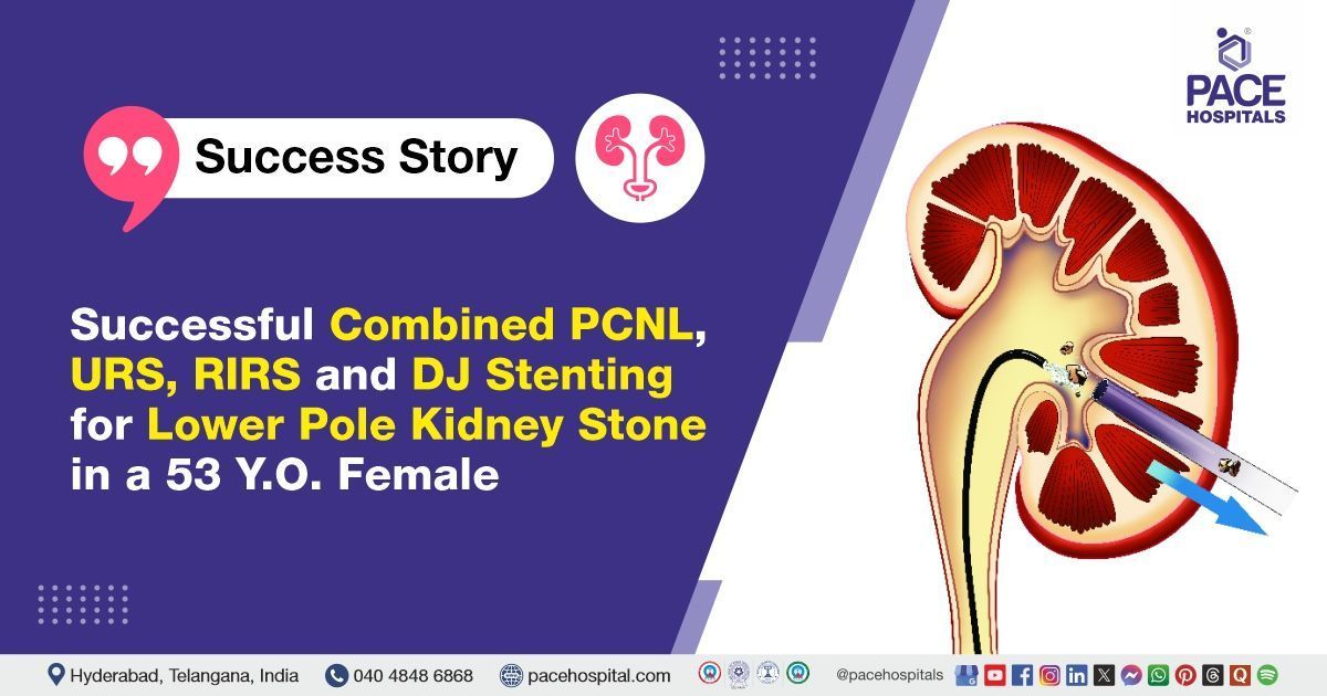
Successful Combined PCNL, URS, RIRS and DJ Stenting for Lower Pole Kidney Stone in a 53-Y.O. Female
PACE Hospitals
The PACE Hospital's expert Urology team successfully performed Left Percutaneous Nephrolithotomy (PCNL) combined with Retrograde Intrarenal Surgery (RIRS), Ureterorenoscopy (URS), and Double J (DJ) stenting in a 53-year-old female patient diagnosed with a left lower pole renal calculus (kidney stone). The procedure was carried out to remove a renal stone, relieve urinary obstruction, and restore normal kidney function.
Chief Complaints
A 53-year-old female patient, with a
body mass index (BMI) of 22.4, presented to the Urology Department at
PACE Hospitals, Hitech City, Hyderabad, with complaints of left flank pain. A detailed clinical evaluation was performed to identify the underlying cause of her symptoms.
Past Medical History
The patient had a known history of type 2 diabetes mellitus and hypertension, for which she was on regular treatment. No known drug allergies were reported.
On Examination
Upon admission, the patient was conscious, coherent, and oriented, and her vital signs were stable. Physical examination revealed mild tenderness in the left flank region on deep palpation, consistent with her presenting symptoms. There was no abdominal distension, guarding, rebound tenderness, or palpable masses. Cardiovascular and respiratory system examinations were within normal limits. No peripheral edema or clinical signs of fluid overload were observed.
Diagnosis
Upon admission to PACE Hospitals, the patient underwent a thorough evaluation by the Urology team, including a detailed clinical examination and review of medical history. She presented with left flank pain, raising suspicion of obstructive uropathy secondary to a renal calculus.
Imaging studies, including X-ray KUB, demonstrated a radiopaque calculus measuring approximately 13 mm in the lower pole of the left kidney. Chest X-ray and echocardiography revealed normal cardiac and pulmonary status.
Laboratory investigations revealed that urine microscopy showed pus cells (8–10/HPF), occasional RBCs, and bacteria; however, urine culture was sterile. Blood tests revealed normal renal function. Serum electrolytes were within normal limits. Complete blood count (CBC) indicated mild neutrophilic leukocytosis and borderline low hemoglobin, with adequate platelets. Liver function tests were essentially regular except for a mild elevation in alkaline phosphatase. Coagulation profile showed a slightly prolonged activated partial thromboplastin time (APTT), while prothrombin time and INR were normal. Glycated hemoglobin (HbA1c) indicated a well-controlled diabetes status. Urine examination showed mild pyuria, hematuria, and occasional bacteria without significant proteinuria or glycosuria.
Based on the confirmed findings, the patient was advised to undergo
kidney Stone treatment in Hyderabad, India, under the expert care of the Urology Department.
Medical Decision Making
After a detailed consultation with Dr. Abhik Debnath, Consultant Laparoscopic Urologist, a comprehensive evaluation was conducted to determine the most appropriate diagnostic and therapeutic approach for the patient, presenting with left flank pain and a confirmed left renal calculus. Based on detailed clinical evaluation and imaging studies, including X-ray KUB, surgical intervention was deemed necessary.
It was determined that the patient had a left renal lower pole calculus (13 mm) responsible for her left flank pain. Left Percutaneous Nephrolithotomy (PCNL) combined with Retrograde Intrarenal Surgery (RIRS), Ureterorenoscopy (URS), and DJ stenting was identified as the most effective intervention to achieve complete stone clearance, relieve obstruction, and improve her overall condition.
The patient and her family were informed about her condition, the procedure, its associated risks, and its potential to alleviate symptoms and enhance her quality of life.
Surgical Procedure
Following the clinical decision, the patient was scheduled for Left Percutaneous Nephrolithotomy (PCNL) combined with Retrograde Intrarenal Surgery (RIRS), Ureterorenoscopy (URS), and DJ stenting in Hyderabad at PACE Hospitals, under the expert care of the urology department.
The procedure involved the following steps:
- Preoperative Preparation and Anesthesia: The patient was taken to the operating theatre and administered general anesthesia to ensure comfort and immobility throughout the procedure. Standard aseptic precautions were observed. She was positioned in the Galdakao-modified supine Valdivia (GMSV) position, allowing simultaneous retrograde and percutaneous access to the left kidney.
- Retrograde Ureterorenoscopy (URS) and RIRS: A ureteroscope was passed through the urethra and bladder into the left ureter. Retrograde Intrarenal Surgery (RIRS) was performed to evaluate the pelvicalyceal system. The anatomy was found to be complex with a bifid collecting system, requiring careful endoscopic navigation to visualise and assess the stone burden.
- Percutaneous Access and Tract Dilation: Under fluoroscopic guidance, a lower calyceal puncture was made to access the left kidney directly. The tract was dilated up to 21 French (Fr) to allow insertion of the nephroscope and other working instruments.
- Stone Fragmentation and Clearance: A 13 mm hard calculus located in the lower pole and infundibulum of the left kidney was identified. Using an 18 Fr nephroscope, the stone was fragmented with pneumatic lithotripsy. Complete stone clearance was achieved, and all fragments were successfully removed. The extracted stone was sent for Fourier Transform Infrared Spectroscopy (FTIR) analysis.
- DJ Stent Placement and Nephrostomy: Following stone clearance, a Double J (DJ) stent was retrogradely placed to ensure optimal postoperative urinary drainage and prevent obstruction. A nephrostomy tube was placed at the end of the procedure for drainage. The surgery was completed without any intraoperative or postoperative complications, and the patient was monitored for recovery.
Postoperative Care
After surgery, the patient was closely monitored to ensure stability and a smooth recovery. Her intraoperative and postoperative courses were uneventful. During her hospital stay, she received intravenous antibiotics to prevent infection, analgesics for pain control, and proton pump inhibitors (PPIs) to protect against gastric irritation. The nephrostomy tube and urinary catheter were removed on postoperative day 2. She was mobilised early and maintained adequate oral intake. The patient was discharged in stable condition with appropriate follow-up instructions.
Discharge Medications
The patient was prescribed a broad-spectrum antibiotic to prevent postoperative infections commonly associated with urological interventions. An analgesic and antipyretic were included to manage post-surgical pain and reduce inflammation. A proton pump inhibitor was advised to minimize gastric acid secretion and protect the stomach lining, especially in the context of multiple concurrent medications. A urinary alkalinizer was included to help maintain optimal urine pH, thereby preventing stone recurrence and easing urinary discomfort.
An alpha-blocker was prescribed to promote relaxation of the urinary tract muscles, aiding in the smooth passage of any residual stone fragments and improving patient comfort with the DJ stent. A nutritional supplement containing antioxidants and coenzymes was also included to support overall urinary tract health and recovery. The patient was advised to continue her regular medications for diabetes mellitus and hypertension.
Advice on Discharge
At the time of discharge, the patient was advised to avoid weight lifting, forward bending, and straining during bowel movements to prevent pressure on the urinary tract during the healing period. Prolonged travel by bus, auto, or two-wheeler was discouraged to minimize discomfort and avoid stent-related irritation. Passage of small stone fragments in the urine is expected following the procedure. Mild flank or suprapubic discomfort, occasional blood in the urine (Hematuria), cloudy appearance of urine, or mild burning sensation during urination may occur during the initial recovery phase. These symptoms are generally self-limiting and are expected to resolve within 7 to 10 days.
Dietary Advice
The patient was advised to maintain a high fluid intake of 3 to 4 litres per day to ensure adequate urine output and help flush out any remaining stone fragments. Inclusion of citrus fruits and juices was recommended to naturally increase urinary citrate levels, which can help prevent future stone formation. A diet low in meat and salt was also advised to reduce the risk of recurrence by minimizing dietary factors that contribute to stone development.
Emergency Care
The patient was informed to contact the emergency ward at PACE Hospitals in case of any emergency or development of symptoms like fever, abdominal pain, bleeding, or vomiting.
Review and Follow-up Notes
The patient was advised to return for a follow-up visit with the Urologist in Hyderabad at PACE Hospitals after one month for removal of the DJ stent, to undergo cystoscopy, and to review the Fourier Transform Infrared (FTIR) stone analysis report.
Conclusion
This case highlights the successful management of a left renal lower pole calculus using combined minimally invasive procedures. The patient had an uneventful recovery with complete stone clearance and a stable postoperative condition. Ongoing follow-up for stent removal and general health management is essential for long-term well-being.
Advanced Endourological Techniques for Complex Kidney Stones
The surgical team, led by the
urologist / urology doctor, opted for a combined minimally invasive approach using RIRS, URS, and PCNL. Although this combination is not routinely required, it was justified because of the patient’s complex bifid collecting system, the presence of a hard lower pole stone, and the need to achieve complete clearance in a single sitting. Standard extracorporeal wave lithotripsy was not considered suitable, as lower-pole stones of this size and hardness often have poor clearance rates. While PCNL alone might have been sufficient, the addition of RIRS and URS allowed better endoscopic navigation within the challenging anatomy, ensuring a thorough and effective stone-free outcome.
Share on
Request an appointment
Fill in the appointment form or call us instantly to book a confirmed appointment with our super specialist at 04048486868
Appointment request - health articles
Recent Articles