Hydrocephalus - Causes, Types, Symptoms, Complications, Treatment
PACE Hospitals
Hydrocephalus definition
Hydrocephalus is a neurological disorder characterized by excessive buildup of cerebrospinal fluid in the ventricles or cavities deep inside the brain. The extra fluid causes these ventricles to expand, which puts extreme pressure on the tissues of the brain.
It is also known as water on the brain and develops gradually as a result of damage or injury, or it may already present at birth or shortly after.
The clear, colourless fluid called cerebrospinal fluid (CSF) surrounds and supports the brain and spine. The body normally generates and absorbs the same quantity of CSF daily. Excessive amount of CSF accumulation can impair brain function, result in brain damage, or could be fatal.
A multidisciplinary team including
neurologists, neurosurgeons, and ophthalmologists,
Pediatrician are necessary for assessing and treating hydrocephalus patients, as numerous specialists are engaged in their care.
Hydrocephalus meaning
Hydrocephalus is a term originated in 1660s, derived from the combination of Greek words:
- "Hydro" meaning water
- "Kephal" (cephal) is a Greek word meaning head.
It means "water on the brain" and describes the medical condition characterized by excessive fluid accumulation within the cranial cavity.
Prevalence of hydrocephalus
The global prevalence of hydrocephalus is estimated to be 85 per 1,00,000 people worldwide, but this figure varies across different age groups. This condition affects 88 individuals per 1,00,000 in children or the paediatric population and 11 individuals per 1,00,000 in adults. The elderly population has a higher prevalence, estimated at 175 individuals per 1,00,000, and over 400 individuals per 1,00,000 above the age of 80.

Types of hydrocephalus
Hydrocephalus is a condition characterized by the build-up of cerebrospinal fluid (CSF) in the brain’s ventricles. It can be classified into many types based on its cause and characteristics. These include:
- Communicating hydrocephalus or non-obstructive hydrocephalus
- Obstructive hydrocephalus or non-communicating hydrocephalus
- Hypersecretory hydrocephalus
- Normal pressure hydrocephalus
Communicating hydrocephalus or non-obstructive hydrocephalus
It is also referred to as non-obstructive hydrocephalus; this type arises when there is reduced cerebrospinal fluid flow but not a completely blocked flow within the brain’s ventricles.
Obstructive hydrocephalus or non-communicating hydrocephalus
The condition known as obstructive hydrocephalus and non-communicating hydrocephalus is caused by blockages in the flow of cerebrospinal fluid (CSF) within the ventricles of the brain.
Normal pressure hydrocephalu
An abnormal accumulation of cerebrospinal fluid (CSF) in the brain's ventricles (cavities) is known as normal pressure hydrocephalus (NPH). It happens when there's a blockage in the normal flow of CSF throughout the brain and spinal cord.
Hydrocephalus ex-vacuo
Hydrocephalus ex-vacuo occurs when brain damage caused by a stroke or injury leads to the shrinking of brain tissues surrounding the ventricles. As these tissues shrink, the ventricles become larger, creating a "hydrocephalus look-alike" condition. It is important to note that this is not a true form of hydrocephalus.
Hypersecretory hydrocephalus
A rare disorder known as hypersecretory hydrocephalus occurs due to the brain producing too much cerebrospinal fluid (CSF). The most common cause is a tumour called plexus papilloma, which is more common in children. Sometimes, in rare cases, it leads to cancer.
Hydrocephalus classification
The hydrocephalus classification explains the initiation of the condition’s, causes and focuses on the onset and progression of the condition.
- Congenital hydrocephalus
- Acquired hydrocephalus.
Congenital hydrocephalus: It is a rare condition where a baby is born with too much buildup of cerebrospinal fluid in the brain. This too much fluid is due to abnormalities between the synthesis and absorption of cerebrospinal fluid.
Acquired hydrocephalus: This is a condition that arises after birth, where the brain’s absorption ability is blocked.

Signs and symptoms of hydrocephalus
The water on the brain symptoms or hydrocephalus symptoms can vary widely from person to person and are primarily influenced by age. Below are some of the clinical features of hydrocephalus:
Symptoms of hydrocephalus in infants
- A rapid increase in head size
- Vomiting
- Seizures
- Sleepiness
- A bulge on the soft spot (fontanel) on the top of the head
- Feeding difficulties
- Irritability
- Unique eye movements (eyes that cannot turn outward or fixated downward ("sunsetting").
Hydrocephalus symptoms in adults
- Headaches
- Vision issues
- Nausea
- Balance problems
- Developmental regression
- Cognitive changes or personality changes, including memory loss.
Hydrocephalus in older adults
- Walking difficulties
- Mental impairment
- A general slowing of movements.
- Loss of bladder control or frequent urination
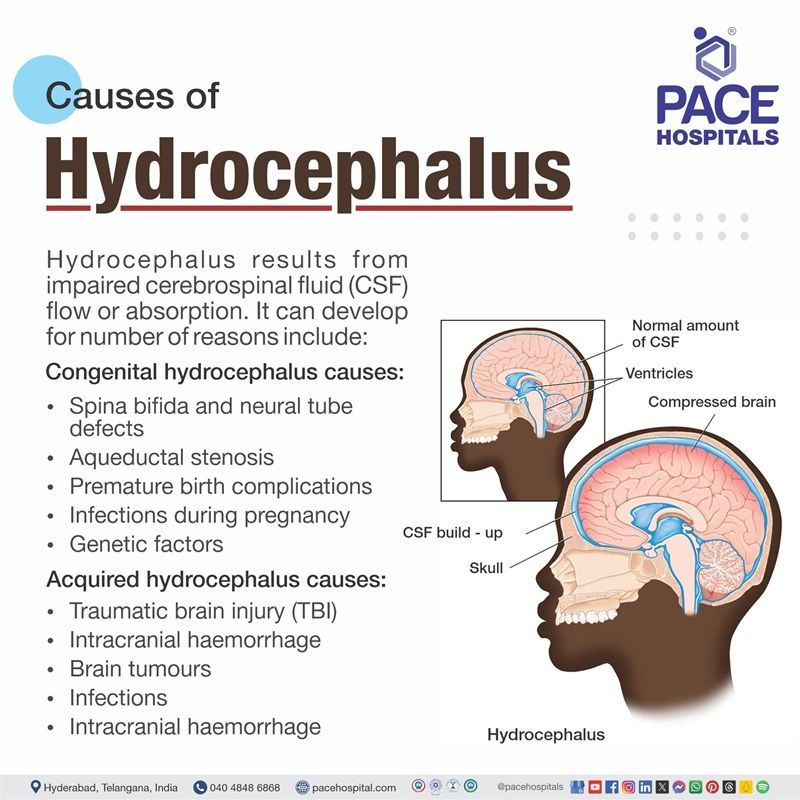
Hydrocephalus causes
Hydrocephalus results from impaired cerebrospinal fluid (CSF) flow or absorption. It can develop for a number of reasons include:
Congenital hydrocephalus causes:
- Spina bifida and neural tube defects
- Aqueduct stenosis
- Premature birth complications
- Infections during pregnancy
- Genetic factors
Acquired hydrocephalus causes:
- Traumatic brain injury (TBI)
- Intracranial haemorrhage
- Brain tumours
- Infections
- Intracranial haemorrhage
Congenital hydrocephalus causes
- Spina bifida and neural tube defects: Spina bifida, also known as a neural tube defect (NTD), leads to hydrocephalus when pregnancy-related myelomeningoceles, a birth abnormality in which a part of the spinal cord and nerves extend through an opening in the back, causes cerebrospinal fluid to flow into the brain.
- Aqueductal stenosis: The most common site connecting the brain's third and fourth ventricles is the narrowing of the Sylvian or Cerebral Aqueduct, where the cerebrospinal fluid blockage takes place and occurs hydrocephalus.
- Premature birth complications: Intraventricular haemorrhage (IVH), which means bleeding in ventricles disturbs the normal flow of cerebrospinal fluid, leads to post haemorrhagic where blood clots cause hydrocephalus in premature infants.
- Infections during pregnancy: Certain maternal or perinatal diseases, including Rubella virus, cytomegalovirus, Herpes simplex type 1 and 2 viruses, toxoplasma, other agents, and cytomegalovirus, may severely damage the placenta; as a result, these viruses can cross the placental barrier and develop hydrocephalus.
- Genetic factors: There is a connection between congenital hydrocephalus in humans and three mutant genes. The most well-known genetic connection is that X-linked hydrocephalus with aqueduct stenosis is caused by mutations in the L1CAM. other mutations and forms are far less understood.
Acquired Hydrocephalus causes
- Traumatic brain injury (TBI): One major consequence that can arise following a traumatic brain injury (TBI) is post-traumatic hydrocephalus (PTH). PTH occurs as an accumulation of improperly drained cerebrospinal fluid (CSF) in the brain. Ventriculomegaly, disruption of brain function or metabolism, delayed clinical recovery, and worsened outcomes of traumatic brain injury.
- Intracranial haemorrhage: Blood clots can obstruct the flow of cerebrospinal fluid in patients suffering from intracerebral haemorrhage (ICH), leading to acute hydrocephalus.
- Brain tumours: The natural flow of CSF may be obstructed by a brain tumour, causing it to accumulate in the brain instead of draining out. This may result in elevated intracranial pressure (ICP) or pressure inside the skull.
- Infections: Conditions like meningitis or ventriculitis can cause inflammation and scarring, disrupting CSF flow or absorption.
- Posterior fossa tumours: When cerebrospinal fluid (CSF) routes are obstructed at the cerebral aqueduct or fourth ventricle level, hydrocephalus results from posterior fossa tumours in these regions, there is no longer any CSF escape, leading to Triventricular hydrocephalus.
In some cases, the causes mentioned above are also regarded as hydrocephalus risk factors.

Complications of hydrocephalus
Untreated hydrocephalus leads to severe complications which affect individuals physical and cognitive development. Some of the complications are listed below:
- Strabismus (where the eyes aren’t straight)
- Gaze palsies (disorders of the brain systems that control eye movement)
- Nystagmus (involuntary, rapid and repetitive movement of the eyes)
- Learning disabilities
- Speech problems
- Memory problems
- Short attention span
- Problems with organizational skills
- Difficulties with physical coordination
- Epilepsy (long-term brain condition with repeated seizures)
Hydrocephalus diagnosis
Based on age, symptoms, and known or suspected abnormalities in the brain or spinal cord, hydrocephalus is diagnosed by a clinical neurological examination, brain imaging, and other procedures. Below are the steps involved in diagnosing hydrocephalus:
The neurological exam may include tests to assess:
- Strength and reflexes of muscles
- Balance and coordination
- Vision, moving the eyes, and hearing
- Mood and mental health
Diagnostic evaluation of hydrocephalus
- Ultrasound: Since ultrasound is an easy test and has low risk, hence it is frequently used by doctors to diagnose babies. Ultrasound is a useful tool for routine prenatal checkups because it can identify hydrocephalus in foetus or unborn babies.
- Magnetic resonance imaging (MRI): By using magnetic resonance imaging (MRI), one can evaluate the CSF flow, identify whether the ventricles are enlarged, and learn more about the brain tissue that surrounds the ventricles. The first test performed to diagnose adults is usually an MRI.
- Computed tomography (CT): Shows ventricle enlargement and potential obstructions.
- Spinal tap (lumbar puncture): A spinal tap, also known as a lumbar puncture, involves putting a needle into the lower back, extracting and analysing a portion of the fluid to allow clinicians to assess CSF pressure and analyse the fluid.
- Intracranial pressure monitoring: A tiny pressure monitor is put into the ventricles or brain to measure the pressure and determine whether or not there has been any brain swelling. To keep oxygenated blood flowing to the brain, a physician may drain the CSF if the pressure is too high.
- Fundoscopic examination: It views the optic nerve present at the backside of the eye using a special device. It might show swelling which indicates increased intracranial pressure due to hydrocephalus.
Hydrocephalus treatment
The cause of hydrocephaly determines the treatment. The standard treatment for hydrocephaly is cerebrospinal fluid shunting, although additional medical therapies might be used either in addition to or instead of shunting. The following are the treatment methods:
Surgical management of hydrocephalus
Congenital hydrocephalus, which affects babies, and acquired hydrocephalus, which affects children or adults, usually require immediate medical attention to relieve pressure on the brain. The rise in pressure will harm the brain if hydrocephalus is not corrected. Shunt surgery or neuro endoscopy is used to treat both acquired and congenital hydrocephalus.
Surgical management of hydrocephalus include:
- Shunt surgery
- Endoscopic Third Ventriculostomy (ETV)
Shunt surgery: A tiny tube known as a shunt is inserted into the brain during shunt surgery. Through the shunt, extra cerebrospinal fluid (CSF) from the brain is sent to another area of the body, typically the stomach. It then gets absorbed into the blood. A valve within the shunt regulates the flow of CSF, preventing it from draining too rapidly. The valve feels like a bump under the scalp's skin.
Endoscopic Third Ventriculostomy (ETV): An endoscopic third ventriculostomy (ETV) is an alternative for shunt surgery. Instead of placing a shunt, the brain surgeon creates a hole in the brain's floor to release the trapped cerebrospinal fluid (CSF) and allow it to reach the surface of the brain, where it can be absorbed.
Although it's not an effective option for everyone, ETV may be considered if obstructive hydrocephalus—a blockage—is the reason for the build-up of CSF in the brain. Avoiding the obstruction, the CSF will be allowed to drain through the hole that exists.
Medical management of hydrocephalus
In cases of hydrocephalus, medical treatment is used to postpone the need for surgery. However, these treatments may not be effective for the long-term treatment of chronic hydrocephaly, but they can help to balance CSF dynamics with some of the hydrocephalus medications commonly used, such as:
- Carbonic anhydrase inhibitors
- Osmotic diuretics
- Glucocorticoids
Carbonic anhydrase inhibitors:
Carbonic anhydrase is a non-competitive reversible inhibitor that catalyses the reaction between carbon dioxide and water, producing carbonate and protons. This decreases the CSF secretion by the choroid plexus.
Osmotic diuretics:
Hydrocephalus is treated with osmotic diuretics such as mannitol and isosorbide, which cause the brain to shrink and lower intracranial pressure (ICP).
Glucocorticoids:
By decreasing the production of cerebrospinal fluid (CSF) and improving its absorption, the class of steroids known as glucocorticoids are useful in the treatment of hydrocephalus. They can also help in the reopening of the CSF pathways in the posterior fossa.
Prevention of hydrocephalus
There's no definitive way to prevent hydrocephalus from getting worse, but there are treatments that can help. If there is concern about the possibility of hydrocephalus and the plan to have further children, the paediatrician treating the baby might advise genetic counselling.
Difference between Hydranencephaly and Hydrocephalus
Hydranencephaly vs Hydrocephalus
Hydranencephaly and hydrocephalus both involves in accumulation of fluid in the brain, but their causes, treatment methods, and prognosis are different as mentioned below:
| Elements | Hydranencephaly | Hydrocephalus |
|---|---|---|
| Causes | Numerous factors, such as exposure to chemicals, genetic abnormalities, and intrauterine issues, can result in hydranencephaly. | Several factors might lead to hydrocephalus, such as birth, trauma, or medical interventions. |
| Treatment | Since hydranencephaly has no known cure, treatment is symptomatic and supportive. | A surgically implanted shunt that transfers fluid from the brain to another area of the body is used to treat hydrocephalus. |
| Prognosis | While patients with hydranencephaly do not improve much through treatment. | Those with severe hydrocephalus can improve based on treatment. |
Frequently Asked Questions (FAQs) on Hydrocephalus
Can hydrocephalus be cured?
Currently, there is no cure for hydrocephalus, but it can be managed by the surgical procedures, which include shunt placement, which drains extra cerebrospinal fluid.
What are hydrocephalus risk factors?
Risk factors for hydrocephalus include malignancies in the brain or spinal cord, infections of the central nervous system, including meningitis caused by bacteria, trauma or stroke resulting in cerebral haemorrhage.
What is the difference between communicating and non-communicating hydrocephalus?
The name “communicating” indicates the fact that CSF can still flow between the ventricles, which remain open. Non-communicating hydrocephalus, also known as obstructive hydrocephalus, is a condition in which one or more tiny channels that connect the ventricles are obstructed by the flow of cerebral spinal fluid.
What causes water on the brain in adults?
Hydrocephalus, which develops in adults, is usually the result of an injury or illness. Possible causes of hydrocephalus include bleeding inside the brain, blood in the brain, meningitis, brain tumours, head injury, and stroke, born with narrowed passageways in the brain.
What is hydrocephalus disease?
Hydrocephalus is a neurological disorder characterized by excessive amount of cerebrospinal fluid storage in the brain's deep ventricles. The extra fluid causes these ventricles to expand, which puts extreme pressure on the tissues of the brain.
What are the types of shunts for hydrocephalus?
Hydrocephalus can be managed by different kinds of shunts, the most widely used are ventriculoperitoneal (VP) shunts, which transfer fluid from the brain's ventricles to the abdominal cavity; ventriculoarterial (VA) shunts, which transfer fluid from the brain's ventricles to a heart chamber; and lumboperitoneal (LP) shunts, which transfer fluid from the lower back to the abdominal cavity.
What are the common methods for hydrocephalus treatment in infants?
The treatment approach often involves multiple specialists, including neurologists, neurosurgeons, and paediatricians, and most of the time it is treated, but children may require ongoing monitoring throughout their lives. Methods include shunts and endoscopic third ventriculostomy used for treating hydrocephalus.
What is the treatment for Normal pressure hydrocephalus?
In order to treat NPH, surgery is typically needed to implant a shunt or tube, which directs extra CSF from the brain ventricles into the abdomen. It is termed as a ventriculoperitoneal shunt.
Is hydrocephalus treatable?
Yes, hydrocephalus is curable with early diagnosis and treatment. The only available treatment for hydrocephalus is brain surgery, however many patients achieve success with one of the following three surgical options, endoscopic Third Ventriculostomy (ETV), shunt Endoscopy, and choroid Plexus Cauterization (CPC) in Endoscopic Third Ventriculostomy (ETV).
What are spina bifida and hydrocephalus?
If a baby's spine and spinal cord do not grow normally in the womb, it results in spina bifida. Excess cerebrospinal fluid (CSF) buildup in the head caused by a disruption in development, flow, or absorption is known as hydrocephalus. A large number of newborns born with spina bifida suffer from hydrocephalus, sometimes known as water on the brain.
What is arrested hydrocephalus?
The type of hydrocephalus known as compensated hydrocephalus, sometimes known as arrested hydrocephalus, may have occurred from birth and may have even been treated throughout early childhood, but it remained mainly compensated (unchanging) and asymptomatic for many years.
Share on
Request an appointment
Fill in the appointment form or call us instantly to book a confirmed appointment with our super specialist at 04048486868

