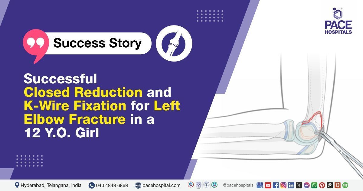
Successful Closed Reduction and K-Wire Fixation for Left Elbow Fracture in a 12 Y.O. Girl
PACE Hospitals
PACE Hospitals’ expert Orthopaedic team successfully performed a Closed Reduction and K-wire Fixation of the left supracondylar humerus in a 12-year-old female patient who presented with complaints of pain, deformity, and swelling of the left elbow following a fall in the playground. The aim of the procedure was to achieve anatomical alignment of the fractured humerus, alleviate pain, restore normal elbow function, and prevent long-term complications such as malunion, neurovascular injury, and joint stiffness.
Chief Complaints
A 12-year-old female patient presented to the Orthopaedic Department at
PACE Hospitals, Hitech City, Hyderabad, with complaints of pain, deformity, and swelling of the left elbow following a fall in the playground. She also had difficulty using her left upper limb due to pain.
Past Medical History
There was no documented history of other chronic conditions, such as
diabetes,
hypertension, or
liver disease, at the time of admission.
On Examination
On examination, the patient was conscious, coherent, and well-oriented to time, place, and person, with stable vital signs. There was no pallor, icterus, lymphadenopathy, clubbing, or cyanosis noted. Local examination of the left elbow revealed swelling, tenderness, and deformity, with a painfully restricted range of motion. Distal neurovascular status was normal.
Diagnosis
Following the clinical examination, the Orthopaedics team at PACE Hospitals conducted a thorough assessment, including a detailed evaluation of the patient's presenting complaints and a focused local examination of the left elbow.
To confirm the diagnosis and assess the extent of the injury, a comprehensive clinical and systemic examination was performed. Initial X-rays of the left elbow revealed a comminuted supracondylar fracture of the humerus on the left side. The patient exhibited significant swelling, tenderness, and deformity at the injury site, with a painful restricted range of motion. Distal neurovascular status was intact. These findings were consistent with a displaced, comminuted supracondylar humerus fracture without intercondylar extension.
Based on the confirmed diagnosis, the patient was advised to undergo Left Humerus Fracture Treatment in Hyderabad, India, under the care of the Orthopaedic Department to ensure anatomical alignment, stabilisation, and optimal recovery.
Medical Decision Making (MDM)
After a detailed consultation with Dr. Raghuram, Consultant Orthopaedic Surgeon, a comprehensive evaluation was performed to determine the most appropriate diagnostic and therapeutic approach. Considering the patient’s history of trauma, clinical presentation of pain, swelling, and deformity in the left elbow, along with restricted range of motion, a focused local examination and radiological assessment were undertaken to formulate an optimal treatment strategy.
Based on the clinical findings and imaging, which confirmed a comminuted supracondylar fracture of the left humerus, it was determined that closed reduction and K-wire fixation were identified as the most suitable surgical intervention to achieve anatomical alignment, maintain fracture stability, reduce pain, and prevent long-term complications such as malunion, deformity, or impaired limb function.
The patient and family members were thoroughly counselled regarding the injury, need for surgery, procedure details, risks, and recovery process. Informed consent was obtained, ensuring their understanding and involvement in the treatment plan.
Surgical Procedure
Following the decision, the patient was scheduled to undergo a Left Supracondylar Humerus Closed Reduction Surgery in Hyderabad at PACE Hospitals, along with K-Wire Fixation under the supervision of the expert orthopaedic Department.
The following steps were carried out during the procedure:
- Preoperative Preparation: The patient was shifted to the operating room after obtaining informed consent from the parents. Under standard aseptic precautions, the patient was placed in a supine position on the operating table. General anaesthesia was administered, and the left upper limb was prepared and draped in a sterile manner. The limb was positioned on a radiolucent hand table to facilitate fluoroscopic guidance.
- Closed Reduction Under Fluoroscopy: Traction was applied along the long axis of the arm, followed by gentle flexion and manipulation to achieve anatomical alignment of the supracondylar fracture fragments. The reduction was confirmed under C-arm fluoroscopy in both anteroposterior and lateral views. Satisfactory alignment and restoration of the Baumann angle and anterior humeral line were ensured.
- Percutaneous K-Wire Fixation: After confirming the reduction, two smooth Kirschner wires (K-wires) were inserted percutaneously from the lateral aspect of the distal humerus across the fracture site under fluoroscopic guidance. In this case, a crossed configuration was avoided to reduce the risk of ulnar nerve injury. Adequate purchase of the wires in the proximal fragment was confirmed in both planes.
- Post-Fixation Assessment and Dressing: Following fixation, the elbow was immobilised in 90° flexion with the forearm in neutral rotation. Stability of the fixation was confirmed under fluoroscopy. Hemostasis was ensured, and the K-wires were bent and cut outside the skin to facilitate later removal. Sterile gauze and crepe bandage were applied over the pin sites, and the arm was placed in a well-padded posterior above-elbow slab.
Postoperative Care
The postoperative period was uneventful. The patient was initially monitored in the recovery room and later shifted to the ward in stable condition. Adequate analgesics, antibiotics, and limb elevation were continued. The parents were counselled regarding pin-site care, the importance of active finger movements, and vigilance for signs of neurovascular compromise. A follow-up was scheduled for clinical and radiological evaluation, with eventual K-wire removal planned after fracture healing.
Discharge Medications
Upon discharge, the patient was prescribed a non-steroidal anti-inflammatory drug (NSAID) for pain relief and inflammation control. Additionally, a calcium and vitamin D supplement was recommended to support bone healing and enhance skeletal strength during the recovery period.
Advice on Discharge
The patient was advised to perform active finger movements to maintain mobility and prevent stiffness. Use of an arm sling was recommended to support and immobilise the injured limb. Limb elevation was suggested to reduce swelling.
Emergency Care
The patient was informed to contact the emergency ward at PACE Hospitals in case of any emergency or development of symptoms such as fever, severe pain, increased swelling, and vomiting.
Review and Follow-up Notes
The patient was advised to return for a follow-up visit with the Orthopaedic Doctor in Hyderabad at PACE Hospitals after one week for further evaluation.
Conclusion
This case highlights the successful management of a complex comminuted supracondylar humerus fracture in a young child through timely surgical intervention and careful postoperative care. It underscores the importance of thorough evaluation, effective communication with the family, and diligent follow-up to ensure optimal recovery and prevent complications.
Managing Pediatric Supracondylar Humerus Fractures
Pediatric supracondylar humerus fractures require prompt and precise management to avoid long-term complications. Accurate diagnosis and appropriate surgical intervention, such as closed reduction and K-wire fixation, are essential for restoring function and minimising deformity. Early treatment helps ensure better outcomes and reduces the risk of complications. Effective communication with the patient’s family is crucial for adherence to rehabilitation and follow-up care. Advances in surgical techniques have significantly improved recovery in these fractures. Overall, comprehensive care led by an orthopaedic doctor / orthopaedic surgeon, is vital for successful management and recovery in pediatric patients.
Share on
Request an appointment
Fill in the appointment form or call us instantly to book a confirmed appointment with our super specialist at 04048486868
Appointment request - health articles
Recent Articles