Pace Hospitals | Best Hospitals in Hyderabad, Telangana, India

पल्मोनोलॉजी

Best Pulmonology Hospital in Hyderabad for all Respiratory Disease Treatment

PACE Hospitals is one of the Best Lungs Hospital in Hyderabad, India, providing holistic and patient centric pulmonary treatment. The team of experienced and skilled pulmonary doctors, interventional pulmonologist have vast expertise in managing all kind of infectious and critical respiratory disease and disorder, including:

  • Bronchitis & Bronchial Asthma
  • Chronic Obstructive Pulmonary Disease (COPD)
  • Pneumonia
  • Tuberculosis (TB)
  • Pulmonary Hypertension
  • Pulmonary Fibrosis, Cystic Fibrosis
  • Lung infections, Lung Cancer
  • Sleep Apnea
  • Pulmonary Embolism
  • Interstitial Lung Disease (ILD)
  • Pleural effusions & Emphysema
  • Acute and chronic respiratory failure
  • Sleep disorders & Pulmonary hypertension
  • Pulmonary Edema
WhatsApp Appointment Call us : 040 4848 6868

Request an appointment

Pulmonology Appointment Enquiry

Why to choose us?

Comprehensive Respiratory Care | lungs hospital near me

Comprehensive Respiratory Care


Providing treatment to a wide range of lung and respiratory disorders, including acute & chronic respiratory diseases and Infections, sleep apnea, COPD, pulmonary fibrosis and lung cancer. 

Advanced State-of-the-art Facility for Pulmonology Diagnostic Tests and Procedures | best pulmonology hospital in Hyderabad

Advanced State-of-the-art Facility


Equipped with advanced and latest diagnostic equipment, robotic and minimally invasive surgical facilities for lung and respiratory disease treatment.

Skilled & Experienced Pulmonary Doctor and Interventional Pulmonologist | lungs doctor near me

Skilled & Experienced Pulmonary Doctor


Team of experienced pulmonary doctor, chest specialist & interventional pulmonologist with vast experience in minimally invasive procedures.

Empathic, Precise & Affordable care


Providing patient-centric, accurate and affordable pulmonary treatment with compassion and a high success rate.

Advanced Centre for Respiratory Disease Treatment in Hyderabad, Telangana

best pulmonology hospital in hyderabad | best lungs hospital in hyderabad | lungs specialist hospital in hyderabad | best pulmonology hospital near me | pulmonologist hospital near me | respiratory hospital near me

The pulmonology department at PACE Hospitals is one of the top lung care centre in hyderabad. The department is staffed with a team of skilled and experienced chest specialist doctors, lung doctors, interventional pulmonologists and multidisciplinary teams who work collaboratively to provide the most effective and up-to-date care possible. They have vast expertise in diagnosing and managing critical conditions related to the respiratory tract and lungs and are well-versed in the latest treatment modalities catering to compassionate, holistic, and precise respiratory disease treatment with a high success rate.


The Department of Pulmonology is equipped with state-of-the-art and cutting-edge facilities that offer a wide range of comprehensive respiratory care, including advanced imaging systems, bronchoscopy suites, High Flow Non-invasive Ventilator (HF-NIV), minimally invasive surgeries and intensive care units to manage a broad spectrum of respiratory conditions, ensuring the personalized and evidence-based treatment plans dedicated to patient's unique needs.

3,12,338

Happy Patients

98,538

Surgeries Performed

684

Medical Staff

2011

Establishment Year

Best Pulmonology Specialist in Hyderabad | Top Lung Doctor

Team of the best pulmonology specialist in Hyderabad, India, having extensive expertise in specialized respiratory care treatment for acute or chronic respiratory tract infections, chronic obstructive pulmonary disease (COPD), interstitial lung disease (ILD), pulmonary fibrosis, lung cancer and respiratory failure, are committed to delivering patient-centric, evidence-based and compassionate respiratory treatment care to the patients. The specialized team of chest specialist doctor, lung doctor, pulmonology Specialist and interventional pulmonologist are highly skilled and apt with the latest and advanced treatment modalities to cater for the utmost treatment care with precision, minimal complications and a high success rate.

Best pulmonology doctor in Hyderabad, India | pulmonology specialist | lungs specialist near me | respiratory specialist near me

Dr. Pradeep Kiran Panchadi

MBBS, DNB (Pulmonary Medicine), FIP (Fellowship in Interventional Pulmonology), CCEBDM

Experience : 11+ years

Consultant Interventional Pulmonologist, Specialist in Bronchoscopy and EBUS

  • Specialist

    He is a specialist in Bronchoscopy, EBUS, and treating Interstitial Lung Diseases (ILD). He has special expertise in EBUS, evaluation of ILDs, and various airway diseases, including Bronchial Asthma, COPD, Bronchitis, and Pneumonia.

  • Expertise

    • Basic bronchoscopy procedures like Bronchoalveolar lavage (BAL), Protected Specimen Brushing (PSB), Transbronchial lung biopsy (TBLB), Transbronchial Needle Aspiration (TBNA).
    • Trained in more than 30 Thoracoscopy, Thoracoscopic Surgery (VATS), and interventional pulmonology procedures, including Convex Endobronchial ultrasound (EBUS), Radial EBUS, Rigid Bronchoscopy
    • Debulking and various airway stenting procedures
    • Foreign body extraction, Cryobiopsy
    • Balloon bronchoplasty, Bronchial thermoplasty

Consultation Timing:

Mon to Sat - 9 am to 6 pm

Location:

PACE Hospitals, Hitech City

Request an appointment

Pulmonology Diseases and Conditions Explained by Drs

Need Help? 


Struggling with the typical respiratory tract, lung or chest-related issues or seeking treatment for pneumonia recovery, tuberculosis (TB), asthma, persistent cough, shortness of breath, recurrent lung infections, unexplained chest Pain, acute or chronic respiratory tract infections, interstitial lung disease or critical pulmonary conditions like respiratory failure, chronic obstructive pulmonary disease (COPD), pulmonary fibrosis or lung cancer, we offer evidence-based solutions tailored to your needs. Our team of skilled and experienced pulmonary doctors and interventional pulmonologists provides comprehensive and compassionate care for geriatric, adult and pediatric patients.

  • What is pulmonology?

    Pulmonology is the branch of medicine that concentrates specifically on the pulmonary (respiratory) system. This system involves the mouth, nose, sinuses, throat, voice box, windpipe, bronchial tubes, lungs, bronchioles, alveoli, pleura, and diaphragm.

  • What are the diseases and conditions treated at pulmonology department?

    Pulmonology departments at any multi-speciality hospital specialize in diagnosing, managing and treating an extensive range of common and critical respiratory conditions impairing the respiratory system, including Lungs, airways, and related organs. Some of the common conditions include:

    • Asthma
    • Chronic Obstructive Pulmonary Disease (COPD
    • Interstitial Lung Disease (ILD
    • Pulmonary Fibrosis
    • Pulmonary Hypertension
    • Lung Cancer
    • Pneumonia
    • Bronchitis
    • Sleep Apnea
    • Tuberculosis (TB)
    • Cystic Fibrosis
    • Pulmonary Embolism
    • Sarcoidosis
    • Occupational Lung Disease
  • What kind of tests is generally offered by pulmonology hospitals?

    During a visit to a pulmonologist, the patient's medical history and symptoms will be discussed before a physical examination is conducted. The pulmonologist may order tests, such as a complete blood picture, imaging tests like a CT scan or Chest X-ray, spirometry, pulmonary function tests, chest ultrasound, bronchoscopy, and sleep studies, to help diagnose and confirm any potential lung disease based on the signs and symptoms.

  • Why do you need to see a pulmonologist?

    If a patient is experiencing trouble breathing or the underlying cause is unknown, they should consult a pulmonologist if they exhibit symptoms such as coughing, chest pain or tightness, fatigue, wheezing, frequent colds, dizziness, or light-headedness. Additionally, those with a family history of lung conditions, a personal account of lung conditions, or exposure to lung irritants should seek medical attention.

  • What are the standard procedures that interventional pulmonologists perform?

    Interventional pulmonologists are skilled professionals who specialise in conducting sophisticated diagnostic and treatment procedures. These procedures include bronchoscopy, thoracentesis, balloon bronchoplasty, cryobiopsy, airway stenting, foreign body extraction, and bronchial thermoplasty.

  • What is the difference between pulmonologist and interventional pulmonologist?

    Interventional pulmonology is a recently developed field within the pulmonology department that involves using endoscopy procedures and other tools to identify and treat diseases in the chest and lungs. Lung specialists (pulmonologists) who have undergone additional training can provide these techniques.


    Interventional pulmonologists use minimally invasive procedures to diagnose and treat both non-cancerous and cancerous diseases of the airways and lungs, making it the most advanced method available.

  • What are the common symptoms of pulmonology disease?

    Pulmonary disease and conditions affecting the lungs and respiratory system more often manifest a common sign and symptoms, which can be mild or severe depending on the progression and the specific respiratory part affected. Some of the expected signs and symptoms felt when the respiratory system is impaired are:

    • Shortness of Breath (Dyspnea)
    • Excessive Mucus Production (Sputum)
    • Chest Pain or Discomfort
    • Frequent Respiratory Infections
    • Coughing Up Blood (Hemoptysis)
    • Bluish Lips or Fingernails (Cyanosis)
    • Unexplained Weight Loss
    • Chronic Cough
    • Wheezing
  • What is the common treatment offered at pulmonology department?

    The pulmonary department caters to comprehensive treatment modalities to manage and cure respiratory conditions. The treatment modalities can be different based on the patients' health conditions and severity. Some of the common approaches to treatment provided at the pulmonology department are:

    • Pharmacotherapy (Medications): Some of the common medications used are Bronchodilators, Inhaled Corticosteroids, Antibiotics, Antifibrotic Agents, Pulmonary Vasodilators
    • Oxygen Therapy
    • Pulmonary Rehabilitation:
    • Bronchoscopy
    • Lung Cancer Treatments
    • Lung Transplantation
    • Vaccinations
    • Smoking Cessation Programs
    • Management of Acute Exacerbations
    • Thoracentesis (Procedure to remove excess fluid from the pleural space in case of pleural effusion)
    • Non-Invasive Ventilation (NIV): This treatment modality uses devices like CPAP (Continuous Positive Airway Pressure) or BiPAP (Bilevel Positive Airway Pressure) to support patients with breathing abnormalities, especially those with sleep apnea or respiratory failure.
  • What is a pulmonary function test (PFT)?

    Pulmonary function tests (PFTs) are a combination of noninvasive tests to evaluate the lung's effective functionality. These tests diagnose and evaluate lung volume and capacity, flow rates, and gas exchanges, helping pulmonologists better understand the causes, progression, and severity of respiratory conditions and decide on appropriate treatment modalities.


    Diagnostic Tests involved in the Pulmonary function tests (PFTs) are as follows:

    • Spirometry: To measure the volume & speed (flow) of air that can be inhaled and exhaled.
    • Lung Volume Measurement: To measure the total amount of air the lungs can hold (total lung capacity) and the volume of air left after a complete exhalation (residual volume).
    • Diffusion Capacity (DLCO): To measure how effectively gases such as oxygen pass from the lungs into the blood.
    • Peak Flow Measurement: To measure the highest speed of exhalation.
    • Oxygen Saturation and Arterial Blood Gases (ABG): To assess the levels of oxygen and carbon dioxide in the blood.
    • Six-Minute Walk Test (6MWT): This Test evaluates patients' exercise tolerance and endurance by measuring the distance they can walk in six minutes.
  • How to book an appointment with the best pulmonologist in hyderbabad?

    Anyone seeking the best pulmonologist near me in locations like Hitech City, Madhapur, Kondapur, Gachibowli, KPHB, or Kukatpally can visit the PACE Hospital's pulmonology Department webpage and fill out the Appointment Form. They can also directly visit the hospital located near the Hi-Tech City metro station or call 04048486868 to book a hassle-free appointment with the top pulmonologist in Hyderabad.

What we treat?


We specialize in treating various pulmonology diseases and conditions affecting the respiratory tract and lungs. From asthma, pneumonia, tuberculosis (TB), chronic obstructive pulmonary disease (COPD), sleep apnea, and pulmonary hypertension to all kinds of acute and chronic upper respiratory tract infections (URTIs), lower respiratory tract infections (LRTIs) and complicated respiratory disorders like pulmonary fibrosis, pulmonary embolism and lung cancer, our team of chest specialist doctor and interventional pulmonologist is committed to cater patient centric comprehensive solutions for all your respiratory health.
respiratory disease treatment in hyderabad | lungs treatment in hyderabad | lung disease treatment in hyderabad |  pulmonology treatment | pulmonary treatment | treatment for lung disease
  • Asthma

    Asthma is a long-term inflammatory lung condition that affects the lungs' airways. The airways become inflamed, narrowed, swelled and produce mucus, making breathing difficult. 


    It is commonly seen in children, but the exact cause of asthma is unknown. As per some research studies, environmental and genetic factors are involved in causing asthma, such as allergies, stress, pets and pests, exercise, genetics, respiratory infections, certain medications, dust mites, smoke, fumes, pollen, and toxins. A person with asthma may experience chest tightness, coughing, wheezing, shortness of breath, and trouble sleeping (because of breathing problems). These symptoms may vary from person to person.

  • Atelectasis

    Atelectasis is a respiratory condition in which the complete or partial part of the lung collapses, resulting in decreased or no gas exchange. It is caused by the physical blockage of air sacs by mucus, a tumour, or an object inhaled accidentally. 


    In addition, it is caused by pressure buildup at the outside of the lungs by fluid or air filled between the lung and chest wall, a deformed bone, scarred lung tissue, pneumonia, or a tumour pressing the airway. It is characterised by shortness of breath, chest tightness, cough, bluish skin or lips, and fast heart rate.

  • Alpha-1 Antitrypsin Deficiency

    Alpha-1 antitrypsin (AAT) deficiency is an inborn condition caused by a mutated gene in both parents that increases the risk of lung and other diseases. AAT is a protein made by the liver that helps to protect the lungs; if it is not delivered enough, the lungs can be easily damaged by smoking and environmental pollution, leading to COPD and cirrhosis, which is more common in children with AAT deficiency.


    It is not possible to prevent it because it is inherited and runs in families, but there are some treatments (augmentation therapy) which can slow down liver damage.

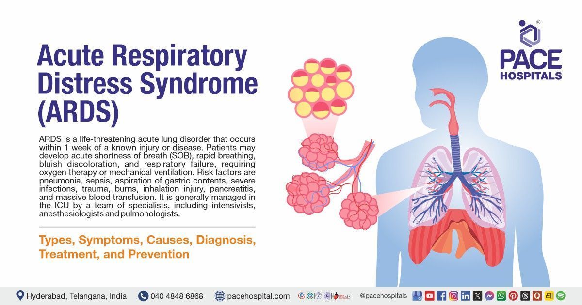
  • Acute Respiratory Disease syndrome (ARDS)

    Acute respiratory distress syndrome (ARDS) is a lung ailment that occurs when the lungs do not function normally due to fluid occupancy in the alveoli, leading to inadequate oxygen levels in the blood. It is a complication of other serious diseases, such as sepsis, pneumonia, and COVID-19. 


    It may develop from a few hours to a few days and worsen quickly. The patient may need to stay in the intensive care unit (ICU) and on a ventilator for breathing. Causes of ARDS are sepsis, accidentally inhaling food or vomit(aspiration), drowning, smoke inhalation, injuries, pneumonia, acute pancreatitis, and Drugs (radiation and chemotherapeutic agents). 

  • Aspiration Pneumonitis and Pneumonia

    Aspiration is the breathing (Inhalation) of gastric or oropharyngeal contents into the lungs. It can cause various syndromes. Aspiration pneumonitis (Mendelson's syndrome), also called chemical pneumonitis, is a short-term lung injury caused by the Inhalation of gastric acid from the stomach (Unintentional gulping of small amounts of gastric contents). Aspiration pneumonia is caused by bacteria that inhabit the oral and nasal pharynx. 


    The signs and symptoms of aspiration pneumonitis and pneumonia range from mildly ill to severely ill. Common symptoms of chemical pneumonitis are wheezing, rapid breathing, irregular heartbeat, fever, and cyanosis. Bacterial aspiration pneumonia may present with symptoms like cough with sputum, fever, difficulty breathing, malaise, and chest pain.

  • Bronchiectasis

    Bronchiectasis is a chronic condition with abnormal widening in bronchi (airways), resulting in the storage of excess mucus. This condition increases the risk of many infections because more mucus is present in the lungs. A cough with more pus and a foul smell of mucus is the prominent symptom. The other symptoms are wheezing, swollen fingertips with clubbed nails (curved nails), difficulty breathing, and chest pain. It will take some years to show the symptoms in patients until they have repeated lung diseases.


    Bronchiectasis is caused by whooping cough and measles. Other lung conditions that can cause this disease are severe pneumonia, tuberculosis, and fungal infections (allergic bronchopulmonary aspergillosis).

  • Bronchiolitis

    Bronchiolitis is a short-term inflammatory condition of the bronchioles with swelling and narrowing, leading to difficulty in breathing. It occurs by a viral infection such as flu, respiratory syncytial virus or RSV and is most commonly seen in young infants and children under age 2; the initial symptoms of bronchiolitis appear similar to a cold, such as blocked or runny nose, sneezing, a cough and low-grade fever. 


    Other signs and symptoms include wheezing, irritability, difficulty feeding or eating, rapid breathing, dehydration, and apnea.

  • Bronchitis

    Bronchitis is a common respiratory condition that develops when the bronchial tubes (airways) become inflamed and swollen. It produces a cough with mucus. Bronchitis can be chronic or acute.


    Acute illness is common and contagious, caused by a virus infection, flu, asthma, allergies, and smoking. Patients may recover within a few days and present with low fever, sore throat, body aches, cough, and runny nose. Chronic bronchitis lasts up to 3 months to years; it is more severe and comes back continuously; the patient may present with a persistent cough with mucus (clear, white, yellow, green), wheezing sound while breathing, and shortness of breath.

  • Blastomycosis

    Blastomycosis is an infectious fungal disease caused by the inhalation (breathing) of the spores of a fungus called Blastomyces, which live in moist soil and decomposing matter (wood, leaves). People who are exposed to wooded areas (such as hunting, camping, and forestry work), moist soil (lakes or rivers), and disturbed soil (excavation digging) have a high chance of getting blastomycosis.


     People with weak immune systems are at an increased risk of developing blastomycosis. Half of the Infected people will show symptoms, which are similar to those of other lung conditions, such as chest pain, cough, weight loss, fatigue, skin bumps, and night sweats. 

  • Bronchopulmonary Dysplasia

    BPD is a severe pulmonary condition that affects newborns(neonates). This condition is caused by underdevelopment of the lungs because of premature birth, infections, continuous mechanical ventilation, supplemental oxygen and inflammation of the lungs before birth.


    This condition has newborns born with breathing problems and less weight (less than 2 pounds) before ten weeks from their due dates and leads to other health problems such as Pulmonary hypertension, delayed speech, gastroesophageal reflux disease (GERD), trouble feeding, delayed speech, learning difficulties, and infections such as sepsis. 

  • Bacterial Pneumonia

    Pneumonia is a common pulmonary condition caused by bacterial, viral or fungal infection. In this condition, the lung's air sacs may be filled with pus, fluid and cellular debris, which causes inflammation and makes it difficult to breathe.


    Common causes for developing bacterial pneumonia are Streptococcus pneumoniae, Pseudomonas, Haemophilus, pneumococcal species, and Klebsiella pneumonia. Patients may present with signs of productive cough with sputum (green or rust or red currant jelly or foul smell), high fever or low fever, rapid breathing, rapid heartbeat, cyanosis, and unstable mental condition.

  • Common cold

    The common cold is an upper respiratory tract infectious disease caused by a virus. It is characterized by inflammation of the membranes that line the throat and nose. Most commonly, it can be caused by the virus rhinovirus. 


    Common cold symptoms may include sneezing, low-grade fever, sore throat, mild hacking cough, tickly throat, watering eyes, runny or blocked nose, headaches and loss of taste and smell. It can be spread through an infected person's sneeze or cough. This condition leads to secondary infections such as bacterial, sinus, and middle ear infections.

  • Cyanosis

    Cyanosis is a condition caused by insufficient oxygen levels. It causes the colour of skin, lips, tongue, ears, under the nails, gums, hands, and feet to turn blue or purplish. Cyanosis is an indicated sign of severe conditions like asthma or pneumonia, croup, and congenital heart disease (these conditions decrease oxygen levels in the blood). 


    Other causes are wearing tight clothes or jewellery, staying in freezing water or water, using beta blockers, having blood clots, and some blood vessel and heart malformations that cause blood to enter the heart without entering the lungs for oxygenation. This type of blood flow is referred to as a shunt.

  • Chronic cough

    Cough is a natural protecting mechanism that eliminates bronchial secretions (mucus) and foreign particles. It is a common symptom of many respiratory diseases. A short-term cough can be manageable by treating the underlying cause, but if it lasts up to 8 weeks or more than eight weeks, it is called a chronic cough. 


    The most common Symptoms of chronic cough are runny nose, heartburn, sore throat, fever, and tickle in the back of the throat (postnasal drip), and severe symptoms include coughing up phlegm or blood, wheezing, overnight sweat, and weight loss. Chronic cough is most commonly caused by respiratory conditions such as asthma, bronchiectasis, bronchitis, COPD, flu, pneumonia, cold and sinusitis, tuberculosis, whooping cough, and fungal infections, and it is a side effect of taking some medications such as ace inhibitors.

  • Chest Infection

    A chest infection is a pulmonary infection that affects the lungs' lower respiratory tract (windpipe and bronchi). It spreads by inhaling air droplets from the infected person's cough or sneeze.


    Chest infections can range from mild to severe. Patients may present with a wet cough with mucus (green or yellow), wheezing, difficulty breathing, chest discomfort, fever, headache, weakness, and muscle aches. Depending on the type, bacterial or viral infection can cause chest infections. For instance, the virus is the main cause of bronchitis, whereas in most cases, bacteria cause pneumonia. 

  • Chronic Obstructive Pulmonary Disease (COPD)

    Chronic obstructive pulmonary disease (COPD) is a group of conditions that cause airflow blockage and tissue damage. Emphysema and chronic bronchitis are the most common conditions associated with COPD, and the patient with COPD presents with both conditions.


    The exact cause is not fully known, as some studies found that smoking is the primary cause of developing COPD. Other causes are air pollution, occupational exposure, breathing dust or chemical fumes, and heredity. Symptoms start with a mild cough and move to recurrent cough and shortness of breath. Severe symptoms include lack of energy, wheezing, chest tightness, chronic cough, frequent colds, and flu. 

  • Cystic Fibrosis

    Cystic fibrosis is a hereditary (inherited) condition caused by two faulty inherited genes that affect the body's tissues, cells, and glands. These genes produce sweat and mucus, which accumulate in the lungs and cause blockages and damage to the airways. 


    It commonly affects the lungs and produces symptoms such as cough with mucus or blood, wheezing, chest pain, dyspnea, and atypical asthma. Management mainly includes lung clearing (airway clearance techniques). Medicines are used to fight infections, help to breathe and improve and maintain lung function.

  • Croup

    Croup is a respiratory infection commonly seen in children. It is characterized by swelling of the larynx and windpipe(airways), resulting in difficulty breathing, a whistling sound, and a barking cough. 


    It can be caused by influenza, adenovirus, enterovirus, and parainfluenza virus. It commonly targets children aged 3 – 5 and is mainly active in the winter and fall months. However, it is rarely caused by bacteria, reflux of stomach particles, or allergies and spreads through direct contact with an infected person.


    Symptoms vary from nose to lungs. They may start mildly, such as runny nose, barking cough, fever, and laryngitis, or moderate and severe, such as difficulty breathing and cyanosis. This condition should worsen at night and disturb the child from sleep.

  • Community acquired pneumonia (CAP)

    Community-acquired pneumonia (CAP) is a common lung infection caused by hospital stays. Moraxella catarrhalis, Haemophilus influenzae, and Streptococcus pneumoniae are the organisms that can cause this condition by inhalation(breathing).


    Patients present with symptoms such as common cold, fever, and cough at early stages with purulent sputum (white, green or yellow-coloured sputum). Severe symptoms include sepsis, lung failure, and acute respiratory distress syndrome. Identification of pathogens plays a vital role in treating this condition. Certain antibiotics will give to a particular pathogen.

  • Coal Worker's Pneumoconiosis

    Coal Worker's Pneumoconiosis, also known as black lung disease, is considered an occupational illness caused by continuous inhalation of coal dust. It is mainly seen in people working at a coal mine. Hence, it is called coal workers' pneumoconiosis (CWP).


    Breathing coal dust leads to dust accumulation in the lungs. The healthy tissue in the lungs tries to remove it, and the immune system fights. In this process, the tissue gets inflammation. Over time, this can result in scarring, known as fibrosis. Patients may present with symptoms such as cough with more phlegm, difficulty breathing, and chest pain. Persistent exposure to dust and smoking can increase the risk of this condition.

    • Diaphragmatic Dysfunction

      The diaphragm is a vital organ as it is the primary inhalation muscle (the muscle that helps expand the thoracic cavity). Its dysfunction is the failure of the force (muscle) itself or phrenic nerve damage, which can be associated with inevitable consequences. 


      Depending on the severity of the dysfunction, the patients can have different signs and symptoms. One may need ventilator support, and some may not show symptoms.

      This condition can be caused by Phrenic nerve damage due to Surgery or trauma, Physical(lesion) compression, or neurological disorders such as diabetic neuropathy, multiple sclerosis, Inflammatory diseases and idiopathy. Its dysfunction can be related to respiratory symptoms such as difficulty in breathing, Intolerance to exercise, and sleep disturbances, and rarely, in severe conditions, may cause death.

    • Dyspnea (Shortness of breath)

      Dyspnea/Shortness of breath is a condition in which one cannot get adequate air into the lungs or feels challenged to take a breath. It is a warning sign for other severe conditions that affect the respiratory system. Lung and heart diseases can be the main cause of shortness of breath. Other causes include allergies, asthma, anxiety, intense physical exercise, high altitude stays, choking, a collapsed lung, and sudden blood loss.


      A person with dyspnea may experience chest tightness, shortness of breath, suffocation, wheezing, palpitations, and cough.

    • Emphysema

      Emphysema is a long-term respiratory condition that occurs in people who often smoke and are exposed to irritants such as chemical fumes. It can be caused when blockage or damage (breakage) happens to the lining of the lungs' air sacs(alveoli), resulting in less oxygen to the bloodstream. The most prevalent cause for developing emphysema is smoking, and other reasons are irritants, Alpha1antitrypsin (AAT) deficiency, exposure to tobacco, weak, compromised immune system, and rare disorders called Marfan syndrome. 


      Emphysema and chronic bronchitis are the most common conditions associated with COPD. Patients may present with symptoms such as difficulty breathing, chronic cough with mucus, wheezing, chest tightness, and Fatigue. 

    • Flu (Influenzae)

      The flu is a contagious lung condition caused by the influenza virus. It is caused by the most common types of influenza viruses (A, B, C). Influenza A and B can cause more severe symptoms, which are seasonal (mainly occur in the winter). Influenza C is not seasonal and doesn't cause severe symptoms.


      Patients may present with symptoms like cough, headache, sore throat, chills, fever, runny nose, body aches, tiredness, vomiting, or diarrhoea. Flu can be cured in less than two weeks. People with certain existing diseases, including asthma, COPD, stroke, diabetes, Weak immune system, and obesity, can increase the risk of severe complications.

    • Herpes Simplex Virus Infection

      Herpes simplex virus is as HSV or herpes; it is prevalent in humans. It is divided into two types: HSV1 AND HSV2; it is contagious, present on the skin and spreads through moist mouth and genitals and sexual contact. Hsv1 Is associated with respiratory disease, mainly in people with compromised immune systems. Initially, symptoms are not noticeable; however, sometimes, they can present with symptoms such as pharyngitis or gingivostomatitis.

    • Hypersensitivity Pneumonitis

      Hypersensitivity pneumonitis is an allergic condition that causes inflammation of the lungs' alveoli (tiny air sacs). It can be caused by repeated inhalation of certain substances (particles) from the environment, such as hardwood dust, contaminated fluids from metalwork, humidifiers, feathers, bird droppings, skin cells, or moulds shed by fur animals (animals having fur). 


      These particles trigger the person's immune system after breathing and cause long-term or short-term inflammation in the lungs' alveoli, leading to breathing difficulty. It may present with symptoms such as cough, chest pain, weight loss, clubbing, abnormal breathing sounds (rales), and flu-like symptoms, including fever, chills, muscle pain, and headaches.

    • Idiopathic Pulmonary Fibrosis

      Idiopathic Pulmonary fibrosis is a serious(potentially) long-lasting lung condition where lung scarring occurs, leading to difficulty breathing where the causing factor is unknown. Usually, it affects persons who have the range of age group between 70 to 75 years old. People who smoke are highly prone to this condition. Symptoms include dry cough, fatigue, shortness of breath, and shortness of breath. If the disease is progressing, the patient may experience clubbing or cyanosis.


      Severe complications of this disease, such as pulmonary hypertension and respiratory failure, can be visible in patients who cannot deliver sufficient oxygen into the blood flow. 


    • Idiopathic Pulmonary Arterial Hypertension

      Idiopathic pulmonary arterial hypertension, also known as pulmonary hypertension, is a rare condition characterized by increased blood pressure (elevated pulmonary artery pressure) in the pulmonary arteries with no exact cause. 


      This condition is diagnosed when the pulmonary artery average blood pressure is higher than 25mmhg during the resting period and higher than 30mm in the active period (exercising). It is serious because repeated increased blood pressure in the pulmonary arteries can strain the heart, resulting in lung and heart problems. Symptoms include dyspnea (difficulty breathing), fatigue, weakness, syncope, heart palpitations, leg swelling, fast pulse, and dizziness. 

    • Interstitial Lung Disease (ILD)

      Interstitial lung disease is a group of long-term lung conditions characterized by scarring and inflammation that make it difficult for the lungs to get adequate oxygen. These diseases include idiopathic pulmonary fibrosis, hypersensitivity pneumonitis, cryptogenic organizing pneumonia (COP) and asbestosis. Symptoms may differ from person to person, and a common link between all diseases is that they all start with inflammation.


      Shortness of breath is a common symptom for all types of interstitial lung disease. Other symptoms include dry cough, tiredness, chest discomfort, loss of appetite, and weight loss. The causative factors include smoking, family history, radiation therapy, exposure to chemicals at the workplace, and certain drugs.

    • Interstitial (Nonidiopathic) Pulmonary Fibrosis

      Non-idiopathic pulmonary fibrosis is a respiratory condition that causes scarring(fibrosis) and lung inflammation, resulting in impaired exchange of gases (O2 and CO2). It is a potential condition that can lead to respiratory dysfunction and death. Early diagnosis and treatment are necessary to avoid the damage.


      It can occur due to numerous factors, such as environmental and occupational exposures to asbestos, silica, and carbon dust, drug-induced lung toxicity, hypersensitivity pneumonitis caused by organic substances, connective tissue diseases, and systemic illnesses. Symptoms may differ from condition to disease. Common symptoms include fever, Church Strauss syndrome, wheezing, and clubbing.

    • Lung Nodules

      A lung nodule is an abnormal (abnormal) growth in the lung. Most of the nodules are noncancerous (benign), but rarely, some nodules are cancerous(malignant) and become a sign of lung cancer. It can be caused due to numerous existing factors, such as tuberculosis or fungal infections, non-infectious inflammation, noncancerous tumours, and if any abnormal growth in the lungs about or larger than 3 cm becomes cancerous. 


      Ninety-five per cent of lung nodules don't show any symptoms. Underlying health issues for this nodule may cause symptoms. For instance, a person who suffers from an infection may present with cough, fever and sickness.

    • Legionnaires' Disease

      Legionnaires' disease is life-threatening pneumonia caused by Inhalation of (breathing) contaminated aerosols of legionella bacteria which are generally found in the freshwaters such as streams, rivers and lakes and also in soil. This bacterium is too small, so it moves inside the tiny water droplets as water vapour and mist, then quickly enters the lungs. Rarely can it spread by drinking contaminated water. 


      Symptoms include shortness of breath, coughing, high temperature, chest pain, and flu-like symptoms. It can be caused by humidifiers, spa pools and bathtubs, air conditioning systems, and unused taps and showers.

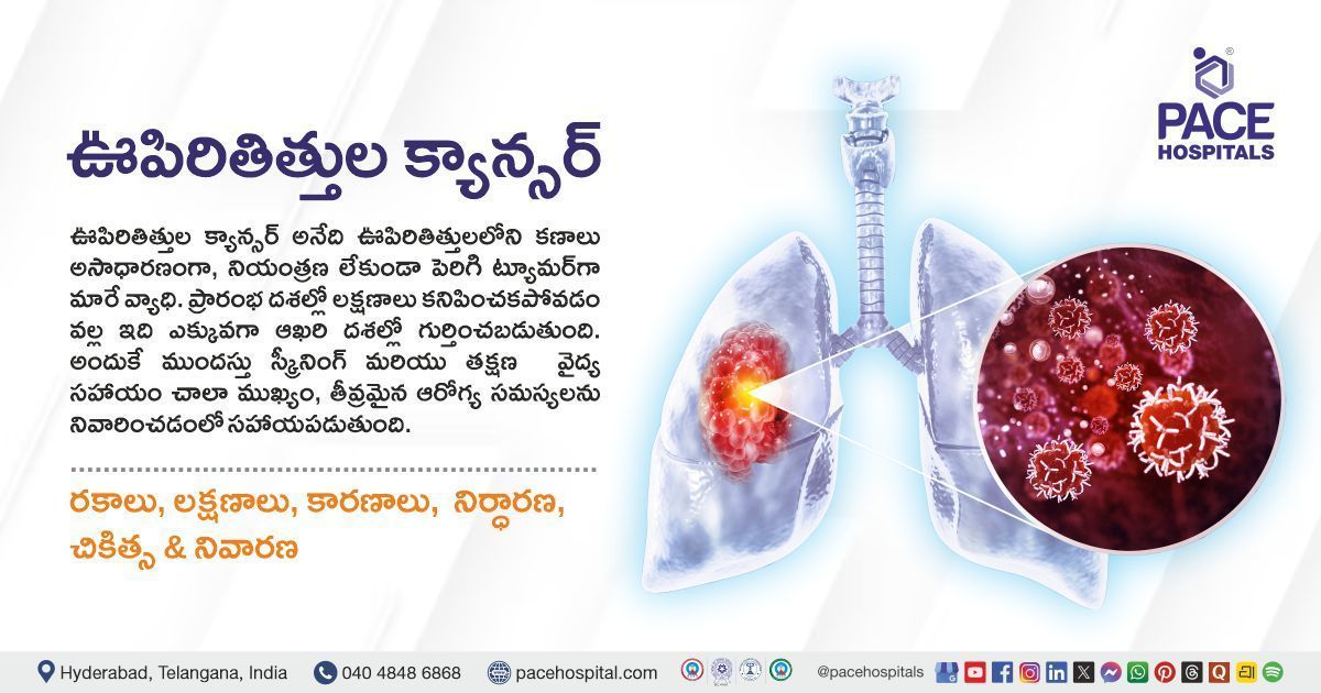
    • Lung Cancer

      Cancer is the unlimited growth of cells. If it occurs in the lungs (bronchi or alveoli), it is called lung cancer. This type of growth is usually seen in bronchi or alveoli. It starts in the lungs and may spread to the other organs in the body or lymph nodes, and cancer from other organs may also come to the lungs.


      Lung cancers are divided into two types: non-small cell (more common) and small cell lung cancer. They grow in different manners, so lung cancer treatment and symptoms depend on the spreading and stages. Symptoms include coughing up phlegm, wheezing, weakness, loss of appetite and weight loss, chest pain, and recurrent respiratory infections. 

    • Lung Abscess

      A lung abscess is a space (cavity) occupied with pus caused by the Inhalation (breathing) of bacteria present in the throat or mouth into the tissue of the lungs, resulting in a bacterial infection. The most prominent symptom of this infection is a productive cough (bloody or pus-like contents) with a foul smell. Other symptoms include chest pain, difficulty breathing, excessive sweat or night sweats, weight loss, and fever.


       Multiple factors, including excessive consumption of alcohol, aspiration pneumonia, anaesthesia, nervous system disease, sedation, weak immune system, and a blocked airway, can cause it.

    • Obesity Hypoventilation Syndrome

      Obesity hypoventilation syndrome (OHS), also known as Pickwickian syndrome, is a respiratory disorder caused by hypoventilation in obese people. Hyperventilation, or overbreathing, is a condition that involves deep and more rapid breathing than normal. This abnormal breathing results in low carbon dioxide in the blood, narrowing blood vessels.


      Patients may present with shortness of breath, dizziness, headaches, depression, fatigue, lack of energy, loud snoring, choking, and pauses in breathing. Researchers have found that a combination of multiple factors, including Excess fat on the neck, chest, and belly, can produce breathing problems. Treatment options include maintaining a healthy weight. 

    • Obstructive Sleep Apnea (OSA)

      Obstructive sleep apnea (OSA) is a respiratory breathing disorder caused by the continuous collapse of part or all of the upper airway while sleeping.


      Symptoms include fatigue, dry mouth, restlessness, snoring, high blood pressure, gastroesophageal reflux disease (GERD), less interest in sex, repeated awakenings at night, loud snoring, gasping, choking, and snorting. OSA can lead to different health conditions if it is not treated, including heart disease, diabetes, stroke, and pulmonary hypertension. 

    • Pneumothorax (Collapsed Lungs)

      Pneumothorax, also known as a collapsed lung, is caused by air between the pleural space (the space between the chest wall and the lung). It occurs due to open lung tissue injury, leading to air leakage into the pleural space. Air accumulation increases the pressure on the outer side of the lung, causing it to collapse.


      These conditions can increase the risk of pneumothorax, such as smoking habit, age group between 15-34, tall man, pregnancy, Marfan syndrome, asthma, COPD, cystic fibrosis, sarcoidosis, and HIV with pneumocystis pneumonia. Symptoms include shortness of breath, chest pain, increased heart rate, rapid breathing, confusion, loss of consciousness, or coma.

    • Pneumonia

      Pneumonia is an infectious disease caused by viruses, bacteria, or fungi. It causes fluid (pus or any liquid) buildup and inflammation of the alveoli or air sacs of the lungs, leading to difficulty breathing. 


      Any age group can get affected by pneumonia, but having the following conditions can improve the high risk of developing pneumonia such as smoking habits, having an age group of 65 and older, children having an age group under 2, and having preexisting medical problems. Symptoms may vary based on the type. However, common symptoms include shortness of breath, fever, sweating, chills, cough with mucus or phlegm, nausea, vomiting, and chest pain during coughing or breathing.

    • Pulmonary Edema

      Pulmonary oedema, also known as lung water or congestion, is an accumulation of fluid in the lungs that causes insufficient oxygen levels and makes breathing difficult.


      Pulmonary oedema can be caused by multiple factors such as congestive heart failure (primary cause), cardiac arrhythmias, pneumonia, kidney failure, sudden hypertension, severe sepsis, exposure to high altitude, drug misuse, inhalation of toxins, near drowning and severe trauma. Symptoms of this condition include wheezing, shortness of breath, rapid weight gain, fatigue, swelling of the lower part of the body, pale skin, breathing difficulties while lying in bed, excessive sweating and anxiety.

      • Pertussis (Whooping cough)

        PERTUSSIS (Whooping cough) is a contagious upper respiratory infection caused by the bacteria Bordetella pertussis. It spreads from person to person through the droplets of cough or sneeze. It is a severe condition in babies. 


        Initial symptoms visible within 7-10 days after infection include mild fever, cough and runny nose, which gradually progresses into a hacking cough followed by a whooping sound that lasts for weeks to months and vomiting. Persons with this infection are highly contagious up to 3 weeks after the cough starts. Common complications of this infection include 68% of babies having apnea, 22% getting pneumonia,2% will get convulsions, 0.6% will have encephalopathy, and 1% will die.

      • Pleurisy

        Pleurisy is characterized by sharp, stabbing chest or shoulder pain caused by the inflammation of the lung's lining (pleura). Hence, it is also known as pleuritis. It can be caused by infection, and the most common is flu (viral infection), bacterial infections such as tuberculosis or pneumonia, and sometimes due to other conditions, including rheumatoid arthritis, chest pain, lung cancer, and pleural tumour. 


        Symptoms include chest pain while coughing, sneezing, breathing deeply and moving the trunk or chest, cough, fever and chills. Treatment options involve managing the underlying cause.

      • Pleural Effusion

        Pleural Effusion, or water on the lung, occurs due to fluid collection in the space (cavity) between the chest cavity or pleural cavity and the lungs. Pleura, called thin membranes, cover the inside of the chest cavity or pleural cavity and outside of the lungs. Usually, some fluid is present to lubricate the lungs, which helps expand during breathing. But in some medical conditions, the excess liquid is going to accumulate. This condition can affect breathing. 


        The result of some medical treatments and other health conditions such as cirrhosis, congestive heart failure, pancreatitis, tuberculosis, asbestos exposure, pulmonary embolism, congestive heart failure and open heart surgery, radiation therapy, and ovarian hyperstimulation can cause it. 


        Symptoms of pleural effusion include shortness of breath, fever, dry cough and chest pain, especially when the patient is deep breathing.

      • Pectus Malformation

        Pectus Malformation, also called a funnel, sunken chest, or hollowed chest, is characterized by an abnormality of the rib cage, with the breastbone (sternum) growing inward, leading to a noticeable sunken appearance in the chest. 


        It is a joint congenital deformity and most commonly affects men. It occurs in adults and children but is most seen in early teens. Due to this abnormality, patients don't have more space in the chest, leading to reduced functioning of the lungs and heart. Symptoms of this condition include fatigue, chest pain, irregular heartbeat, decreased stamina during exercise, self-esteem issues, and clinical depression. 

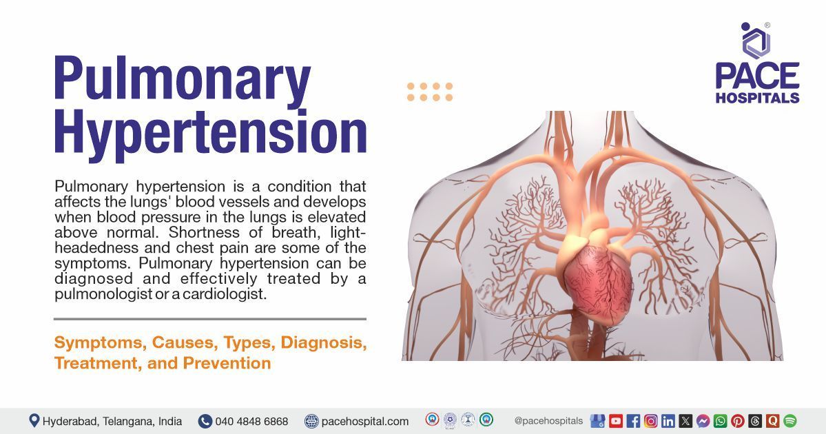
      • Pulmonary Hypertension

        Pulmonary hypertension is an illness caused by increased blood pressure in the lungs. It makes it more challenging to work for the heart than usual to circulate the blood into the lungs. It can progressively damage the heart and produce symptoms such as chest pain, light-headedness, swelling, palpitations, faintness, and chest pain.


        Worldwide, 50% of pulmonary hypertension cases are noticed without any particular cause. The most common type of pulmonary hypertension occurs in left-side heart disease.  Initial symptoms of this condition include fatigue and breathing difficulties, and severe symptoms can include chest pain, ankle, foot or abdomen swelling, palpitations, and dizziness.

      • Pulmonary Embolism

        A pulmonary embolism is a blood clot (blockage) that forms in the lungs' blood vessels. It occurs when a chunk (blood clot) moves from another body part, such as the leg or arms, into the veins of the lung. The embolism restricts blood flow to the lungs, leading to decreased oxygen levels and increased blood pressure in the pulmonary arteries.


        Causes of pulmonary embolism include injury to a vein, cardiovascular disease, changes in blood clotting factors (elevated clotting factors due to hormone replacement or some cancers), and blood accumulation due to inactivity from more extended periods such as bed rest or surgery. Symptoms of this condition include sudden shortness of breath, chest pain (which worsens during breathing), anxiety, irregular heartbeat, sweating, and coughing.

      • Pulmonary Fibrosis

        Pulmonary fibrosis is a serious(potentially) long-lasting lung condition in which lung scarring occurs, leading to difficulty breathing. It usually affects people between the ages of 70 and 75.


        People who smoke are highly prone to this condition. Symptoms include dry cough, fatigue, shortness of breath, and shortness of breath. If the disease is progressing, the patient may experience clubbing or cyanosis.


        Severe complications of this disease, such as pulmonary hypertension and respiratory failure, can be visible in patients who cannot deliver sufficient oxygen into the blood flow. 

      • Pulmonary Hamartoma

        Pulmonary hamartoma is a benign (noncancerous) lung tumour composed of tissue components, including epithelium, cartilage, muscle, fat and bone. It is the most common noncancerous respiratory growth of the lung in adults.


        Pulmonary hamartoma symptoms are not noticeable; it can appear only when one can go for chest imaging tests for other conditions. However, some patients can present with complaints such as continuous coughing or wheezing, bronchial obstruction, dyspnea (difficulty breathing) and hemoptysis (coughing up of blood).

      • Pulmonary Stenosis

        Pulmonary stenosis is a typically noncancerous (benign) condition in many patients. It is a stiffening of the pulmonary valve caused by a defect, resulting in an obstruction to flow. 


        This condition is usually inborn (congenital), noncancerous (benign) and diagnosed in children with curable treatments. Most of the patients don't show any symptoms. Still, some patients may present with associated fatigue or dyspnea on exertion (difficulty breathing during physical exercise), but rarely angina or sudden cardiac arrest is seen. Treatment options include balloon angioplasty or open-heart surgery.


      • Pneumoconiosis

        Pneumoconiosis is an array of interstitial lung diseases caused by inhalation of some specific kinds of dust particles, resulting in lung damage. It is an occupational disease caused by exposure to environmental particles in the workplace, such as asbestos, silica and coal mine dust, leading to illnesses called asbestosis, silicosis, and coal workers' pneumoconiosis. 


        Developing these conditions will take many years, but in severe cases, they may lead to lung disability and premature death. Some dust particles, such as iron, kaolin, talc, aluminium, antimony, barium, mica, and graphite, can cause other pneumoconiosis types. Depending on the severity, symptoms include cough, shortness of breath, and phlegm.

      • Pulmonary Alveolar Proteinosis

        Pulmonary alveolar proteinosis is an uncommon disorder caused by the accumulation of surfactants, such as proteins, fats, and other substances, in the lungs' alveoli (air sacs), which leads to clogging.


        This condition is divided into three types: autoimmune pulmonary alveolar proteinosis (the most common), secondary Pulmonary alveolar proteinosis, which occurs due to exposure to toxins or any other medical condition, and congenital pulmonary alveolar proteinosis, which occurs due to genetic defects. It commonly affects men more than women; about 70% of patients who have this disease are smokers.


        Symptoms of this condition include cough, fever, chest pain, weight loss, low oxygen levels, shortness of breath and clubbing.

      • Respiratory Failure

        Respiratory Failure is a life-threatening condition. The human respiratory system is usually responsible for gas exchange (oxygen in and carbon dioxide out), and the inability to execute either or both tasks leads to respiratory Failure.

        • It is divided into two types. When the respiratory system cannot provide enough oxygen to the body, it is called type 1 respiratory failure, leading to low oxygen levels (hypoxemia). 
        • When the respiratory system cannot remove enough carbon dioxide from the body, it is called type 2 respiratory failure, leading to increased carbon dioxide production (hypercapnia).

        Symptoms of respiratory failure include difficulty breathing, rapid breathing, exhaustion, heart palpitations, bluish skin, nails, or lips (cyanosis), pale skin, and excessive sweating. 

      • Respiratory Syncytial Virus (RSV)

        Respiratory Syncytial virus (RSV) is a contagious, seasonal infection affecting the respiratory system. It involves the lungs' small airways, affecting both adults and children. However, children who are under two years old are more prone to developing this infection.


        Most people can recover within a week or two. Symptoms of this infection include fever, runny nose, coughing, sneezing, and wheezing.

      • Recurrent Respiratory Papillomatosis:

        Recurrent respiratory papillomatosis is an infectious disease caused by the human papillomavirus (HPV). It is characterised by the development (growth) of noncancerous papillomas (tumours) in the air passages leading from the mouth and nose to the lungs. They usually grow around vocal cords, are located in the larynx, and sometimes spread to other structures such as the trachea and bronchi. These tumours may be different in size, grow quickly, and often need surgical removal.


        Two forms of human papillomavirus, HPV 6 and HPV 11, can cause it. Symptoms of this condition include loss of voice or hoarse voice, chronic cough, difficulty breathing, and chronic cough.

      • Sarcoidosis

        Sarcoidosis is an inflammatory illness of unknown cause that affects multiple body organs, most commonly the lymph nodes and lungs. It is characterized by the formation of abnormal nodules or masses (granulomas) with inflamed tissues in specific organs.


        Symptoms of this condition may differ based on the area, and lung symptoms include wheezing, dry cough, difficulty breathing, and chest pain.

      • Severe Acute Respiratory Syndrome (SARS)

        Severe acute respiratory syndrome (SARS) is an infectious respiratory condition caused by the SARS-associated coronavirus (SARS-CoV). It was first found in February 2003 in China and spread to other countries. It is defined as an airborne virus that spreads through droplets of saliva. It can cause upper respiratory problems in humans. 


        Signs and symptoms of this infection include headache, fever higher than 100.4 degrees, body aches, chills, sore throat, cough, breathing difficulties, pneumonia and diarrhoea (in 10% to 20% of people). No vaccine is available to treat SARS, but preventive measures must be taken to avoid it.

      • Tracheal Stenosis

        Tracheal stenosis is a condition that occurs due to inflammation or scarring of the trachea, leading to the trachea being narrower and making it difficult to breathe air. Lungs are not developed fully in premature babies, which leads to relying on breathing tubes for a long time, which might have the chance of developing tracheal stenosis.


        Acquired or congenital tracheal stenosis are the two forms. Acquired is the most common condition affecting adults and children, and Congenital refers to a severe disease present at birth. Symptoms of tracheal stenosis include persistent cough, wheezing, difficulty breathing, sleep apnea, persistent asthma, and chest congestion. Children may present with noisy breathing, difficulty breastfeeding, choking, and difficulty breathing.

      • Tuberculosis (TB)

        Tuberculosis (TB) is a common infectious condition caused by the bacterium called Mycobacterium tuberculosis, it usually attacks the lungs, but TB bacteria can enter any part of the body, including the spine, brain and kidney. Everyone who is infected with TB bacteria is not sick.


        It spreads through the tiny air dropMycobacterium tuberculosis. It usually attacks the lungs, but the TB bacteria can enter any part of the body, including the spine, brain, and kidneyslets produced by the infected person's coughing, sneezing or spitting. Symptoms may vary depending on the area. Common symptoms of tuberculosis in the lungs include coughing up blood or sputum, chest pain, a bad cough (lasts up to more than three weeks), and other symptoms include loss of appetite, weight loss, chills, fever, night sweats, weakness or fatigue. 

      • Silicosis

        Silicosis is an occupational lung disease caused by the continuous inhalation(breathing) of small crystalline silica dust particles. It is usually found in specific stones, rocks, sands, and clays. Working with these substances can produce very tiny dust, which can be easy to inhale. Once it enters the lungs, it causes inflammation, hardening, and scarring of lung tissue.


        Mining, construction, glass manufacturing and road repair are the sites where one can have a higher risk of developing silicosis. Initial Symptoms include trouble breathing (early symptoms) and persistent cough, and later symptoms include weight loss, swollen legs, chest pain, fever, weakness, shortness of breath and blue lips.

      • Pulmonary Vascular Disease

        Pulmonary vascular disease encompasses different conditions that affect the blood vessels, which lead to the middle of the heart and the lungs. Most pulmonary vascular disease conditions can cause the prominent symptom of shortness of breath.


        Causes and symptoms of pulmonary vascular disease differ in which lung blood vessel is affected. These conditions are divided into several forms: pulmonary arterial hypertension, pulmonary venous hypertension, pulmonary embolism, and chronic thromboembolic disease.

        Symptoms of pulmonary vascular disease include chest pain or fainting, shortness of breath, rapid heartbeat, and oedema.

      Patient Testimonials


      Patient with COPD, pneumonia, and acute respiratory failure treated successfully with medications and supportive treatment.

      Pulmonology Diagnostic Tests and Procedures Performed


      We provide comprehensive pulmonology diagnostic tests to carry out evidence-based treatment. Our advanced and latest screening approach examines all kinds of abnormalities and impairments in the upper and lower respiratory tract, lungs, and chest region. This results in early detection and precise evaluation, enabling our pulmonology doctors and interventional pulmonologists to make an informed decision to proceed with the appropriate treatment modalities and surgical procedures.
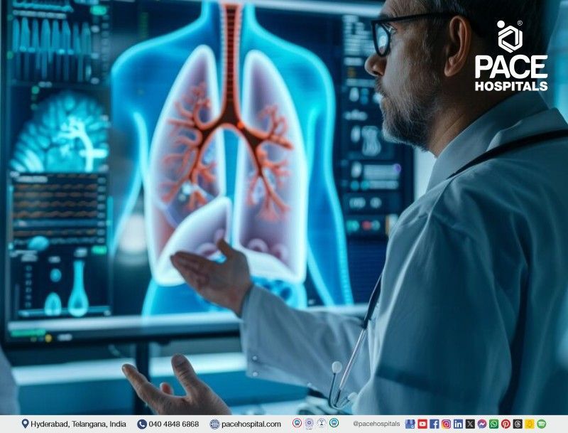
      pulmonology diagnostic tests | pulmonology procedures | pulmonology surgery | interventional pulmonology procedures

      1. Chest X-ray: It is a painless and fast imaging technique to find the structures in and around the patient's chest. The amount of radiation is significantly less, so one cannot worry about taking this test. This test helps find the diseases such as tuberculosis, emphysema, heart failure, pneumonia and lung cancer. Healthcare practitioners may use this imaging test to see how well the treatments are working in patients and to find the complications after some specific surgeries or procedures.


      2. Chest MRI: A chest MRI scan uses magnets, radio waves, and computers to create images of the structures within a patient's chest. This procedure can be done in a hospital or imaging centre. The healthcare provider may inject a contrast dye into the patient's arm veins to highlight the blood vessels and heart. It can diagnose diseases such as blood vessel problems, cystic fibrosis, pulmonary embolism, pulmonary hypertension, tumour or pleural disorders and abnormal lymph nodes. 


      3. Chest Ultrasound: This medical test employs high-frequency sound waves to provide a comprehensive overview of the lungs. It detects fluid accumulation in or around the lungs, including the heart, trachea, oesophagus, thymus, aorta, and lymph nodes. Additionally, it is beneficial in assessing the blood flow to the chest organs. It can help diagnose the conditions such as pneumonia, pleural effusion, pneumothorax and pulmonary oedema.


      4. Computerized Tomography (CT) or Positron Emission Tomography (PET) Scans: Both are advanced imaging tests and can be used to find issues such as cancers (e.g., solitary pulmonary nodule and non-small cell lung cancer). CT scan is a sequence of X-rays that have been taken from various (different) angles that give a complete picture of the lungs. In a PET scan, a special dye will use to see the body parts.


      5. Pulmonary Angiogram: It is a technique that concentrates on pulmonary arteries (blood vessels that connect the heart and lungs), present with an X-ray of the blood vessels. A notable contrast dye is injected into the patient’s blood vessels and used to find dangerous blood clots within or around the lungs called pulmonary embolism.


      6. Pulmonary Function Test (PFTs): Pulmonary function tests are a type of non-invasive examination that assesses the functionality of the lungs without the need for intrusive instruments. These tests measure various lung parameters, including capacity, volume, gas exchange, and flow rates.


      7. Spirometry: The test is simple and commonly used. The patient simply inhales and exhales through a tube, and the pulmonologist measures the amount of air exhaled after a deep breath. This helps determine the patient's lung capacity and the speed at which they can exhale air. This test identifies conditions such as chronic obstructive pulmonary disease (COPD), which can affect the amount of air the lungs can hold.


      8. Pulse Oximetry: Measuring the oxygen levels in the blood is done through pulse oximetry. This method is usually performed by placing a clip on the fingertip, but it can also be done on the toes, feet, or ears. The procedure is painless, and the results are shown as a percentage. If a patient's pulse oximetry reading is less than 90%, they may require oxygen.


      9. Lung Volume Test: Body plethysmography, also known as lung volume measurement, is a test that accurately estimates the amount of air the patient's lungs can hold. This test helps to assess the air volume in the lungs.


      • Tidal Volume (VT): It determines the amount of air taken in or breathed out during a patient's regular breathing.
      • Minute Volume (MV): It refers to the total amount of air that is exhaled in a single minute.
      • Vital Capacity (VC): It measures the amount of air that can be exhaled after inhaling as much air as possible.
      • Functional Residual Capacity (FRC): This test calculates the amount of air that left in patient lungs after normal exhalation.
      • Inspiratory Reserve Volume (IRV): It refers to the amount of air a patient can inhale forcefully after taking a normal tidal volume.
      • Expiratory Reserve Volume (ERV): It refers to the volume of air that a person can exhale forcefully after breathing out the regular tidal volume.
      • Residual Volume (RV): It is used to determine the amount of air that remains in a patient's lungs after they have exhaled as much as possible.
      • Total Lung Capacity (TLC): It refers to the maximum volume of air that the lungs can hold when they are completely filled.
      • Forced Vital Capacity (FVC): It refers to the amount of air that is exhaled forcefully and rapidly following a maximal inhalation.
      • Forced Expiratory Volume (FEV): It measures the amount of air that is exhaled during the first three seconds of a Forced Vital Capacity (FVC) test.
      • Forced Expiratory Flow (FEF): The Forced Expiratory Flow (FEF) is the average airflow rate during the middle half of the Forced Vital Capacity (FVC).
      • Peak Expiratory Flow Rate (PEFR):  It refers to the maximum speed at which a person can exhale air from their lungs.
      • Inspiratory Capacity (IC): It can be calculated by adding up the four primary lung volumes, which are TV, IRV, expiratory reserve volume (ERV), and residual volume (RV).
      • Vital Capacity (VC): The vital capacity of a patient is the total amount of air exhaled after taking in a full breath. This can be calculated by adding together the tidal volume, inspiratory reserve volume, and expiratory reserve volume. The formula for vital capacity is VC = TV + IRV + ERV.
      • Function Residual Capacity (FRC): refers to the air that remains in the lungs after a typical exhalation. The residual capacity of the lungs, also known as FRC, refers to the air that remains in the lungs after a regular exhalation.


      10. Gas Diffusion Study: This test measures the amount of oxygen and other gases that are transferred from the lungs to the blood.


      11. Cardiopulmonary Exercise Test (CPET): This assessment evaluates the functionality of the patient's heart, lungs, and muscles while engaging in physical activity. The patient will ride a bicycle or walk on a treadmill with attached machines, and a healthcare practitioner will monitor blood pressure, blood oxygen levels, and heartbeat.


      12. Fractional Exhaled Nitric Oxide (FENO) Tests: This test determines the nitric oxide level in the patient's breath. Elevated nitric oxide levels indicate lung inflammation, which can cause breathing difficulties. The patient must exhale into a tube connected to a portable device to conduct the test.


      13. Arterial Blood Gas Test: This medical examination assesses the quantity of oxygen and carbon dioxide in the bloodstream. Usually, the blood sample is collected from the patient's wrist artery, where the pulse is monitored.


      14. Bronchial Provocation Test: The bronchial provocation test is a helpful tool for identifying asthma and determining its severity. In this test, the patient is asked to inhale a specific medication that causes the airways to narrow. Then, the patient must undergo a spirometry test multiple times while the pulmonologist records readings to measure the extent of the airway narrowing during an asthma attack.


      15. Respiratory Muscle Function Test: This measures the pressure generated by the breathing muscles when the patient inhales or exhales.


      16. Sputum Cytology: Sputum cytology is a simple, non-invasive, and safe diagnostic process that identifies and counts the cell types and its morphology to assess the normal and diseased characteristics of the mucosa. This method aids in the identification of abnormal cells, inflammatory cells (lymphocytes, neutrophils, eosinophils, mast cells), bacteria, and hyphae/spores of fungi in the sputum. It helps in diagnosing lung cancer.


      Pulmonology Procedures Performed:


      1. Bronchoscopy: It is a procedure that looks inside the lung airways and is also used to find the causative factor of a lung problem. It can recognize signs of infection, tumours, bleeding, excess mucus in airways and lung blockages. The pulmonologist will slide a small tube attached to a camera at the end of the airways. By looking into the camera, they can see the passages inside, such as blood, mucus or tumours. A medicine to sleep, or a drug, should be given to numb air passages before performing this procedure.


      • Flexible Bronchoscopy: Bronchoscopy is one of the most common procedures in pulmonology. It entails the insertion of a flexible endoscope through the nose or mouth and into the airways of the lungs to detect any abnormalities. The bronchoscope also has a small tool passage at the end, which allows the pulmonologist to perform various procedures.
      • Rigid Bronchoscopy: A lengthy, rigid bronchoscope (a metallic tube) is inserted into the patient's primary airways and trachea during this procedure. This enables the pulmonologist to employ more advanced surgical instruments and methods. The process necessitates general anaesthesia, just like a surgical operation.
      • Ballon Bronchoscopy:  A pulmonologist inserts a deflated balloon into a narrowed and abnormal section of the airways. They then inflate the balloon with water, which helps to enlarge the airway and reduce symptoms. This procedure can be done before placing an airway stent to help widen a bronchus.
      • Electromagnetic Navigational Bronchoscopy: An electromagnetic navigational bronchoscopy uses GPS-like technology to examine a patient's lungs more precisely than a common bronchoscopy. It allows healthcare professionals to explore a larger area of the lungs. During the procedure, a long, flexible tube with a light at the end, known as a bronchoscope, is guided down the patient's throat and into their lungs to inspect them. Biopsies can also be taken inside the patient's lungs to determine if additional treatment is necessary. This procedure is often recommended when abnormal tissue, known as nodules, is detected during a CT scan or chest X-ray.
      • Robotic Bronchoscopy: This specific bronchoscopy method allows pulmonologists to control the flexible bronchoscope tube using a controller accurately. The instrument is smaller than standard bronchoscopes and helps access hard-to-reach areas of the lungs that cannot be easily reached with a traditional bronchoscope. This procedure is usually carried out on patients with lung nodules or biopsies (masses in the lungs) to determine whether they are cancerous.


      2. Chest Tube Procedure: Pulmonologists use the chest tube procedure to drain fluid or blood, inflate a collapsed lung, or deliver medications. The procedure involves inserting a thin plastic tube into the pleural cavity (space) to identify conditions such as emphysema, pneumothorax, pleural effusion, and hemothorax.


      3. Mediastinoscopy: A mediastinoscopy is a surgical procedure involving a mediastinoscope to examine the mediastinum, the space between the lung lobes behind the breastbone. The mediastinoscope contains a small camera to provide a visual of the area. However, healthcare practitioners must create a small hole in the chest to insert the device. This procedure is typically performed to identify signs of various cancers and to diagnose conditions like inflammation, sarcoidosis, thymoma, and cancer affecting the bronchi or other structures in the mediastinum.


      4. Pleural Biopsy: The lungs are surrounded by two layers of membranes, known as the pleural cavity. Sometimes, certain illnesses can lead to fluid accumulation in the area between the lungs and the pleura. If this occurs, pulmonologists may suggest a test to check for any fluid build-up. This test involves using a specialized biopsy needle to extract a small piece of pleura. Its purpose is to diagnose infections, cancers, or other diseases.


      5. Oxygen Therapy: Some individuals with breathing disorders may be unable to obtain sufficient oxygen on their own. In such cases, they may need oxygen therapy or supplemental oxygen. This is usually caused by underlying lung conditions that prevent oxygen absorption, such as pneumonia, asthma, heart failure, upper airway obstruction, trauma, severe asthma, and chronic obstructive pulmonary disease (COPD).


      6. Bronchoalveolar Lavage: During bronchoscopy, a process is performed where sterile water is injected into lung segments using a bronchoscope. This process is called bronchoalveolar lavage. The resulting fluid is then suctioned back and sent for testing. This procedure can aid in diagnosing bleeding, infection, cancer, and other medical conditions.


      8. Airway Stent (Bronchial Stent): When cancer or certain other conditions affect a bronchus (airway tube), it can become compressed or narrow, resulting in coughing, breathing difficulties, and the pneumonia. A pulmonologist can use a bronchoscope to insert a wire mesh stent into the affected airway to alleviate these symptoms. This stent expands to open up the airway and relieve constriction.


      9. Pleuroscopy: Interventional Pulmonologist creates a small opening in the chest wall and inserts a pleuroscope, a type of endoscope, into the chest cavity. The pleuroscope is navigated around one side of the lung and chest wall, enabling the diagnosis of certain lung lining conditions (pleura) and providing visibility of the outer edge of the patient's lung. However, this procedure cannot be done through bronchoscopy.


      10. Lung Biopsy or Lymph Node: During a bronchoscopy procedure, a pulmonologist may collect a small piece of tissue from the lungs or a nearby lymph node to examine for infection, sarcoidosis, cancer, or other conditions. This is done using forceps or needles through the bronchoscope to obtain a specific tissue sample. The collected tissue (biopsies) can accurately identify cancer spreading in patients with lung cancer or other types of cancer.


      11. Thoracoscopy: A thoracoscopy is a medical procedure that utilizes a thin, flexible tube with a light and attached camera at the end to examine the cavity within the chest and the surface of the lungs. It is performed to identify the underlying causes of lung problems, such as coughing up blood and difficulty breathing, and to obtain tissue samples (biopsy) from abnormal lung tissue, the chest wall, lymph nodes, or the pleura (lining of the lung). This procedure is commonly used for patients who have lung cancer and mesothelioma.


      12. Thoracentesis: A pulmonologist conducts thoracentesis to address pleural effusion, which is the accumulation of fluid around the lungs. During this procedure, a needle is inserted into the chest wall, and a plastic catheter is attached to it. The excess fluid is then suctioned out from the pleura. After the procedure, the catheter is removed and properly disposed of.


      13. Pleurodesis: For patients experiencing recurring pleural effusions, a medical procedure called pleurodesis is often recommended. During this procedure, a pulmonologist makes an incision in the chest wall and inserts a plastic tube into the chest cavity. An irritating chemical is then sprayed around the lung, causing the inflamed pleura to stick tightly to the chest wall, preventing the re-accumulation of fluid around the lung. 


      14. Indwelling Pleural Catheter: A pleural catheter is used as an alternative to pleurodesis for treating recurrent pleural effusion (fluid around the lung). This procedure involves the insertion of a plastic catheter under the skin and into the chest cavity, with its tip placed inside. As fluid builds up around the lung, a healthcare professional can use sterile supplies to drain the indwelling pleural catheter at home.


      15. Endobronchial Ultrasound (EBUS): This interventional procedure involves using a bronchoscope, a thin tube with a bright light inserted through the patient's mouth and down to the windpipe and bronchi. The bronchoscope has a small camera that allows a pulmonologist to visually examine the airways, blood vessels, lungs, and lymph nodes on an ultrasound monitor. Additionally, a needle is attached to the bronchoscope to extract fluid and tissue samples from the patient's lungs and surrounding lymph nodes. This procedure is referred to as transbronchial needle aspiration.


      16. Percutaneous Tracheostomy: Percutaneous dilational tracheostomy is a frequently used medical procedure for critically ill patients. It is typically performed on those experiencing short-term respiratory failure and requiring prolonged mechanical ventilation. This procedure involves creating a surgically generated airway that remains open with a breathing or tracheostomy tube inserted directly into the trachea through a neck incision. A flexible bronchoscope is necessary during the procedure to ensure proper positioning of the oral endotracheal tube.


      17. Bronchoscopic Cryobiopsy: A new procedure has been developed for identifying lung diseases by taking a sample of lung tissue. It involves using a bronchoscope with a flexible cryoprobe tool to reach inside the lung. The process can be done with a specialized cryoprobe or cryotherapy spray in the airway. The cryoprobe works by freezing tissue to collect a sample that can be evaluated further.


      18. Bronchoscopic Cryotherapy: Cryotherapy is a medical procedure that utilizes extreme cold to freeze and eliminate abnormal cells. Pulmonologists often use this technique to treat certain types of cancers, including lung cancer, by inducing tissue necrosis or cellular death. This is accomplished using a specialized cryoprobe attached to a bronchoscope or administering a cryotherapy spray. Cryotherapy is also referred to as cryoablation. 

      Bronchoscopic cryotherapy can address various medical conditions, such as foreign body removal, endobronchial biopsy, transbronchial biopsy, and the treatment of benign and malignant central airway obstruction.


      19. Bronchoscopic Thermoplasty: Thermoplasty is a respiratory procedure designed for individuals with severe asthma who do not respond to medication. During a bronchoscopy, a pulmonologist uses a heat probe to apply heat to the airway walls. This heat eliminates a layer of smooth muscle responsible for the constriction that leads to asthma symptoms.


      20. Lobectomy: A lobectomy may be necessary when one part of a lung is damaged or diseased. This surgical procedure removes the affected lobe, leaving the remaining healthy tissue to function normally. Conditions requiring a lobectomy include tuberculosis, emphysema, lung abscess, fungal infections, and lung cancer.


      21. Pneumonectomy: Pneumonectomy is a surgical procedure involving removing one of a patient's lungs due to trauma, cancer, or other diseases. The most common reason for this procedure is lung cancer, and other conditions may include congenital lung disease, traumatic lung injury, tuberculosis, and bronchiectasis. Usually, healthcare professionals aim to remove some lung tissue.


      22. Bullectomy: This type of surgical procedure is performed to remove lung air pockets. The lungs contain various tiny air sacs known as alveoli that can grow into larger spaces called bullae when they are damaged. Bullae are significant lung air pockets that can develop due to certain medical conditions like emphysema and COPD. Pulmonologists may suggest removing bullae when they cause breathing difficulties or other health problems.


      23. Lung Volume Reduction Surgery (LVRS): Patients suffering from severe emphysema or end-stage chronic obstructive pulmonary disease may undergo lung volume reduction surgery (LVRS) to improve their lung capacity, breathing ability, and overall quality and quantity of life. This surgery can be performed using open sternotomy, video-assisted thoracoscopic surgery, or thoracotomy.


      24. Pulmonary Endarterectomy: Pulmonary thromboendarterectomy is a complicated surgical procedure that involves removing scar tissue and old blood clots from the pulmonary arteries in the lungs. The procedure helps to eliminate the blockages, which restores normal blood pressure and blood flow in the lungs while reducing the strain on the right side of the heart. By removing these clots, the heart and lungs can function better, leading to a general improvement in well-being.


      25. Pulmonary Wedge Resection: It is a surgical procedure to remove a wedge-shaped small piece of tissue from the lung. It is typically done for individuals with early-stage lung cancer, metastatic tumours, bullous lung disease, tuberculosis, and lung nodules or lesions. This procedure is most effective when the cancer or disease is identified early and only affects a small area.

      Health and Diseases Information

      Pneumonia symptoms & Causes | Pneumonia treatment in India | bacterial pneumonia
      के हिसाब से Pace Hospitals 6 दिसंबर 2024
      Pneumonia is a lung infection that inflames air sacs, causing cough, fever, and difficulty breathing. Explore its types, bacterial and viral causes, risk factors like age or health conditions, symptoms, treatment options, and prevention strategies.
      Cystic fibrosis symptoms & Causes | what is cystic fibrosis​ | cystic fibrosis​ treatment in India
      के हिसाब से Pace Hospitals 23 नवंबर 2024
      Cystic fibrosis is a genetic disorder that causes thick, sticky mucus to accumulate in the body, leading to breathing and digestive challenges. Discover its symptoms, causes, risk factors, complications, and prevention methods.
      Bronchitis Symptoms & Causes | Bronchitis treatment in India | what is bronchitis​
      के हिसाब से Pace Hospitals 15 नवंबर 2024
      Bronchitis is a condition that causes inflammation in the lungs, leading to persistent coughing and difficulty breathing. Learn about Bronchitis symptoms, causes, treatment options, and prevention techniques.
      Asthma Symptoms, Causes, Types, Diagnosis &Treatment | Asthma treatment in India
      के हिसाब से Pace Hospitals 17 अक्तूबर 2024
      Asthma is a chronic respiratory condition that causes airway inflammation, making breathing difficult. Learn about the various types of asthma, its root causes, how it’s diagnosed, and the most effective treatment options available to help manage symptoms and prevent flare-ups.
      Obstructive sleep apnea symptoms and causes | Obstructive sleep apnea treatment in India
      के हिसाब से Pace Hospitals 16 अक्तूबर 2024
      Obstructive sleep apnea (OSA) is a serious sleep disorder characterized by repeated interruptions in breathing during sleep. Discover its symptoms, causes, risk factors, complications, and treatment options in this comprehensive guide.
      Interstitial lung disease causes & symptoms  | Interstitial lung disease treatment in India | ILD
      के हिसाब से Pace Hospitals 10 अगस्त 2024
      Interstitial lung disease refers to a group of conditions causing lung scarring and impaired breathing. Discover key symptoms, causes, preventive steps, and treatment options for optimal respiratory care.
      Show More

      Why choose PACE Hospitals?

      • A Multi-Super Speciality Hospital.
      • NABH, NABL, NBE & NABH - Nursing Excellence accreditation.
      • State-of-the-art Liver and Kidney transplant centre.
      • Empanelled with all TPA’s for smooth cashless benefits.
      • Centralized HIMS (Hospital Information System).
      • Computerized health records available via website.
      • Minimum waiting time for Inpatient and Outpatient.
      • Round-the-clock guidance from highly qualified pulmonologists.
      • Standardization of ethical medical care.
      • 24X7 Outpatient & Inpatient Pharmacy Services.
      • State-of-the-art operation theaters.
      • Intensive Care Units (Surgical and Medical) with ISO-9001 accreditation.
      Share by: