Successful Minimally Invasive L2-L3 Lumbar Discectomy Relieves Compressive Myelopathy in a 59-Y.O. Male
PACE Hospitals
PACE Hospitals’ expert Neurosurgery team successfully performed a Minimally Invasive L2-L3 Lumbar Discectomy on a 59-year-old male patient diagnosed with compressive myelopathy. The aim of the procedure was to relieve pressure on the spinal cord and nerve roots, alleviate neurological symptoms, and restore the patient’s mobility and quality of life.
Chief Complaints
A 59-year-old male patient with a
body mass index (BMI) of 21 presented to the Neurosurgery Department at
PACE Hospitals, Hitech City, Hyderabad, with complaints of weakness predominantly in the right lower limb for the past four days.
Past Medical History
The patient had no known history of hypertension or diabetes. The absence of these comorbid conditions was considered clinically favorable, as it had minimized the risk of intraoperative and postoperative complications and had supported a smoother, more stable recovery in this case.
On Examination
On examination, the patient was conscious, coherent, and cooperative, with stable vital signs. Gait was unsteady due to noticeable weakness in the right lower limb. Motor strength was reduced on the right side, while the left lower limb was normal. Sensory assessment showed decreased sensation over the right L2-L3 dermatome. Deep tendon reflexes were exaggerated on the right side, accompanied by a positive Babinski sign. Cardiac examination revealed normal heart sounds without murmurs. Abdominal examination was unremarkable, with a soft, non-tender abdomen and no palpable organomegaly or masses.
Diagnosis
Upon admission, the patient underwent a comprehensive clinical evaluation along with the patient's medical history and detailed diagnostic investigations conducted by the Neurosurgery team.
To support clinical findings, laboratory and special investigations were carried out as part of the preoperative assessment. Complete blood count (CBC), liver function tests (LFTs), renal function tests (RFTs), serum electrolytes, and coagulation profile, including PT, INR, bleeding time, and clotting time, were all within normal limits, indicating no systemic abnormalities or bleeding risk. A 2D echocardiogram showed normal cardiac chambers and valves with tachycardia and grade I left ventricular diastolic dysfunction. Chest X-ray revealed aortic arch calcification and mild bronchovascular markings.
Imaging studies included MRI of the thoracolumbar spine, which revealed diffuse disc bulges at multiple levels with a significant disc extrusion at L2-L3 causing severe spinal canal stenosis, moderate bilateral lateral recess stenosis, and neural foramen compromise. MRI brain showed ischemic microangiopathy changes without acute intracranial abnormalities. Viral screening was negative, and lipid profile showed borderline triglycerides and elevated LDL cholesterol.
Based on the confirmed diagnosis, the patient was advised to undergo
Compressive Myelopathy Treatment in Hyderabad, India, under the expert care of the Neurosurgery Department.
Medical Decision Making
After a detailed consultation with Dr. U L Sandeep Varma, Neurosurgeon, and Dr. Seshi Vardhan Janjirala, Cardiologist, a comprehensive preoperative cardiovascular risk assessment and overall evaluation were performed to determine the most diagnostic and therapeutic approach. Considering the patient’s presentation with right lower limb weakness for 4 days, with clinical suspicion of compressive myelopathy and concerns about spinal canal stenosis, neural foramen compromise, and risk of neurological deterioration.
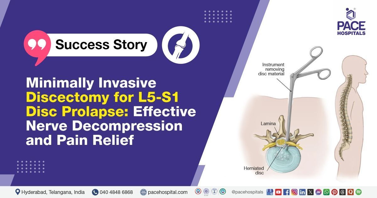
Further evaluation with MRI of the thoracolumbar spine revealed a diffuse disc bulge with extrusion at L2-L3, causing severe spinal canal stenosis and moderate to severe bilateral lateral recess stenosis. Laboratory investigations, including CBC, liver and renal function tests, serum electrolytes, and coagulation profile, were all within normal limits, indicating no systemic contraindications for surgery. Based on these findings, it was determined that minimally invasive lumbar discectomy at L2-L3 was identified as the most effective medical approach to decompress neural elements, relieve symptoms, and prevent further neurological decline.
The patient and his family were informed about the diagnosis, surgical procedure, associated risks, expected benefits such as symptom relief and improved mobility, and postoperative care and follow-up plans to ensure optimal recovery and monitoring.
Surgical Procedure
Following the decision, the patient was scheduled to undergo L2-L3 Minimal Invasive Lumbar Discectomy Surgery in Hyderabad at PACE Hospitals, under the expert supervision of the Neurosurgery Department.
The procedure involved the following steps:
- Preparation and Positioning: Under strict aseptic precautions, the patient was prepped and draped. The surgical level (L2-L3) was localised using fluoroscopy.
- Incision and Exposure: A vertical midline incision was made over the L2-L3 interlaminar space. The incision was deepened carefully to expose the lamina.
- Laminotomy and Ligamentum Flavum Excision: Laminotomy at L2 was performed, and the ligamentum flavum was excised to access the spinal canal.
- Identification and Decompression: The dural sac and bilateral L3 nerve roots were identified. Severe compression of the dura was observed due to extruded disc material at L2-L3, which was completely removed.
- Closure and Hemostasis: After ensuring the roots and dura were lax and pulsatile, hemostasis was secured, and the wound was closed in layers.
Postoperative Care
The postoperative period remained uneventful. The patient was initially shifted to the ICU for overnight monitoring and subsequently shifted to a regular room. Intravenous antibiotics, proton pump inhibitors, analgesics, and other supportive treatments were administered. The patient showed symptomatic improvement and was hemodynamically stable, hence prepared for discharge with detailed post-discharge care instructions and follow-up recommendations.
Discharge Medications
The patient was prescribed a corticosteroid for reducing inflammation and swelling, along with a gastroprotective agent to prevent gastrointestinal irritation. A muscle relaxant was included to alleviate muscle spasms and discomfort. Calcium and Vitamin D supplements were given to support bone health and aid recovery. Lastly, an anticoagulant was prescribed to prevent blood clots and reduce the risk of thromboembolic complications post-surgery.
Advice on Discharge
The patient was advised to undergo a Bone Mineral Density (BMD) scan of three sites, including the spine, hip, and forearm. Additionally, X-rays of the dorso-lumbar and lumbo-sacral spine are to be performed in both anteroposterior (AP) and lateral views. The patient was advised to continue with prescribed physiotherapy and bladder training. Strict adherence to limb physiotherapy was recommended along with the use of DVT stockings as part of the recovery plan.
Emergency Care
The patient was informed to contact the emergency ward at PACE Hospitals in case of any emergency or development of symptoms such as fever, abdominal pain ,wound drainage and vomiting.
Review and Follow-up Notes
The patient was advised to follow up with the Neurosurgeon in Hyderabad at PACE Hospitals after 1 month or earlier if needed. A review with the Cardiologist was scheduled after 2 weeks. Additionally, the patient was instructed to consult the Rheumatologist within 1 month or sooner if required, and to get all relevant scan reports.
Conclusion
This case highlights the successful diagnosis and surgical management of compressive myelopathy in a patient with right lower limb weakness. A minimally invasive lumbar discectomy at L2-L3 was performed following confirmatory imaging. The patient showed stable postoperative recovery with planned follow-up and rehabilitation.
Multidisciplinary Approach in Spinal Cord Compression
This case highlights the critical role of a multidisciplinary evaluation in diagnosing and managing acute neurological symptoms. Initial brain imaging was inconclusive, emphasizing the need for a detailed spinal MRI to identify the cause, severe disc extrusion causing spinal canal stenosis. Prompt surgical intervention by a neurosurgeon / neurosurgery doctor using a minimally invasive technique ensured effective decompression and symptom relief. The collaboration between cardiology, neurology, and neurosurgery specialists was essential for comprehensive care, demonstrating that timely, coordinated management improves patient outcomes in complex spinal conditions.
Share on
Request an appointment
Fill in the appointment form or call us instantly to book a confirmed appointment with our super specialist at 04048486868
Appointment request - health articles
Recent Articles