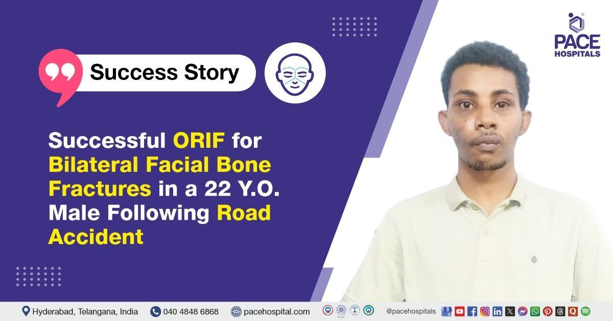
Successful ORIF for Bilateral Facial Bone Fractures in a 22-Y.O. Male Following Road Accident
PACE Hospitals
PACE Hospitals’ expert Oral and Maxillofacial and orthopedic Surgery teams successfully performed Open Reduction and Internal Fixation (ORIF) of Facial Fractures along with Debridement and Suturing of a Laceration on the Left Leg in a 22-year-old male patient who presented with bilateral facial bone fractures and a deep laceration on the left leg following a road traffic accident (bike skid). These procedures were performed to stabilise the fractured facial bones, restore function and appearance, and ensure proper healing of the leg wound.
Chief Complaints
A 22-year-old male patient with a body mass index (BMI) of 20 presented to the Plastic and Reconstructive Surgery Department at PACE Hospitals, Hitech City, Hyderabad, with complaints of bilateral facial bone fractures and a laceration on the left leg following a road traffic accident (bike skid). The patient also had a history of ear and nose bleeding immediately after the accident. Initial emergency care was provided at a private hospital after which the patient reported to PACE Hospitals for further management.
Past Medical History
There was no known history of drug allergies, chronic illnesses, or previous surgeries relevant to the presenting condition.
On Examination
The patient was conscious, coherent, and cooperative. General condition was stable, and vital signs were within normal limits. There were no abnormal findings such as pallor, icterus, cyanosis, lymphadenopathy, clubbing, or pedal edema. Local examination revealed a 12x4 cm laceration over the anterior aspect of the left leg and superficial abrasions over the right knee. Gross facial asymmetry was noted due to edema in the upper and middle thirds of the face.
Bilateral periorbital edema with ecchymosis and subconjunctival hemorrhage were present in both eyes. Eye movement was slightly restricted on the right side. A saddle-shaped deformity of the nose was observed. Tenderness and step deformities were present in the left frontozygomatic and right infraorbital regions. The root of the nose was tender and unstable, with depression noted in the right cheek region. Intraoral examination revealed deranged occlusion with fractured tooth number 13 and a mobile tooth number 12.
Diagnosis
Following the clinical examination, the Oral and Maxillofacial and orthopaedic Surgery teams conducted a thorough assessment, including a detailed review of the patient’s medical history and a focused evaluation of his facial and leg injuries.
To confirm the diagnosis and determine the extent of bone and soft tissue damage, the patient underwent detailed clinical and radiological evaluations, including a CT scan of the face. The imaging revealed fractures involving the bilateral nasal bone, the lateral wall of the left orbit, the bilateral medial orbital wall, and the right infraorbital rim. Additional fractures were seen involving the anterior, medial, and lateral walls of the right maxillary sinus. The CT scan also showed patchy opacification of the bilateral maxillary, ethmoidal, and sphenoidal sinuses, suggestive of associated soft tissue swelling or hemorrhage. These findings confirmed the presence of complex midface trauma requiring surgical intervention.
Based on the confirmed diagnosis, the patient was advised to undergo
Facial bone fracture treatment along with Leg Laceration treatment in Hyderabad, India, under the care of the Oral and Maxillofacial and orthopaedic Surgery teams, to restore both function and appearance.
Medical Decision Making
After a detailed consultation with Dr. B. Arvind, Consultant Oral and Maxillofacial Surgeon, and cross-consultations with Dr. Raghuram, Orthopaedic Surgeon, and Dr. U L Sandeep Varma, Consultant Neurosurgeon for pre-operative clearance, a comprehensive evaluation was carried out to determine the most appropriate diagnostic and therapeutic approach for the patient. The surgical team conducted a thorough assessment, including a detailed review of the patient’s medical history and a focused physical examination of the facial and leg injuries.
Clinical and radiological evaluations confirmed the presence of multiple facial fractures and soft tissue injuries resulting from a road traffic accident involving a bike skid.
It was determined that the patient had bilateral facial bone fractures, including fractures of the nasal bones, orbital walls, infraorbital rim, and maxillary sinus walls, along with a 12x4 cm laceration on the anterior aspect of the left leg. Given the extent of skeletal and soft tissue damage, Open Reduction and Internal Fixation (ORIF) of facial fractures, along with wound debridement and suturing of the left leg laceration, were identified as the most effective interventions to stabilise the facial structures, restore function, and ensure optimal wound healing.
The patient and his family were thoroughly counselled regarding the severity of the injury, the surgical procedure, potential risks, and the importance of the procedure to restore function.
Surgical Procedure
Following the decision, the patient was scheduled to undergo Open Reduction and Internal Fixation (ORIF) of facial fractures, along with Debridement and Suturing of the left leg laceration, in Hyderabad at PACE Hospitals, under the supervision of the expert Oral and Maxillofacial and orthopaedic Surgery teams.
The following steps were carried out during the procedure:
- Preoperative Preparation: After a detailed clinical evaluation, a pre-anesthetic check-up was completed. Necessary clearances were obtained from the Neurosurgery and Ophthalmology departments to ensure the patient was fit for surgical intervention under general anesthesia.
- Anesthesia and Surgical Access: Standard aseptic protocols were followed for patient preparation and draping. To aid in intraoperative stabilization of the maxillofacial fractures, arch bars were placed on the maxilla and mandible.
- Surgical access to the fracture sites was achieved through the following incisions: Right vestibular incision, Right subciliary incision and left lateral brow incision.
- Fracture Reduction and Fixation: After exposure of the affected facial bones, intermaxillary fixation (IMF) was performed to establish and maintain proper occlusion. The fracture sites were anatomically reduced and internally fixed using titanium plates and screws. The fixation was achieved as follows: the frontozygomatic region was stabilized with a 2 mm, 2-hole plate secured with two 2 mm × 8 mm screws; the right infraorbital rim was fixed with a 1.5 mm orbital plate using three 1.5 mm × 6 mm screws; the zygomaticomaxillary buttress was reconstructed with a 2.5 mm, 6-hole plate and three 2.5 mm × 8 mm screws; and the nasomaxillary buttress was stabilized with a 2 mm, 4-hole plate using two 2 mm × 6 mm screws.
- Hemostasis and Closure: After fixation, hemostasis was achieved. The surgical wounds were then closed in layers using appropriate suture materials. Deep tissue closure was performed with 3-0 Vicryl sutures, while the skin and mucosal layers were closed using 4-0 Prolene and 4-0 Monocryl sutures to ensure optimal healing and minimal scarring. A closed reduction was also carried out for the nasal bone fractures, and an external nasal splint was applied to maintain alignment and support the healing process. The closure was done carefully, prioritising both structural stability and aesthetic outcome.
- Management of Leg Laceration: Simultaneously, an orthopaedic surgeon performed wound debridement and suturing for the 12x4 cm laceration on the anterior aspect of the left leg. The wound was cleaned, debrided, and closed in layers to prevent infection and support healing.
Postoperative Care
The post-operative period was uneventful, and the patient remained hemodynamically stable. During his hospital stay, he was treated with intravenous antibiotics, analgesics, and supportive medications. He was later deemed fit for discharge with prescribed medications and instructions for follow-up and wound care.
Discharge Medications
Upon discharge, the patient was prescribed oral antibiotics, analgesics, and anti-inflammatory drugs to manage infection, pain, and inflammation. Gastric protectors were given to prevent stomach irritation, along with supplements to support healing. Anti-allergy medication was included to prevent respiratory allergies. Nasal drops, antibiotic eye drops, and antiseptic mouthwash were prescribed for symptom relief and infection prevention. Steam inhalation was recommended for respiratory support, and an antiemetic was provided if vomiting occurred. A topical antibiotic ointment was advised for wound care.
Advice on Discharge
The patient was advised to follow a soft diet for 1 month to aid recovery and to take medications regularly. The patient was advised to avoid strenuous activity and to maintain wound hygiene. Follow-up is scheduled after 3 days for dental pack removal and dressing change.
Emergency Care
The patient was informed to contact the emergency ward at PACE Hospitals in the event of any emergency or the development of symptoms such as fever, abdominal pain, vomiting, swelling, pain at the surgical site, or signs of allergic reaction.
Review and Follow-up Notes
The patient was advised to return for a follow-up consultation at the Dentist after 3 days for pack removal, with a prior appointment. Additionally, a review was scheduled with the Orthopaedic doctor after 3 days for dressing change and wound care.
Conclusion
This case highlights the successful management of bilateral facial bone fractures with leg laceration through open reduction, internal fixation, and wound debridement with suturing. The patient’s postoperative course was stable and without complication. Close monitoring and continued care have been planned to support long-term health outcomes.
Role of Multidisciplinary Collaboration in Trauma Care
The management of this complex maxillofacial trauma case exemplified the importance of multidisciplinary collaboration. Preoperatively, Neurosurgery was consulted to rule out skull base or intracranial involvement, while Ophthalmology evaluated orbital wall fractures, restricted eye movements, and subconjunctival hemorrhage to ensure ocular safety. Intraoperatively, the Oral and Maxillofacial Surgery team performed open reduction and internal fixation of multiple facial fractures, orthopaedic doctor / orthopaedic surgeon managed the deep leg laceration with debridement and suturing, and the Anesthesia team ensured safe airway control and hemodynamic stability throughout.
This coordinated, team-based approach not only allowed simultaneous management of facial fractures and extremity injuries in a single surgical setting but also ensured comprehensive trauma care, optimising functional recovery, aesthetic restoration, and postoperative outcomes for the patient.
Share on
Request an appointment
Fill in the appointment form or call us instantly to book a confirmed appointment with our super specialist at 04048486868
Appointment request - health articles
Recent Articles