Atrial Septal Defect: Symptoms, Causes, Treatment & Prevention
PACE Hospitals
Written by: Editorial Team
Medically reviewed by: Dr. Shriniwas Rajamouli Dussa - Consultant Cardiothoracic and Vascular Surgeon
Overview | Prevalence | Types | Symptoms | Causes | Risk Factors | Complications | Diagnosis | Treatment | Prevention | ASD vs VSD | PFO vs ASD | FAQs | When to consult a Doctor
Atrial septal defect definition
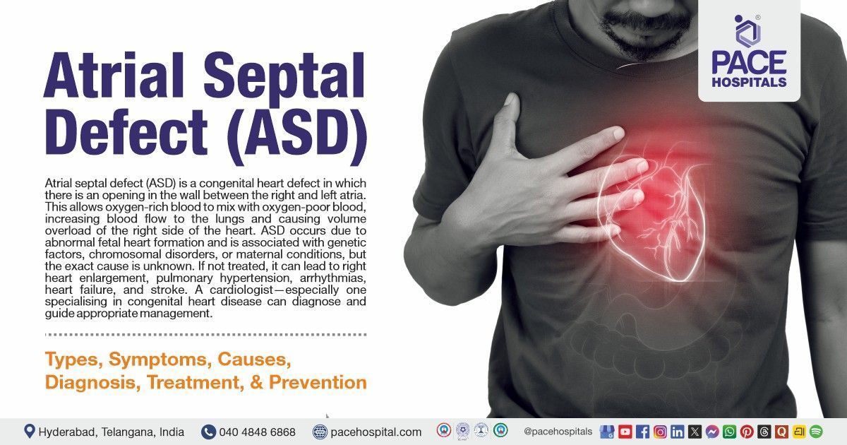
Atrial septal defect (ASD) is a congenital heart defect with an abnormal opening in the atrial septum (the wall that separates the right and left atria of the heart). This opening allows oxygen-rich blood from the left atrium to mix with oxygen-poor blood in the right atrium, increasing blood flow to the lungs and leading to volume overload of the right side of the heart. Small ASDs often cause no symptoms. Moderate to large ones lead to shortness of breath, quick tiring, repeated lung infections in kids, heart flutters, poor exercise ability, and leg swelling.
An atrial septal defect (ASD) results from abnormal development of the heart during fetal growth. Contributing factors include genetic influences, chromosomal conditions such as Down syndrome, and maternal factors like diabetes or rubella infection. In many cases, however, the exact cause is still unknown. If left untreated, ASD can lead to complications such as right heart enlargement, pulmonary hypertension, atrial arrhythmias, heart failure, and an increased risk of stroke due to paradoxical embolism (clot bypass).
A cardiologist, particularly one specialising in congenital heart disease, can accurately diagnose and guide appropriate management.
ASD meaning
The term atrial septal defect is derived from Latin and Greek words:
- “Atrial” means “large hall" or "chamber," referring to the heart's upper chambers.
- “Septal” means “a dividing wall” or “partition.”
- “Defects” meaning “a failure, deficiency, or lack.”
Thus, an atrial septal defect means a deficiency or opening in the wall that separates the upper chambers of the heart.
ASD Prevalence
ASD prevalence Worldwide
Worldwide, atrial septal defect is one of the most frequent congenital heart diseases, with recent epidemiological studies estimating its birth prevalence at approximately 1.6 per 1,000 live births. While the reported prevalence was low in the past, it has increased significantly over the last 50 years due to improvements in imaging technologies. ASD generally accounts for 10% to 15.4% of all CHD cases diagnosed at birth and becomes even more prominent in the adult population, representing around 25% to 30% of all congenital heart defects identified in adulthood.
ASD prevalence in India
In India, atrial septal defect (ASD) is a major contributor to the congenital heart disease (CHD) burden, affecting both pediatric and adult populations significantly as of 2026. Among the roughly 8 to 10 out of every 1,000 live births affected by CHD in India, significant ASD (defects greater than 5mm) has a specific birth prevalence estimated at 0.4 to 0.6 per 1,000 live births.
")
Types of ASD
Atrial septal defect (ASD) can occur in different forms depending on the location and nature of the opening in the atrial septum. These types vary in severity, abnormalities, and clinical implications, making accurate classification necessary for diagnosis and management. The following are atrial septal defect type:
- Ostium secundum ASD
- Ostium primum ASD
- Sinus venosus ASD
- Coronary sinus ASD
Ostium secundum ASD: Ostium secundum atrial septal defect is the common type of ASD (70-75 % of cases). It occurs in the central portion of the atrial septum at the site of the fossa ovalis (fetal foramen remnant). It results from the incomplete closure of the foramen ovale after birth. The size of the defect can vary widely and can influence symptom severity.
Ostium primum ASD: Ostium primum atrial septal defect is located in the lower portion of the atrial septum, near the atrioventricular valves. It arises due to abnormal development of the endocardial cushions and is often associated with mitral or tricuspid valve defects.
Sinus venosus ASD: Sinus venosus atrial septal defect occurs near the junction where the superior or inferior vena cava enters the right atrium (5-10% of cases). Sinus venosus type ASDs are commonly associated with partial anomalous pulmonary venous return, in which one or more pulmonary veins drain abnormally. Because of their location and related anomalies, these defects are managed surgically rather than with device closure.
Coronary sinus ASD: Coronary sinus atrial septal defect is a rare form that results from the partial or complete absence of the wall between the coronary sinus and the left atrium. This leads to abnormal inter-atrial shunting through the coronary sinus and is frequently associated with other congenital cardiac abnormalities.

ASD Symptoms
Atrial septal defect signs and symptoms may vary depending on the size of the defect, the amount of blood shunting, and the patient's age. Generally, small ASDs may remain asymptomatic for many years, while moderate to large ASDs usually produce symptoms that become more noticeable over time. The following are the ASD signs and symptoms:
Common symptoms of ASD in children
- Poor weight gain or failure to thrive
- Frequent respiratory infections
- Easy fatigability (tiredness), exercise intolerance
Common symptoms of ASD in adults and adolescents
- Dyspnea (difficulty breathing)
- Palpitations due to atrial arrhythmias (e.g., atrial fibrillation)
- Recurrent lung infections
- Swelling of legs or ankles (in advanced stages)
Poor weight gain or failure to thrive: In children, a left-to-right shunt allows oxygenated blood to flow from the left atrium to the right atrium, increasing the workload on the heart and lungs. This raises metabolic demands, making feeding tiring and energy use inefficient, which can lead to poor weight gain and failure to thrive.
Frequent respiratory infections: ASD increases blood flow to the lungs due to continuous left-to-right shunting. This excessive pulmonary blood flow leads to congestion in lung tissues, making them more susceptible to infections. Over time, the lungs become less efficient at clearing secretions and pathogens, as a result, children often experience repeated respiratory infections.
Easy fatigability (tiredness) exercise intolerance: Children with ASD tend to tire easily because their hearts must pump extra blood to handle the abnormal circulation. The right side of the heart becomes volume overloaded, and oxygen delivery to the body may be relatively inefficient during activity. Even mild physical exertion (like playing, feeding, or crawling) can cause rapid tiredness, reduced stamina, and the need for frequent rest compared to healthy children.
Dyspnea (difficulty breathing): In adults, long-standing left-to-right shunting causes chronic over-circulation of blood to the lungs. This leads to increased pressure in the pulmonary arteries (pulmonary hypertension) and reduced lung compliance. As a result, the lungs and heart are less efficient during physical activity, causing difficulty breathing initially with exertion and, in more advanced cases, even at rest.
Palpitations due to atrial arrhythmias: Chronic volume overload from ASD causes dilation of the right atrium (and often left atrium). This stretching of atrial tissue disrupts normal electrical conduction pathways, predisposing adults to atrial arrhythmias such as atrial fibrillation or atrial flutter. Patients commonly experience palpitations, irregular heartbeat, dizziness, or episodes of near syncope.
Recurrent lung infections: In adults and adolescents, chronic congestion due to excess blood flow to the lungs can increase susceptibility to repeated respiratory tract infections, including bronchitis and pneumonia.
Swelling of legs or ankles (in advanced stages): If ASD remains untreated, chronic volume overload weakens the right heart, leading to right-sided heart failure. This causes systemic venous congestion with fluid accumulation in the legs and ankles, resulting in peripheral edema, which is a late sign of advanced disease. It may also indicate Eisenmenger syndrome if cyanosis develops.

Atrial Septal Defect Causes
Congenital atrial septal defect is a heart abnormality that results from the incomplete formation of the interatrial septum during fetal development. The causes of ASD are primarily related to genetic factors and disruptions in normal cardiac development, though environmental factors during pregnancy may also contribute. Most cases are sporadic (idiopathic), but familial patterns occur in 10-15%. The etiology of atrial septal defect is discussed below:
- Genetic or chromosomal abnormalities
- Associated congenital syndromes
- Maternal/environmental factors
- Associated cardiac conditions
Genetic or chromosomal abnormalities: ASD can be caused by genetic mutations or chromosomal abnormalities that disrupt normal heart development in early fetal life. Key genes include NKX2.5, GATA4, and TBX5. The atrial septum forms between the 4th and 6th weeks of gestation, and abnormalities in genes that regulate cardiac septation can prevent the atrial wall from fully closing. Conditions involving chromosomal abnormalities often affect multiple organ systems, and the heart is commonly involved.
Associated congenital syndromes: ASD is commonly seen as part of broader congenital syndromes in which abnormal embryological development affects the heart and other structures. Syndromes like Down syndrome, Holt-Oram syndrome, Noonan syndrome and Treacher Collins syndrome can result in incomplete formation of the atrial septum. These children often have additional physical, developmental, or systemic abnormalities along with the heart defect.
Maternal or environmental factors: Certain maternal and environmental influences during pregnancy can increase the risk of ASD. Poorly controlled maternal diabetes, infections during early pregnancy, alcohol consumption, smoking, exposure to certain medications, or environmental toxins can interfere with normal fetal heart development.
Associated cardiac conditions: ASD may also develop alongside other congenital cardiac abnormalities. Defects that affect heart valves, pulmonary veins, or ventricular structures can alter normal blood flow patterns during development, indirectly impairing atrial septal formation.

Risk Factors for Atrial Septal Defect
While atrial septal defect (ASD) is a congenital heart anomaly, several risk factors can increase the likelihood of its development. These factors do not directly cause an atrial septal defect but may interfere with normal heart formation during fetal development, increasing the risk of ASD. The following are risk factors of atrial septal defect:
- Family history
- Maternal diabetes
- Advanced maternal age
- Maternal obesity
- Alcohol consumption or smoking during pregnancy
- Use of certain medications
- Viral infections in the mother
- Premature birth or low birth weight
Family history: Having a family history of heart defects can increase the chance of a child being born with an ASD. This is because certain inherited genes, such as NKX2-5 and TBX5, can affect the normal development of the wall between the heart's chambers. Children with a close relative who has a heart defect are at higher risk.
Maternal diabetes: If a mother has poorly controlled gestational diabetes mellitus, then the baby is exposed to high blood sugar. This can affect the normal development of the heart, including the wall between the heart's chambers, increasing the risk of an atrial septal defect.
Advanced maternal age: Pregnancy at an advanced maternal age is linked to a higher risk of chromosomal abnormalities and genetic alterations. These alterations can impair proper cardiac septation during embryonic development.
Maternal obesity: Maternal obesity (BMI >30) is linked to chronic inflammation, insulin resistance, and altered metabolic states during pregnancy. These factors can adversely affect fetal organ development, including the heart, and increase the risk of incomplete formation of the atrial septum.
Alcohol consumption or smoking during pregnancy: Alcohol and tobacco exposure during pregnancy have toxic effects on the developing fetal heart. Alcohol can disrupt cardiac cell migration and growth, while smoking reduces oxygen delivery to fetal tissues.
Use of certain medications: Medications such as antiepileptics, retinoids, or selective serotonin reuptake inhibitors (SSRIs) can disrupt folate metabolism or neural crest migration, which is important for atrial septation.
Viral infections in the mother: If a mother gets rubella or certain other viral infections, especially during the first three months of pregnancy, it can affect the baby's heart development. The infection can interfere with the formation of the heart's walls, increasing the risk of congenital heart defects.
Premature birth or low birth weight: Premature infants (before 37 weeks) and those with low birth weight (under 2.5 kg) may be more likely to have an atrial septal defect. Incomplete cardiac maturation and disrupted fetal growth patterns can contribute to delayed or abnormal development of the atrial septum.

ASD Complications
If left untreated or undiagnosed for a long period, it can lead to several cardiovascular and systemic complications. These arise due to prolonged abnormal blood flow between the heart chambers, increased pressure in the pulmonary circulation, and strain on the right side of the heart. The following are the atrial septal defect complications:
- Atrial dysrhythmias
- Pulmonary arterial hypertension
- Right-sided congestive heart failure
- Transient ischemic attack (TIA)
- Endocarditis (heart infection)
- Stroke
- Eisenmenger syndrome (late complication)
Atrial dysrhythmias: Long-term left-to-right shunting causes chronic volume overload and atrial dilatation, particularly in the right atrium. This stretching of atrial muscle fibres alters normal electrical conduction pathways, increasing the risk of atrial dysrhythmias such as atrial fibrillation (AF) and atrial flutter.
Pulmonary arterial hypertension: Excessive blood flow through the hole into the lungs raises pressure in the lung arteries over time. This damages the lung vessels, making it harder for the heart to pump blood to the lungs and leading to shortness of breath during activity.
Right-sided congestive heart failure: ASD causes chronic volume and pressure overload, which progressively weakens the right ventricle. Systemic venous congestion arises when the right side of the heart loses its ability to pump blood properly. This causes symptoms of right-sided congestive heart failure, such as peripheral edema, abdominal distention, liver congestion, and fatigue.
Transient ischemic attack (TIA): An ASD allows abnormal communication between the right and left atria, enabling blood clots from the venous system to bypass the lungs and enter systemic circulation—a phenomenon known as paradoxical embolism. This usually needs transient right-to-left shunting (e.g., during Valsalva or elevated right atrial pressure). When such a clot temporarily blocks cerebral blood flow, it can cause a transient ischemic attack, producing short-lived neurological symptoms such as weakness, speech difficulty, or vision loss.
Endocarditis (heart infection): Abnormal blood flow patterns across the atrial septum may damage the endocardial lining, creating a surface where circulate bloodborne bacteria can adhere and multiply.
Stroke: ASD significantly increases the long-term risk of stroke, particularly in adults who are associated with atrial dysrhythmias or paradoxical embolism. Blood clots may form in dilated atria due to irregular heart rhythms or pass directly from the right to the left side of the heart through the defect.
Eisenmenger syndrome (late complication): It develops when untreated ASD leads to irreversible pulmonary hypertension over decades. Long-term left-to-right shunting damages lung vessels, eventually reversing flow to right-to-left with cyanosis, clubbing, and heart failure. Closure becomes contraindicated, median survival post-diagnosis is ~3 years with PAH therapy.
ASD Diagnosis
The diagnosis of an ASD is essential because this congenital heart defect often remains clinically silent during early life and may go undetected until adolescence or adulthood. Early and accurate diagnosis helps identify the presence, size, and type of the defect, assess the degree of left-to-right shunting, and evaluate the impact. Atrial septal defect diagnosis can be done during and after birth.
The following are the steps commonly included in the diagnostic criteria for atrial septal defect:
Prenatal diagnosis of atrial septal defect (before birth)
- Routine antenatal ultrasound
- Fetal echocardiography (18–24 weeks of gestation)
Postnatal diagnosis of atrial septal defect (after birth)
- Medical history
- Physical examination
- Primary imaging studies
- Transthoracic echocardiogram (TTE) with colour Doppler
- Electrocardiography (ECG)
- Chest X-ray
- Advanced and confirmatory tests
- Transesophageal echocardiogram (TEE)
- Cardiac computed tomography (CT)
- Magnetic resonance imaging (MRI) scan
- Cardiac catheterisation
- Exercise testing (for functional assessment in select symptomatic cases)
Atrial Septal Defect Treatment
The treatment of atrial septal defect depends on the size of the defect, the severity of symptoms, the age of the patient, and the presence of complications. Management is broadly classified into non-pharmacological, pharmacological, and surgical/interventional care. Below are the ASD treatment options:
Non-pharmacological management of atrial septal defect
- Observation and monitoring (for small, asymptomatic defects)
- Regular follow-up
- Lifestyle modifications and counselling
- Early detection and timely closure
- Patient/family education on symptom recognition
Pharmacological management of atrial septal defect
- Diuretics
- Anti-arrhythmic drugs
- Anticoagulants or antiplatelet agents
- Pulmonary vasodilator
Note: Medications do not close the defect but manage symptoms and complications.
Surgical management of atrial septal defect
- Percutaneous transcatheter atrial septal defect closure
- Surgical atrial septal defect closure (open-heart surgery)
Why Choose PACE Hospitals?
Expert Super Specialist Doctors
Advanced Diagnostics & Treatment
Affordable & Transparent Care
24x7 Emergency & ICU Support

Prevention of Atrial Septal Defect
While most ASDs can't be completely prevented, as they are congenital. There are certain preventive measures and risk reduction strategies during pregnancy that may help lower the likelihood of its occurrence. These measures focus on optimising maternal health, avoiding harmful exposures, and ensuring appropriate prenatal care to support normal fetal heart development. The following are the preventive tips:
Primary prevention (before and during pregnancy)
- Preconception counselling
- Optimal maternal health
- Avoidance of harmful substances
- Vaccinations
- Safe medication use
- Healthy nutrition and weight management
Secondary prevention (early detection and monitoring)
- Routine antenatal screening
- Postnatal screening and follow-up
Tertiary prevention (prevention of complications)
- Timely closure of significant ASDs
- Regular cardiology follow-up
Preconception counselling: This helps identify women who may have a higher risk of having a child with an atrial septal defect. During counselling, family history of congenital heart disease, maternal illnesses, genetic risks, and medication use are reviewed.
Optimal maternal health: Maintaining good maternal health before and during pregnancy is important for normal fetal cardiac development. Proper control over chronic conditions like pregestational diabetes, hypertension, maternal infections, and thyroid disorders ensures stable metabolic and hormonal conditions in early pregnancy.
Avoidance of harmful substances: Exposure to alcohol, tobacco, smoking, and recreational drugs during pregnancy interferes with fetal cell growth and oxygen delivery. These substances can disrupt cardiac tissue formation during the critical early weeks of gestation when the atrial septum develops.
Vaccinations: Certain viral infections, such as rubella during early pregnancy, can disrupt normal cardiac development. Vaccination before pregnancy protects the mother from such diseases, thereby safeguarding the fetus.
Safe medication use: Certain medications taken during early pregnancy can have teratogenic effects and alter fetal heart morphogenesis. Safe medication use, under medical supervision, is encouraged to prevent exposure to drugs that may interfere with atrial septal formation.
Healthy nutrition and weight management: Adequate maternal nutrition provides essential nutrients required for fetal organ development, including the heart. Maintaining a healthy body mass index (BMI) reduces inflammation, insulin resistance, and metabolic stress during pregnancy. Balanced nutrition, supplemented with folic acid, supports proper cardiac tissue development and lowers the risk of congenital heart defects.
Routine antenatal screening: Routine prenatal screening helps detect congenital impairments early. Early detection permits continuous monitoring and rapid decision-making.
Postnatal screening and follow-up: This helps to confirm the presence and severity of ASD after birth. Regular follow-up allows the doctor to monitor defect size, cardiac chamber enlargement, and pulmonary pressures.
Timely closure of significant ASDs: Early interventional or surgical closure of hemodynamically significant ASDs prevents chronic left-to-right shunting. By eliminating abnormal blood flow, timely closure reduces the risk of complications.
Regular cardiology follow-up:
Lifelong cardiology follow-up is essential even after ASD closure. Regular evaluations help detect late complications such as arrhythmias, residual shunts, or pulmonary hypertension at an early stage.
Difference between Atrial and Ventricular Septal Defect
ASD vs VSD
Atrial and ventricular septal defects are the common congenital heart defects, which are characterised by abnormal openings in the heart's septum. Although both involve shunting of blood between chambers, but they are different in their anatomical locations, hemodynamic effects, symptoms, complications, and management. Understanding these differences is essential for accurate diagnosis and appropriate treatment. Below are the key differences between ASD and VSD:
| Parameters | Atrial septal defect | Ventricular septal defect |
|---|---|---|
| Definition | An abnormal opening in the septum between the right and left atria | An abnormal opening in the septum between the right and left ventricles |
| Location in the heart | Upper chambers (atria) | Lower chambers (ventricles) |
| Pressure difference | Low-pressure shunt | High-pressure shunt |
| Hemodynamic effect | Volume overload of the right atrium and right ventricle of the heart | Volume overload of the lungs and the left ventricle |
| Common symptoms | Symptoms include breathlessness on exertion, fatigue, and palpitations | Symptoms include rapid breathing, poor feeding, sweating, and failure to thrive |
| Risk of arrhythmias | Common in adulthood | Less common |
| Overall severity | Usually milder in early life | Often more severe in early life |
Difference between Patent Foramen Ovale and Atrial Septal Defect
PFO VS ASD
Patent foramen ovale (PFO) and atrial septal defect (ASD) are both conditions involving an opening in the wall (septum) between the right and left atria of the heart. Although they may appear similar, they differ in how they develop, their structural characteristics, their effect on blood flow, and their clinical significance. Understanding these differences helps in choosing appropriate evaluation and management strategies. The table below highlights key differences between patent foramen ovale and atrial septal defect:
| Parameters | Patent foramen ovale (PFO) | Atrial septal defect (ASD) |
|---|---|---|
| Definition | Patent foramen ovale is a persistent, flap-like opening in the interatrial septum that fails to seal completely after birth. | Atrial septal defect is a true structural defect where part of the atrial septum is missing, creating a permanent opening. |
| Cause / Origin | It results from incomplete closure of the normal fetal foramen ovale, which usually seals soon after birth. | It is a congenital heart defect caused by abnormal development of the atrial septum during fetal life. |
| Nature of the Opening | The opening is usually small and covered by a tissue flap, which can open transiently under certain pressure conditions. | The opening is a fixed hole without a flap and remains continuously open. |
| Hemodynamic Impact | It usually has little effect on heart function and does not cause chamber enlargement in most individuals. | It can lead to significant volume overload of the right atrium and ventricle, causing chamber enlargement over time. |
| Symptoms | Most individuals are asymptomatic, though it may be associated with unexplained stroke or migraine in some cases. | Symptoms may include shortness of breath, fatigue, recurrent respiratory infections, or heart failure if large and untreated. |
Frequently Asked Questions (FAQs) on Atrial Septal Defect
What is an atrial septal defect?
ASD is a congenital condition in which there is an opening in the wall, called the septum that separates the two upper chambers of the heart (atria). This opening allows oxygen-rich blood to mix with oxygen-poor blood. Over time, this causes extra blood flow to the lungs and strain on the right side of the heart. Many people have no symptoms early in life, but complications can develop later if untreated.
Is atrial septal defect genetic?
Atrial septal defect can have a genetic component, but it is not always inherited. Many studies show that genetic changes affecting heart development can increase risk when there is a family history of congenital heart disease. However, many cases occur without any known genetic cause. Environmental factors during pregnancy also play an important role. Hence, ASD usually results from a multifactorial combination of genetic and non-genetic factors.
What is secundum atrial septal defect?
Secundum ASD is the most common type of ASD. It occurs in the central part of the atrial septum, where normal closure needs to happen before birth. This type causes no symptoms in childhood and is frequently detected later in life during routine tests.
Can an atrial septal defect cause death?
Atrial septal defect rarely causes death in early life, especially when it is mild. However, studies show that untreated moderate to large ASDs can lead to serious complications over time. These complications can become life-threatening in adulthood. Early diagnosis, regular follow-up, and timely closure greatly reduce long-term risks and improve life expectancy.
Can ASD cause heart failure?
Yes, atrial septal defect can cause heart failure if it is left untreated for many years. A persistent hole allows excess blood to flow from the left to the right side of the heart, resulting in chamber enlargement and weakening. Over time, this strain may result in right-sided heart failure, increased lung pressure, and reduced heart efficiency.
Can Atrial Septal Defect in Pregnancy Affect the Baby?
Yes, Atrial septal defects (ASDs) during pregnancy can potentially affect the baby, especially if the defect is large or untreated. Small ASDs usually do not cause significant issues, but larger defects may place extra strain on the mother's heart, which could indirectly impact the baby's health.
Potential Effects on the Baby:
- Fetal growth restriction: Occurs when the baby receives less oxygen and nutrients because of problems in the mother’s circulation.
- Preterm birth: Significant strain on the mother’s heart can increase the risk of early labor, potentially leading to a baby being born before full term.
- Congenital heart defects: Slightly higher, but rare, risk in the baby.
- Mitigation through monitoring: Regular cardiac check-ups during pregnancy help minimize these risks.
Is ASD the same as autism?
No, ASD does not mean autism. Atrial septal defect, or ASD, refers to a hole in the wall between the upper heart chambers present at birth. Autism spectrum disorder shares the same letters but describes a brain development issue affecting social skills, communication, and behaviour.
What to avoid if someone has a hole in their heart?
For individuals with atrial septal defect, avoiding activities that place sudden strain on the heart, such as extreme exertion without medical advice. Smoking, excessive alcohol intake, and untreated high blood pressure need to be avoided, as they worsen heart stress. Certain medications and long-distance air travel may require medical clearance.
What are atrial septal defects and ventricular septal defects?
Both atrial and ventricular septal defects are congenital heart defects involving openings in the heart's internal walls. In atrial septal defect, the opening is between the upper chambers, while in Ventricular Septal Defect, the opening is between the lower chambers.
What is an iatrogenic atrial septal defect?
An iatrogenic atrial septal defect is a small hole created in the wall between the upper heart chambers as an unintended result of a medical procedure. It most often occurs after catheter-based heart treatments, such as procedures for irregular heart rhythm. Many iatrogenic ASDs are small and close on their own within weeks or months.
How common is a sinus venosus atrial septal defect?
Sinus venosus ASD is an uncommon form of defect that accounts for about 5–6% of all atrial septal defects worldwide. It is less common than the secundum type and is often associated with abnormal drainage of lung veins into the heart. Because symptoms may be mild in childhood, diagnosis is frequently delayed until adolescence or adulthood, especially in settings without routine heart screening.
What are the symptoms of an atrial septal defect in adults?
Atrial septal defect may remain silent for years. When symptoms appear, the patient generally complains of shortness of breath during exertion, easy fatigue, frequent respiratory infections, and reduced exercise tolerance. Some adults develop palpitations due to abnormal heart rhythm, leg swelling, or chest discomfort.
Can a person with ASD live a normal life?
Yes, many people with atrial septal defects can live a normal life after successful closure. Individuals with small ASDs or those who undergo timely closure usually have normal life expectancy and quality of life.
Can an atrial septal defect close on its own?
Yes, some atrial septal defects (especially small secundum types) can close on their own, especially small defects detected in infancy. Many small ASDs gradually shrink and close during early childhood without treatment. However, moderate to large defects usually do not close naturally and may require medical monitoring or closure.
When to consult a doctor for an atrial septal defect?
Consult a doctor for an atrial septal defect if there is ongoing breathlessness, easy fatigue, or poor exercise tolerance. Signs that indicate a need for medical assistance are:
- Shortness of breath during activity or at rest
- Frequent chest infections, especially in children
- Palpitations or irregular heartbeat
- Swelling of legs or ankles
- Poor growth or weight gain in children
If these symptoms persist, it is best to see an atrial septal defect doctor for accurate diagnosis and monitoring. Seek urgent care for severe breathlessness, chest pain, loss of consciousness, or a bluish discoloration of the skin, as these symptoms can signal serious complications. A cardiologist can provide the proper ASD treatment to manage symptoms and reduce long-term risks.
Share on
Request an appointment
Fill in the appointment form or call us instantly to book a confirmed appointment with our super specialist at 04048486868
Appointment request - health articles
Recent Articles