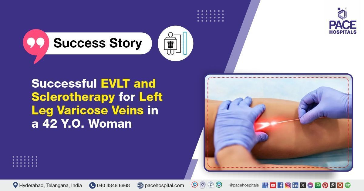
Successful EVLT and Sclerotherapy for Left Leg Varicose Veins in a 42-Year-Old Woman
PACE Hospitals
PACE Hospitals’ Expert Interventional Radiology Team successfully performed Endovenous laser therapy (EVLT) and sclerotherapy for varicose veins in the left lower limb of a 42-year-old female patient, who presented with dilated veins and swelling in the left leg. The procedure was performed to relieve symptoms such as pain, swelling, heaviness, and discomfort associated with varicose veins, and to prevent further complications and enhance the patient's mobility and quality of life.
Chief Complaints
A 42-year-old woman with a
Body Mass Index (BMI) of 22.4 presented to the Interventional Radiology Department at
PACE Hospitals, Hitech City, Hyderabad, with complaints of dilated veins and swelling in her left leg. She had been experiencing progressive discomfort, heaviness, and occasional pain in the affected limb over several months, which had started to interfere with her daily activities and mobility.
Past Medical History
The patient had no known history of drug allergies or chronic illnesses. The absence of comorbid conditions was considered clinically favorable, as it minimized the risk of intraoperative and postoperative complications and supported a smoother, more stable recovery.
On Examination
Upon admission to PACE Hospitals, the patient was hemodynamically stable. General examination revealed that she was moderately built and nourished, with no signs of systemic illness. Cardiovascular and respiratory examinations were unremarkable, with normal heart sounds (S1, S2) and adequate bilateral air entry. Abdominal examination was normal, with a soft and non-tender abdomen. Local examination of the left lower limb revealed visibly dilated superficial veins, consistent with varicosities, along with mild swelling.
Diagnosis
Following the clinical examination, the Interventional Radiology team conducted a comprehensive assessment, which included a detailed review of the patient’s medical history and a focused clinical evaluation. A color Doppler ultrasound was performed to assess venous reflux, valve competence, and the extent of venous dilation.
The imaging revealed incompetence of the superficial venous system, which is more pronounced in the left lower limb. Based on the patient's symptoms, physical findings, and Doppler results, a provisional diagnosis of varicose veins in both lower limbs (left greater than right) was established.
Based on the confirmed diagnosis, the patient was advised to undergo varicose veins treatment in Hyderabad, India, under the care of the Interventional Radiology Department, to ensure comprehensive management of her condition.
Medical Decision Making
After a thorough evaluation by Dr. Lakshmi Kumar CH, Interventional Radiologist, a comprehensive assessment of the patient’s clinical condition and color Doppler was performed. Considering the patient’s symptoms, functional limitations in daily activities, and the confirmed diagnosis of Varicose veins of both lower limbs (left greater than right), surgical intervention (Varicose vein surgery, or phlebectomy) was deemed necessary.
The patient and her family were thoroughly counselled about the nature of the condition, the planned surgical procedure, potential risks, and the necessity for surgery.
Surgical Procedure
Following the decision, the patient was scheduled to undergo Endovenous laser therapy (EVLT) and sclerotherapy surgery in Hyderabad at PACE Hospitals, under the supervision of the expert Interventional Radiology Department. The following steps were carried out during the procedure:
- Anesthesia Administration: Spinal anesthesia was administered to provide regional pain relief, followed by the infiltration of tumescent anesthesia along the targeted veins to minimize discomfort and reduce the risk of thermal injury to surrounding tissues.
- Endovenous Laser Ablation:
- The left great saphenous vein (GSV), extending from just above the ankle to approximately 6 cm proximal to the saphenofemoral junction (SFJ), was accessed and ablated using a radial laser fiber.
- The left anterior accessory saphenous vein (AASV) in the upper thigh was also identified and ablated using the same technique.
- Additionally, the left small saphenous vein (SSV), extending from the apex of the calf to about 4 cm proximal to the popliteal fossa, was treated using the radial laser fiber.
- Foam Sclerotherapy: Selected varicosities in the left leg were treated using foam sclerotherapy with anionic surfactant, effectively targeting the residual superficial varicosities.
- Haemostasis and Dressing: Hemostasis (process of stopping bleeding) was meticulously secured. A compression dressing was then applied using crepe bandages to support the treated veins and minimize post-procedure complications such as bruising or swelling. The surgery was completed successfully without intraoperative complications.
Postoperative Care
The postoperative period was uneventful, and the patient was closely monitored in the ICU to ensure a stable recovery. Intravenous antibiotics, analgesics, anticoagulants, and other supportive treatments were given. The patient demonstrated symptomatic improvement, remained hemodynamically stable, and showed no signs of complications. She was subsequently prepared for discharge with comprehensive post-discharge care instructions and follow-up recommendations.
Discharge Medications
Upon discharge, the patient was prescribed a course of oral medications, including antibiotics to prevent infection, analgesics for pain relief, antipyretics to manage any potential fever, and anticoagulants to reduce the risk of thromboembolic events. Additional supportive treatments were also advised as part of the post-discharge care plan.
Advice on Discharge
The patient was given specific instructions to avoid crossing the legs, prolonged standing or sitting for more than 30 minutes, and lifting weights over 5 kg for one week. She was also advised to wear above-knee Class II compression stockings during the daytime for a duration of two months to support venous return and aid in recovery.
Emergency Care
The patient was informed to contact the Emergency ward at PACE Hospitals in case of any emergency or development of symptoms like fever or leg swelling or severe leg pain or chest pain or breathlessness.
Review and Follow-Up Notes
The patient was advised to return for a follow-up visit with the Interventional Radiologist in Hyderabad at PACE Hospitals, after 14 days. During this visit, her recovery would be assessed to evaluate the treatment outcome and determine if any further management was necessary.
Conclusion
This case highlights the successful use of Endovenous laser therapy (EVLT), also known as Endovenous laser ablation, and sclerotherapy in treating varicose veins in the lower left limb. The procedure significantly improved the patient’s symptoms, demonstrating the effectiveness of minimally invasive methods in managing chronic venous disease.
The Essential Role of Color Doppler Ultrasound in Varicose Vein Diagnosis and Management
Color Doppler ultrasound plays a vital role in the diagnosis and management of varicose veins by providing a non-invasive method to visualize blood flow and detect vascular abnormalities. It assists in identifying incompetent veins, determining the cause of poor circulation, and locating areas of reversed or obstructed blood flow, thereby guiding the interventional radiologist /interventional radiology doctor, in appropriate treatment planning. Additionally, Color Doppler enables assessment of the severity of the condition, allowing for the development of effective treatment strategies. It is also valuable for monitoring treatment outcomes and detecting any recurrence of varicose veins. By identifying potential issues early, Color Doppler helps prevent complications such as deep vein thrombosis (DVT).
Share on
Request an appointment
Fill in the appointment form or call us instantly to book a confirmed appointment with our super specialist at 04048486868
Appointment request - health articles
Recent Articles