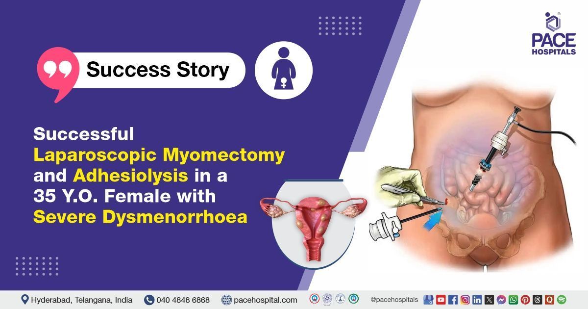
Successful Laparoscopic Myomectomy and Adhesiolysis in a 35 Y.O. Female with Severe Dysmenorrhoea
PACE Hospitals
PACE Hospitals’ expert Gynaecology team successfully performed a Laparoscopic Myomectomy with Adhesiolysis on a 35-year-old female patient diagnosed with multiple fibroids in the uterus and adenomyosis. She had presented with on-and-off heavy menstrual bleeding and severe dysmenorrhoea. The procedure aimed to remove the fibroids, relieve the patient’s symptoms, and preserve fertility.
Chief Complaints
A 35-year-old female patient with a
body mass index (BMI) of 21.2 presented to the Gynaecology Department at
PACE Hospitals, Hitech City, Hyderabad, with complaints of on-and-off heavy menstrual bleeding and severe
dysmenorrhoea (painful menstruation) not relieved by medication.
Past Medical History
The patient had no significant past medical history and no known drug or food allergies. She was nulliparous (P0L0), with no history of previous deliveries or surgical procedures related to fertility or sterilisation.
On Examination
On general examination, the patient was conscious, coherent, and cooperative. Vital signs were stable. Systemic examination revealed normal air entry bilaterally on respiratory assessment and normal heart sounds (S1 and S2) on cardiovascular examination. Per abdominal examination showed a soft, non-tender abdomen. Breast examination was normal, with no lumps detected. On per speculum and per vaginum examination, the cervix was bulky and high up, with no fornical tenderness noted.
Diagnosis
Upon admission to PACE Hospitals, the patient was assessed by the Gynaecology team. Clinical evaluation revealed a bulky uterus with multiple fibroids and features of adenomyosis. She presented with intermittent heavy menstrual bleeding and severe dysmenorrhoea that were not controlled with medication.
Ultrasound imaging confirmed multiple fibroids measuring 5×4 cm at the fundus, 3×3 cm on the right anterolateral side, and 3×2 cm on the left posterolateral side. Preoperative blood tests showed mild anemia with normocytic normochromic red blood cells and mild neutrophilic leucocytosis. Renal function and electrolyte levels were within normal limits.
Based on these findings, the patient was advised to undergo
Uterine Fibroid Treatment in Hyderabad, India, under the expert care of the Gynaecology Department, ensuring comprehensive evaluation and management tailored to the patient's needs.
Medical Decision Making
After a detailed consultation with Dr. Mugdha Bandawar, Obstetrician, Gynecologist, in collaboration with Dr. Suresh Kumar S, Surgical Gastroenterologist GI and HPB Oncologist, a multidisciplinary approach was taken to determine the most appropriate diagnostic and therapeutic strategy for the patient. Considering her nulliparous status, alongside the clinical findings of a bulky uterus with multiple fibroids and ultrasound evidence supporting adenomyosis, the team thoroughly assessed the extent of the pathology and potential surgical implications.
Further evaluation, including physical examination and laboratory investigations, confirmed the need for surgical intervention. and the presence of adhesions between the uterus and bladder, it was determined that Laparoscopic Myomectomy with Adhesiolysis was identified as the most effective surgical approach to relieve symptoms, preserve fertility, and prevent further complications associated with fibroids and chronic pelvic pain.
The patient and her family members were counselled regarding the diagnosis, the planned procedure, associated risks, expected benefits, and the potential for significant improvement in quality of life, after which informed consent was obtained.
Surgical Procedure
Following the decision, the patient was scheduled to undergo Laparoscopic Myomectomy Surgery in Hyderabad at PACE Hospitals, along with Adhesiolysis, under the expert supervision of the Gynaecology Department.
The following steps were carried out during the procedure:
- Preoperative Preparation and Diagnostic Laparoscopy: Under general anesthesia, pneumoperitoneum was established, and laparoscopic ports were inserted. Initial diagnostic laparoscopy revealed a bulky uterus with multiple fibroids. These included a 5×4 cm fibroid arising from the uterine fundus, a 3×3 cm fibroid located on the right anterolateral wall, and a 3×2 cm fibroid on the left posterolateral wall. Bilateral fallopian tubes and ovaries appeared normal with no evidence of adnexal pathology. Mild flimsy adhesions were identified between the uterus and the bladder.
- Adhesiolysis: Adhesions between the uterus and bladder were carefully dissected using a combination of blunt and sharp dissection techniques. This restored the normal anatomical relationship and ensured safe access to the fibroids and uterine surface for myomectomy.
- Laparoscopic Myomectomy: Each identified fibroid was enucleated laparoscopically. The uterine serosa was incised over the fibroids, and the myomas were carefully dissected out. Hemostasis was achieved using bipolar cautery, and the uterine muscle was sutured to restore the integrity of the uterine wall.
- Pelvic Assessment: Following fibroid removal, a thorough pelvic inspection was conducted. No other abnormalities were found, and the remaining peritoneal cavity was normal in appearance. The uterus remained bulky post-myomectomy, indicative of underlying adenomyosis.
- Final Irrigation and Port Closure: The pelvic cavity was irrigated with saline and inspected for active bleeding. After confirming complete hemostasis, the laparoscopic instruments were removed. The abdominal ports were closed using absorbable sutures, and sterile dressings were applied.
Postoperative Care
The postoperative period was uneventful. During the hospital stay, the patient was initially managed in the Surgical Intensive Care Unit (SICU) for close observation and was later shifted to the ward after becoming hemodynamically stable. Supportive care included administration of intravenous antibiotics, analgesics, antipyretics, antiemetics, proton pump inhibitors, and intravenous fluids.
The patient tolerated oral intake well, achieved adequate pain control, and was gradually mobilized. Postoperative ultrasound screening revealed no evidence of collections, ascites, pleural effusion, or bowel dilatation. Throughout the stay, the patient remained stable and was discharged in satisfactory general condition with instructions for follow-up.
Discharge Medications
At the time of discharge, the patient was prescribed a short course of oral antibiotics and anti-inflammatory medications to manage postoperative infection risk and inflammation. Proteolytic enzymes were advised to aid in reducing postoperative swelling. A proton pump inhibitor was included to prevent gastric irritation associated with medication use. The patient was also prescribed nutritional supplements to support overall health, as well as aid in postoperative recovery. A topical antibiotic ointment was recommended for local wound care.
Advice on Discharge
The patient was advised to avoid heavy lifting and strenuous physical activity during the postoperative recovery period to prevent stress on the surgical site. She was also advised to avoid prolonged sitting and strenuous coughing. She was recommended to follow a regular, balanced diet as tolerated. Additionally, the use of an abdominal binder was suggested to provide support and comfort during the healing process. The patient was encouraged to maintain a comfortable lifestyle and follow all postoperative care instructions for optimal recovery.
Emergency Care
The patient was informed to contact the emergency ward at PACE Hospitals in case of any emergency or development of symptoms like fever, abdominal pain, or vomiting.
Review and Follow-up Notes
The patient was advised to return for a follow-up appointment with the Gynecologist in Hyderabad at PACE Hospitals after 10 days.
Conclusion
This case highlights the successful laparoscopic management of multiple
uterine fibroids and adenomyosis in a patient presenting with heavy menstrual bleeding and severe dysmenorrhoea. A multidisciplinary approach, involving both gynaecology and surgical gastroenterology expertise, facilitated safe surgical outcomes, including ureteric protection through intraoperative ureteroscopy. The patient experienced an uneventful postoperative recovery, with no complications reported. She was discharged in stable condition, with appropriate follow-up advice.
Role of Ultrasound in Postoperative Assessment
Postoperative ultrasound screening is essential for detecting complications such as fluid collections, ascites, or bowel dilatation, along with the mild distension of the gallbladder with sludge, which provides valuable information for ongoing patient management. Ultrasound's non-invasive nature allows for repeated assessments without additional patient risk, making it an ideal tool for routine postoperative evaluations. The ability to visualize the surgical site and surrounding structures aids in early identification of potential issues, enabling timely interventions when necessary.
Furthermore, ultrasound can guide clinicians in making informed decisions regarding the need for further imaging or therapeutic procedures. Its role in postoperative care underscores its importance in enhancing patient outcomes through effective monitoring and management. A Gynaecologist / Gynaecology doctor can utilise ultrasound to monitor healing, assess for complications, and plan for any necessary follow-up treatments.
Share on
Request an appointment
Fill in the appointment form or call us instantly to book a confirmed appointment with our super specialist at 04048486868
Appointment request - health articles
Recent Articles