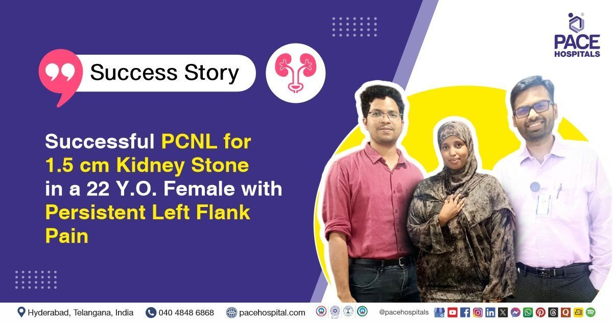
Successful PCNL for 1.5 cm Kidney Stone in a 22 Y.O. Female with Persistent Left Flank Pain
PACE Hospitals
The PACE Hospital's expert Urology team successfully performed a Left Percutaneous Nephrolithotomy (PCNL) with Double J Stent (DJS) Placement on a 22-year-old female patient diagnosed with a left renal calculus. The procedure was aimed at removing the kidney stone, relieving urinary obstruction, and restoring normal urinary flow.
Chief Complaints
A 22-year-old female patient with a
body mass index (BMI) of 21.5 presented to the Urology Department at
PACE Hospitals, Hitech City, Hyderabad, with a three-month history of left flank pain.
Past Medical History
The patient had no known history of hypertension or diabetes. The absence of these comorbid conditions was considered clinically favorable, as it had minimized the risk of intraoperative and postoperative complications and had supported a smoother, more stable recovery in this case.
On Examination
On examination, the patient was in a stable general condition with normal vital signs. Cardiovascular and respiratory system examinations were normal. Abdominal examination was normal except for tenderness in the left flank. The genitourinary system was abnormal due to left flank tenderness. Nervous and musculoskeletal system examinations were normal.
Diagnosis
Upon admission to PACE Hospitals, the patient underwent a comprehensive evaluation by the Urology team, including a detailed clinical examination and review of medical history. She presented with left flank pain for three months, raising suspicion of a left renal calculus.
Imaging studies, including non-contrast computed tomography (NCCT), KUB and ultrasound of the abdomen and pelvis, revealed a non-obstructing left renal calculus measuring approximately 1.5 cm at the upper pole. The right kidney was normal. Bilateral ovaries were mildly bulky with multiple small peripheral follicles, consistent with polycystic ovarian morphology. Chest X-ray and ECG were unremarkable.
Laboratory investigations showed mild neutrophilic leukocytosis on complete blood count (CBC) and hematuria on urine analysis. Renal and liver function tests, coagulation profile, and serum electrolytes were within normal limits. Vitamin D deficiency was noted. Other metabolic parameters, including blood sugar, TSH, Vitamin B12, and glycosylated hemoglobin, were within normal limits.
Based on the confirmed findings, the patient was advised to undergo
kidney Stone treatment in Hyderabad, India, under the expert care of the Urology Department.
Medical Decision Making
After a detailed consultation with Dr. K Ravichandra and Dr. Abhik Debnath Consultant Urologists, a comprehensive evaluation was conducted to determine the most appropriate diagnostic and therapeutic approach for the patient, presenting with left flank pain of 3 months’ duration. Based on detailed clinical evaluation and imaging studies, including CT, KUB and ultrasonography of the abdomen and pelvis, surgical intervention was deemed necessary.
It was determined that the patient had a left renal calculus measuring 1.5 cm responsible for her symptoms. Left Percutaneous Nephrolithotomy (PCNL) combined with double J stenting (DJS) was identified as the most effective intervention to achieve complete stone clearance, relieve symptoms, and prevent potential obstruction. Concurrently, medical management was advised for Vitamin D deficiency and polycystic ovarian morphology.
The patient and her family members were informed about her condition, the planned procedures, associated risks, and their potential to alleviate symptoms and improve her quality of life.
Surgical Procedure
Following the clinical decision, the patient was scheduled for a Left Percutaneous Nephrolithotomy (PCNL) with Double J Stenting (DJS) surgery in Hyderabad at PACE Hospitals, under the expert care of the Urology Department.
The procedure involved the following steps:
- Patient Positioning and Preparation: The patient was placed in the prone position to allow optimal access to the left kidney. Standard aseptic preparation and draping of the flank and back region were performed to maintain a sterile surgical field.
- Percutaneous Access (Supracostal Puncture): Using imaging guidance, a supracostal puncture was made to access the upper pole of the left kidney. The tract was carefully dilated to accommodate the surgical instruments necessary for stone removal.
- Stone Visualization and Fragmentation: A 1.5 cm calculus located in the left kidney was identified during the procedure. The stone was completely fragmented using lithotripsy, ensuring full clearance of the calculus from the renal collecting system.
- Placement of Double J Stent (DJS): A 5 French, 26 cm Double J stent was inserted into the ureter to ensure proper drainage and prevent post-operative obstruction. Correct placement of the stent was confirmed before proceeding further.
- Completion and Closure: The surgical site was reviewed to confirm complete stone clearance and correct stent placement. Hemostasis was ensured, and the site was dressed appropriately.
Postoperative Care
The procedure was uneventful. During her hospital stay, she received intravenous antibiotics to prevent infection, analgesics for pain control, and proton pump inhibitors (PPIs) to reduce gastric acid secretion and protect the stomach lining. She was discharged in stable condition with appropriate instructions for follow-up care.
Discharge Medications
Upon discharge, the patient was prescribed a broad-spectrum antibiotic to prevent or treat potential postoperative infections, an analgesic for pain management, a muscle relaxant to relieve urinary tract or procedural discomfort, and a proton pump inhibitor to reduce gastric acid secretion and protect the stomach lining.
Advice on Discharge
The patient was advised to adhere to a low-salt, low-oxalate, and low-uric-acid diet to support kidney health and prevent stone recurrence.
Emergency Care
The patient was informed to contact the emergency ward at PACE Hospitals in case of any emergency or development of symptoms such as fever, hematuria, or dysuria.
Review and Follow-up Notes
The patient was advised to return for a follow-up visit with the Urologist in Hyderabad at PACE Hospitals after 2 days and again after two weeks for stent removal.
Conclusion
This case highlights the successful management of a left renal calculus through left percutaneous nephrolithotomy (PCNL) with DJ stenting. The intervention effectively achieved complete stone clearance and relieved obstruction. The patient was discharged in stable condition with advice for dietary modifications, medications, and follow-up for stent removal.
Integrating Dietary and Metabolic Counselling for Stone Recurrence Prevention
Dietary modification is essential in preventing recurrent renal calculi, and this process is best guided through collaboration between the urologist, nephrologist, and clinical dietitian. The
urologist/urology doctor identifies stone type, evaluates metabolic risks, and directs personalised prevention strategies. Together, the team formulates tailored diets such as low salt, low oxalate, and low uric acid plans while correcting deficiencies like low
vitamin D. Patients are educated on hydration, dietary triggers, and long-term kidney protection. Regular counselling and follow-up reinforce healthy habits, reduce recurrence risk, and support sustained renal health.
Share on
Request an appointment
Fill in the appointment form or call us instantly to book a confirmed appointment with our super specialist at 04048486868
Appointment request - health articles
Recent Articles