Acute Respiratory Distress Syndrome: Symptoms, Causes & Treatment
PACE Hospitals
Written by: Editorial Team
Medically reviewed by: Dr. Pradeep Kiran Panchadi - Consultant Pulmonologist, Specialist in Bronchoscopy and EBUS
Overview | Epidemiology | Types | Pathophysiology | Symptoms | Causes | Risk Factors | Complications | Diagnosis | Treatment | Prevention | ARDS vs Pneumonia | FAQs | When to consult a Doctor
ARDS definition
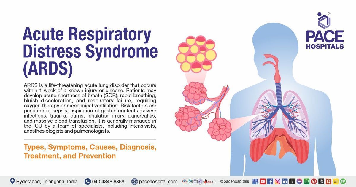
Acute Respiratory Distress Syndrome (ARDS) is a life-threatening acute lung disorder that occurs within 7 days of a known injury or disease. It is caused by sudden injury to the lungs, which increases the permeability of the alveolar-capillary membrane, allowing fluid to leak into the alveoli. This process leads to severely impaired gas exchange and low blood oxygen levels. Patients may develop acute shortness of breath, rapid breathing, bluish discoloration, and respiratory failure, requiring oxygen therapy or mechanical ventilation.
ARDS usually develops as a complication of serious illness or trauma. Common risk factors include pneumonia, sepsis, and aspiration of gastric contents, severe infections, trauma, burns, inhalation injury, pancreatitis, and massive blood transfusion. Most affected patients are already hospitalized or critically ill when ARDS develops. If not treated promptly, ARDS can result in multi-organ failure, prolonged ventilator dependence, and death.
ARDS is managed in the intensive care unit by a team of specialists, including intensivists, pulmonologists, and anesthesiologists. Treatment focuses on addressing the cause and providing oxygenation and lung support through respiratory therapies.
According to the Berlin definition (2012), ARDS is diagnosed when acute respiratory failure develops within one week of a known clinical insult, with bilateral lung opacities on chest imaging not fully explained by effusions or cardiac causes. The condition must be associated with impaired oxygenation, defined as a PaO₂/FiO₂ ratio of ≤300 mmHg while receiving a minimum positive end-expiratory pressure (PEEP) of at least 5 cm H₂O.
ARDS meaning
ARDS stands for acute respiratory distress syndrome and was first described in 1967 by American doctors Ashbaugh, Bigelow, Petty, and Levine in The Lancet.
Word Origins and Meanings:
- Acute: From Latin acutus, meaning “sharp”; refers to sudden onset.
- Respiratory: From Latin respirare, meaning “to breathe”; relates to breathing.
- Distress: From Old French destresse, meaning “constraint” or “severe difficulty.”
- Syndrome: From Greek syndromē, meaning “running together”; refers to a group of related symptoms.
ARDS Epidemiology
ARDS epidemiology worldwide
Acute respiratory distress syndrome (ARDS) is an important cause of critical illness worldwide, occurring in about 10.4% of patients admitted to intensive care units (ICU). In general population, about 13 to 23 individuals per 100,000 develop ARDS every year, however the rate is much more heightened among patients on mechanical ventilation. Mortality remains high, depending on disease severity and quality of care, usually between 30% and 50%. During the COVID-19 pandemic, the number of ARDS cases increased, with 32.2% of infected patients worldwide, emphasising its major impact on public issue.
ARDS epidemiology in India
In India, precise national data on ARDS incidence are limited, but hospital and ICU-based studies provide important insights. ARDS occurs in about 11.4% of mechanically ventilated patients in tertiary care centres. The overall mortality rate for ARDS patients in India is reported to be approximately 41.8%, which is comparable to global figures. Unique to India, infectious diseases such as scrub typhus, malaria, and pneumonia are frequent causes of ARDS. In pediatric populations, acute respiratory distress syndrome in children (ARDS) has a prevalence of about 9.9% in ICUs, with mortality rates reaching up to 51.4%.

Types of ARDS
ARDS can be classified into different types based on severity, cause of lung injury, and disease phase. Understanding these types helps guide treatment and predict patient outcomes. The following are the different types of ARDS:
ARDS Based on Severity (Berlin Definition)
The Berlin definition of ARDS classifies the condition based on the severity of hypoxemia using the PaO₂/FiO₂ ratio, which is assessed at the minimum level of positive end-expiratory pressure. This severity-based classification helps in treatment decision-making and prognosis assessment.
- Mild acute respiratory distress syndrome: PaO₂/FiO₂ between 200 and 300 mmHg
- Moderate acute respiratory distress syndrome: PaO₂/FiO₂ between 100 and 200 mmHg
- Severe acute respiratory distress syndrome: PaO₂/FiO₂ less than 100 mmHg
ARDS Based on the Cause of Lung Injury
ARDS can also be categorised according to whether lung injury occurs directly within the lungs or indirectly due to systemic inflammation.
- Direct (Pulmonary) ARDS: This type results from a primary insult to the lung tissue, predominantly affecting the alveolar epithelium. Common causes include pneumonia, aspiration of gastric contents, inhalation injury, pulmonary contusion, and near-drowning.
- Indirect (Extrapulmonary) ARDS: This form develops due to systemic inflammatory conditions that secondarily damage the pulmonary capillary endothelium. Common causes include sepsis, severe trauma, acute pancreatitis, massive blood transfusion, and major burns.
ARDS Pathophysiology
Acute respiratory distress syndrome pathophysiology is characterised by extensive damage to the alveolar-capillary membrane, resulting in inflammation, fluid accumulation in the lungs, and compromised oxygen exchange. Regardless of the triggering event, such as sepsis, trauma, aspiration (inhalation of foreign material into the airways and lungs), or severe infection, these mechanisms disrupt normal lung function and lead to respiratory failure.
Alveolar-Capillary Barrier Damage: The initial injury damages both the endothelial cells of the pulmonary capillaries and the epithelial cells lining the alveoli. This disrupts the normal barrier that maintains fluid balance and avoids leakage.
Increased Permeability & Edema Formation: Damage to these barriers elevates microvascular permeability, allowing protein-rich fluid to leak into the interstitial and alveolar spaces. This proteinaceous edema fluid accumulates in the lungs, impairing oxygen diffusion and reducing lung compliance.
Inflammatory Cascade: Pro-inflammatory cytokines (e.g., TNF-α, IL-1, IL-6) are released in response to injury, recruiting neutrophils and other immune cells. Activated neutrophils release reactive oxygen species and proteases that worsen endothelial and epithelial damage.
Diffuse Alveolar Damage (DAD): The characteristic histological lesion in ARDS is diffuse alveolar damage, which progresses through phases:
- Exudative phase: Edema, hyaline membrane formation, and acute inflammation
- Proliferative phase: Type II pneumocyte proliferation for early repair
- Fibrotic phase: Collagen deposition and fibrosis in some cases
Surfactant Dysfunction & Alveolar Collapse: Injury to type II alveolar cells decreases surfactant production, elevates surface tension, and causes atelectasis (alveolar collapse), further impairing gas exchange.
Gas Exchange Abnormalities: The combination of alveolar flooding, loss of functional alveoli, and ventilation–perfusion mismatch causes refractory hypoxemia unresponsive to basic oxygen therapy.
Systemic Inflammatory Effects: Inflammation can spill over into the bloodstream, contributing to systemic organ dysfunction and multi-organ failure in severe cases.

Acute Respiratory Distress Syndrome Symptoms
ARDS symptoms develop rapidly after a triggering illness or injury, leading to severe breathing difficulties and low oxygen levels. Early recognition of symptoms is crucial for prompt treatment. The following are the ARDS signs and symptoms:
Symptoms of ARDS:
- Shortness of breath
- Rapid, shallow, or laboured breathing
- Hypoxemia (low blood oxygen levels)
- Cyanosis (bluish lips or fingertips)
- Tachycardia (fast heartbeat)
- Chest tightness or discomfort
- Confusion or altered mental status
- Low blood pressure with signs of organ dysfunction
- Progression to respiratory failure in advanced stages
Shortness of breath: ARDS causes shortness of breath that often worsens rapidly, as inflammation and fluid buildup in the lungs make oxygen transfer difficult. This symptom can escalate within hours, signalling acute respiratory distress that may require urgent intervention.
Rapid, shallow, or laboured breathing: The stiff lungs cannot expand properly, forcing the body to compensate with fast, shallow breaths. Accessory muscles of the neck and chest may be used, indicating severe respiratory effort.
Hypoxemia (low blood oxygen levels): This is the hallmark of ARDS. Fluid-filled alveoli prevent efficient oxygen transfer into the bloodstream, sometimes making supplemental oxygen insufficient. This can impair organ function if left uncorrected.
Cyanosis (bluish lips or fingertips): Critically low oxygen levels cause tissues to turn bluish, especially in the lips and fingertips. This visible sign indicates critically low oxygen delivery to the body.
Tachycardia (fast heartbeat): The heart rate increases as a compensatory response to deliver more oxygen to tissues. Persistent tachycardia increases cardiac workload and may worsen cardiac strain if hypoxia continues.
Chest tightness or discomfort: Increased work of breathing, use of accessory muscles, and lung inflammation can produce a sensation of chest tightness, even though ARDS is not caused by cardiac ischemia.
Confusion or altered mental status: The brain is extremely sensitive to hypoxia. Low oxygen and high carbon dioxide levels affect brain function, leading to confusion, restlessness, drowsiness, or difficulty concentrating.
Low blood pressure with signs of organ dysfunction: ARDS often occurs as part of a systemic inflammatory response or sepsis. Capillary leakage and vasodilation reduce effective circulating volume, leading to hypotension and impaired perfusion of organs such as the kidneys and liver.
Progression to respiratory failure in advanced stages: In advanced ARDS, lung function may fail, requiring mechanical ventilation to maintain oxygenation. Without proper management, this can lead to multi-organ failure and become life-threatening.

ARDS Causes
Acute respiratory distress syndrome (ARDS) arises from a variety of pulmonary and extrapulmonary conditions. These causes may be direct or indirect clinical events that initiate lung injury and activate an inflammatory cascade, resulting in alveolar–capillary membrane damage and severe hypoxemia. The causes include:
Direct (Pulmonary) Causes
- Pneumonia (bacterial, viral, including COVID-19)
- Aspiration of gastric contents
- Inhalation of toxic fumes or smoke
- Pulmonary contusion
- Chest trauma
- Drowning-related lung injury
- Radiation or drug-induced lung injury
Indirect (Extrapulmonary) Causes
- Sepsis and septic shock
- Major trauma (including fat embolism)
- Massive blood transfusion (TRALI)
- Acute pancreatitis
- Burns
- Drug overdose
- Cardiopulmonary bypass
- Increased intracranial pressure
Direct (Pulmonary) causes
Pneumonia (bacterial, viral, including COVID-19): Infection of the lung parenchyma causes intense alveolar inflammation and epithelial injury. This increases alveolar–capillary permeability, leading to fluid accumulation in the lungs, impaired gas exchange, and low blood oxygen levels.
Aspiration of gastric contents: Inhalation of acidic gastric material causes chemical pneumonitis with direct damage to alveolar epithelium. This triggers an inflammatory cascade resulting in diffuse alveolar injury.
Inhalation of toxic fumes or smoke: Toxic gases and smoke cause direct thermal and chemical injury to the airways and alveoli. This leads to epithelial necrosis, inflammation, and increased pulmonary capillary permeability.
Pulmonary contusion: Blunt chest trauma causes hemorrhage and edema within lung tissue. The resulting inflammation and fluid accumulation interfere with oxygen diffusion and may progress to ARDS, especially in severe trauma.
Chest trauma: Severe chest injuries can disrupt normal lung structure and blood vessels. This damage, combined with inflammation and reduced oxygen delivery, increases the ARDS risk.
Drowning-related lung injury: Water aspiration into the lungs destroys the surfactant and alveolar lining. This causes alveolar collapse, irritation, and decreased oxygenation.
Radiation or Drug-Induced Lung Injury: Radiation therapy and certain medications can cause direct toxic injury to lung cells. In severe cases, this results in inflammation, increased vascular permeability, and acute lung failure consistent with ARDS.
Indirect (extrapulmonary) causes
Sepsis and Septic Shock: Systemic infection provokes widespread inflammatory response. Inflammatory mediators circulating in the blood increase lung capillary permeability, causing fluid leakage into the air sacs and diffuse lung injury.
Major Trauma (Including Fat Embolism): Severe trauma releases inflammatory mediators into the circulation. Fat emboli from bone fractures may enter the lungs, block blood vessels, and trigger inflammation that can lead to ARDS.
Massive Blood Transfusion (TRALI): Transfusion-related acute lung injury occurs due to immune-mediated activation of white blood cells within the lung circulation. This causes capillary leak and acute non-cardiogenic pulmonary edema.
Acute Pancreatitis: Inflammatory enzymes and mediators released from the pancreas enter the circulation. These substances damage the lung blood vessels, increasing permeability and leading to lung edema and ARDS.
Burns: Severe burns cause systemic inflammation and increased capillary leak throughout the body. Fluid shifts and inflammatory mediators contribute to pulmonary edema and ARDS.
Drug Overdose: Drug overdose can lead to slowed breathing, loss of airway protection, aspiration, or direct lung toxicity. These factors can trigger severe lung inflammation and ARDS.
Cardiopulmonary Bypass: During major heart surgery, blood contact with artificial surfaces activates inflammatory pathways. This systemic inflammatory response may damage lung capillaries, leading to ARDS.
Increased Intracranial Pressure: Severe brain injury may cause neurogenic pulmonary edema. Sympathetic overactivity increases pulmonary vascular pressure and permeability.

Risk Factors for ARDS
Risk factors are patient characteristics or clinical settings that increase the likelihood of developing ARDS after an inciting event. Not all patients with risk factors develop ARDS, and up to 20% of cases have no identifiable risk factor. Risk factors of ARDS include:
- Advanced age
- Female gender (noted mainly in trauma patients)
- Cigarette smoking
- Chronic alcohol use
- Genetic susceptibility
- Severe systemic illness (high APACHE score)
- Aortic vascular surgery
- Cardiovascular surgery
- Traumatic brain injury
- Massive fluid resuscitation
- Immunosuppression
Advanced age: Older adults have reduced lung repair mechanisms, reduced immune system, and increased comorbid conditions makes them more susceptible to severe inflammatory lung injury. Studies showed that advanced age is associated with higher incidence, severity, and mortality in ARDS patients.
Female gender: Some trauma studies have shown higher ARDS incidence in women, possibly related to hormonal influences on inflammation and immune response. However, gender differences in ARDS risk remain complex and may vary by patient population.
Cigarette smoking: Smoking harms the airway epithelium, weakens pulmonary immune defenses, and changes alveolar macrophage activity, increasing susceptibility to inflammatory lung injury. Epidemiological statistics indicate that smoking increases the likelihood of ARDS in at-risk patients.
Chronic alcohol use: Chronic alcohol abuse is a well-established independent risk factor for ARDS; it impairs alveolar epithelial and immune cell functions and increases oxidative stress. Alcohol use also prolongs mechanical ventilation and ICU stay in ARDS patients.
Genetic susceptibility: Genetic variations can influence individual inflammatory responses and endothelial integrity, affecting susceptibility to ARDS. Certain polymorphisms are associated with increased risk of lung injury, although more research is needed.
Severe systemic illness (high APACHE score): Higher severity-of-illness ratings, such as the APACHE II, suggest systemic organ dysfunction and inflammation, which are significantly linked to ARDS progression. Severity scores are used clinically to identify people who are more likely to develop ARDS.
Aortic vascular surgery: Major vascular procedures can cause systemic inflammation, damage to the endothelium, and ischemia-reperfusion changes. These issues increase the permeability of pulmonary capillaries and lead to fluid leaking into the lungs. As a result, patients who have aortic surgery face a greater risk of developing ARDS after the procedure.
Cardiovascular surgery: Cardiac surgery, particularly cardiopulmonary bypass surgery activates inflammatory pathways and systemic cytokine release. This systemic response contributes to endothelial injury and increases ARDS risk after surgery.
Traumatic brain injury: Severe head injury induces a neurogenic inflammatory response and sympathetic activation that increases pulmonary vascular permeability. This cascade heightens susceptibility to ARDS, especially in multiply injured patients.
Massive fluid resuscitation: Large volumes of intravenous fluids can increase hydrostatic pressure in the pulmonary circulation and worsen lung edema. Excessive fluid administration may also impair clearance of fluid from the alveoli. In critically ill patients, aggressive early resuscitation is associated with an increased risk of ARDS.
Immunosuppression: Immunosuppression, whether due to underlying disease or medical therapy, reduces the body's ability to control infections and inflammation. This can lead to persistent lung injury, delayed healing, and increased susceptibility to severe inflammatory responses. As a result, immunosuppressed patients are at higher risk of developing ARDS in critical illness.
ARDS Complications
Complications of acute respiratory distress syndrome (ARDS) can affect the lungs, other organs, and overall patient recovery. Mechanical ventilation, prolonged ICU stay, and systemic inflammation are major contributors to these adverse outcomes. Common complications of ARDS include:
Respiratory Complications
- Barotrauma
- Pulmonary fibrosis
- Prolonged mechanical ventilation
- Post-extubation airway issues
Infectious Complications
- Nosocomial infections
- Ventilator-associated pneumonia (VAP), line sepsis, urinary tract infection
- Antibiotic resistance
Systemic and Other Complications
- Multi-organ failure
- Deep venous thrombosis (DVT)
- Muscle weakness and ICU-acquired weakness
- Renal failure
- Psychological effects (PTSD, anxiety, depression)
ARDS Diagnosis
Acute respiratory distress syndrome diagnosis involves recognizing the sudden onset of breathing difficulties and lung changes that are not caused by heart disease. Using ARDS diagnostic criteria helps identify the severity and ensures timely treatment to improve patient outcomes.
The following are the steps commonly included in the acute respiratory distress syndrome diagnosis:
- Medical history
- Physical examination
- Laboratory tests
- Arterial blood gas (ABG)
- Blood tests: CBC, metabolic panel, cultures (blood, urine, sputum)
- Infection markers: PCR, antigen tests
- Autoimmune panels (done when no clear cause is found)
- Imaging studies
- Chest X-ray
- Chest computed tomography scan
- Lung ultrasound (optional)
- Excluding Heart-Related Lung Fluid
- Echocardiography and clinical exam
- Pulmonary artery wedge pressure <18 mmHg (if measured)
- Applying Berlin Definition (2012) ARDS Diagnostic Criteria
- Optional Additional Tests
- Bronchoscopy for infection evaluation
- Serologic or autoimmune testing if no clear cause
- Hemodynamic monitoring in complex cases
ARDS Treatment
Acute respiratory distress syndrome (ARDS) is a life-threatening condition marked by severe hypoxemia and diffuse lung injury. The acute respiratory distress syndrome treatment aims to support oxygenation, prevent further lung damage, and address the underlying cause through ventilation strategies, fluid management, nutritional support, and selective pharmacologic or adjunctive therapies.
Below are the ARDS treatment options:
Management of the Underlying Cause
- Early broad-spectrum antibiotics for suspected sepsis or pneumonia
- Antiviral or antifungal therapy when indicated
- Source control (e.g., drainage of abscess, control of bleeding, surgery for trauma)
- Discontinuation of offending drugs
- Hemodynamic stabilization with intravenous fluids and vasopressors
- Blood transfusion if clinically indicated
- Early correction of the triggering event prevents further lung injury.
Respiratory Support (Core of ARDS Treatment)
A. Oxygen Therapy (Mild ARDS)
- Nasal cannula
- Face mask
- High-flow nasal oxygen (HFNO)
Used when oxygen levels are low but the patient can breathe independently.
B. Non-Invasive Ventilation (Selected Cases)
- CPAP (Continuous Positive Airway Pressure)
- BiPAP (Bilevel Positive Airway Pressure)
Used cautiously in mild ARDS with close monitoring.
C. Invasive Mechanical Ventilation (Moderate–Severe ARDS)
- Endotracheal intubation
- Lung-protective ventilation strategy:
- Low tidal volume (6 mL/kg predicted body weight)
- Plateau pressure ≤ 30 cm H₂O
- Appropriate PEEP titration
- Driving pressure monitoring
- Permissive hypercapnia (pH ≥ 7.20 if tolerated)
- Target SpO₂ 88–95%
Mechanical ventilation is the main life-saving therapy in ARDS.
D. Advanced Ventilatory Strategies (Severe ARDS)
- Prone positioning ≥16 hours/day (PaO₂/FiO₂ <150)
- Short-term neuromuscular blockade for ventilator synchrony
- Recruitment maneuvers (selected cases)
- Airway pressure release ventilation (selected centers)
E. Extracorporeal Support (Rescue Therapy)
- Venovenous Extracorporeal Membrane Oxygenation (ECMO)
- Extracorporeal CO₂ removal (in specialized centers)
Used when oxygenation fails despite optimal ventilation.
Pharmacological Therapy
A. Adjunctive ARDS Therapy
- Corticosteroids in selected moderate–severe cases
- Short-term neuromuscular blocking agents
- Inhaled pulmonary vasodilators (e.g., nitric oxide) as rescue therapy
B. Hemodynamic & Organ Support
- Vasopressors for shock
- Sedatives and analgesics for ventilated patients
- Diuretics in fluid-overloaded patients
Not routinely recommended:
- High-frequency oscillatory ventilation (HFOV)
- Routine surfactant therapy in adults
- Routine beta-agonists
Procedural & Long-Term Support
- Central venous and arterial line placement for monitoring
- Tracheostomy (for prolonged ventilation)
- Dialysis (if acute kidney injury develops)
- Nutritional tube placement (if prolonged feeding required)
Supportive ICU Care
- Conservative fluid strategy after stabilization of shock
- Early enteral nutrition (within 24–48 hours if feasible)
- Glycemic control
- Deep vein thrombosis (DVT) prophylaxis
- Stress ulcer prophylaxis (when indicated)
- Ventilator-associated pneumonia (VAP) care bundle
- Pressure area care
- Early physiotherapy and mobilization
Why Choose PACE Hospitals?
Expert Super Specialist Doctors
Advanced Diagnostics & Treatment
Affordable & Transparent Care
24x7 Emergency & ICU Support

ARDS Prevention
Prevention of ARDS focuses on reducing the risk of lung injury in vulnerable patients and preventing progression in those with early respiratory compromises. Key strategies include:
- Early treatment of underlying causes
- Lung-protective ventilation strategies
- Conservative fluid management
- Prevention of aspiration
- Restrictive blood transfusion practices
- Infection prevention measures
- Avoidance of toxic exposures
- Optimal management of comorbid conditions
- Early nutritional and supportive care
Early treatment of underlying causes: Prompt control of sepsis, pneumonia, trauma, and aspiration reduces the inflammatory cascade that leads to ARDS.
Lung-protective ventilation strategies: Use of low tidal volume ventilation (≈6 mL/kg predicted body weight) and limiting plateau pressures (<30 cm H₂O) prevents ventilator-induced lung injury in high-risk or critically ill patients.
Conservative fluid management: Avoiding fluid overload and using a conservative fluid strategy after initial resuscitation reduces pulmonary edema and improves lung function.
Prevention of aspiration: Head-end elevation, safe feeding practices, and airway protection in high-risk patients help prevent aspiration-related lung injury.
Restrictive blood transfusion practices: Limiting unnecessary transfusions and careful monitoring reduces the risk of transfusion-related acute lung injury (TRALI), a known ARDS trigger.
Infection prevention measures: Strict hand hygiene, early appropriate antibiotics, and vaccination reduce severe infections that commonly lead to ARDS.
Avoidance of toxic exposures: Reducing exposure to tobacco smoke, toxic gases, and environmental pollutants lowers risk of direct lung injury.
Optimal management of comorbid conditions: Effective control of chronic diseases (e.g., diabetes, alcohol misuse, cardiac disease) decreases susceptibility to severe systemic inflammation.
Early nutritional and supportive care: Early enteral nutrition and maintenance of physiological stability support immune function and reduce complications.
Difference between ARDS and Pneumonia
ARDS vs Pneumonia
Acute respiratory distress syndrome (ARDS) and pneumonia are serious lung conditions that impair breathing, but they differ significantly in cause, pathophysiology, severity, and management. Pneumonia is primarily an infection of the lung tissue, while ARDS is a severe inflammatory response of the lungs that can result from many direct or indirect injuries. Below are the key differences between ARDS and pneumonia:
| Feature | ARDS | Pneumonia |
|---|---|---|
| Definition | Acute respiratory distress syndrome is a severe, sudden-onset condition caused by widespread injury to the alveoli, leading to leakage of fluid into the air sacs, reduced lung compliance, and acute respiratory failure | Pneumonia is an inflammatory condition of the lung parenchyma caused by infection, resulting in alveolar filling with pus, fluid, and inflammatory cells |
| Etiology | ARDS develops secondary to a serious underlying illness like sepsis, severe trauma, aspiration of gastric contents, pancreatitis, burns, or severe pneumonia | Pneumonia is caused by infectious agents including bacteria, viruses, fungi, or parasites, often acquired from the community or hospital |
| Lung involvement | Diffuse and bilateral lung disease affects both lungs uniformly, resulting in substantial loss of functioning alveoli. | Usually affects a specific lung segment or lobe, though severe cases may involve multiple lobes or both lungs |
| Oxygenation | Causes profound hypoxemia due to shunting and impaired gas exchange that does not respond well to supplemental oxygen | Hypoxemia occurs due to alveolar consolidation but usually improves with oxygen therapy and treatment of infection |
| Radiologic findings | Chest X-ray or CT scan shows bilateral, diffuse pulmonary infiltrates not explained by heart failure or fluid overload | Imaging typically shows lobar consolidation, patchy infiltrates, or interstitial patterns depending on the causative organism |
| Management approach | Managed primarily with supportive care including lung-protective mechanical ventilation, prone positioning, careful fluid management, and treatment of the underlying cause | Treated mainly with appropriate antimicrobial therapy, along with supportive care such as oxygen, fluids, and fever control |
Frequently asked questions (FAQs) on ARDS
What is acute respiratory distress syndrome?
Acute respiratory distress syndrome (ARDS) is a severe, life-threatening lung condition in which intense inflammation causes fluid to build up in the air sacs (alveoli), resulting in very low blood oxygen levels and stiff, poorly functioning lungs. It usually develops within hours to days after a serious underlying illness or trauma, such as sepsis, pneumonia, or COVID-19. It often requires treatment in an intensive care unit (ICU) under the care of a pulmonologist or critical care (intensivist) physician, frequently with mechanical ventilation support.
Is acute respiratory distress serious?
Yes, acute respiratory distress syndrome (ARDS) is serious. It is a life-threatening form of acute respiratory failure that typically occurs in critically ill patients and often requires ICU care, high levels of oxygen support, and frequently mechanical ventilation. Mortality remains significant: under the Berlin definition, reported hospital mortality rises with severity (about 27% in mild, 32% in moderate, and ~45% in severe ARDS).
How does sepsis cause ARDS?
Sepsis can lead to acute respiratory distress syndrome (ARDS) by triggering an excessive and uncontrolled inflammatory response throughout the body. During sepsis, large amounts of inflammatory substances are released into the bloodstream, which damage the lining of the lung blood vessels and air sacs. The lung tissue becomes damaged and leaky, causing fluid and proteins to gather in the air sacs. Damage to the lungs causes fluid and proteins to collect in the air sacs. This alleviates oxygen in the blood, makes the lungs stiff, and may result in worsening breathing, often resulting in respiratory failure that needs intensive care and mechanical ventilation.
Is ARDS contagious?
No. ARDS is not a contagious condition. It develops as a reaction to a serious medical problem or injury rather than from exposure to another person. In ARDS, widespread inflammation causes the lungs to fill with fluid, reducing the amount of oxygen that can enter the bloodstream. Although infections such as pneumonia or sepsis can lead to ARDS, the lung syndrome itself does not spread from person to person. Only the illness that triggered ARDS, if it is infectious, can be passed on to others, not ARDS itself.
Can ARDS be reversed?
Yes, ARDS can often be reversed, especially when treated early with prompt intensive care, including oxygen therapy and mechanical ventilation in an ICU.
In many people, the lung swelling and fluid buildup gradually reduce once the main trigger (such as sepsis, pneumonia, or aspiration) is controlled and breathing support is given. Most survivors gradually recover much of their lung function, though this process can take several weeks to months. Individuals who experienced severe ARDS or a prolonged stay in the ICU may continue to have fatigue, shortness of breath during physical activity, or muscle weakness for an extended period.
Can ARDS lead to heart failure?
Yes, ARDS can affect the heart, but it does not directly cause heart failure in the usual sense.
In ARDS, the lungs are badly inflamed and filled with fluid, which lowers oxygen levels in the blood. Low oxygen and high pressure inside the lungs can place extra strain on the right side of the heart, making it work harder to pump blood through the lungs. In severe cases, this strain can lead to temporary right-sided heart dysfunction. ARDS can also worsen existing heart disease or trigger heart problems in critically ill patients.
With proper treatment and recovery from ARDS, heart strain often improves. Permanent heart failure is uncommon unless there is pre-existing heart disease or prolonged severe illness.
What is the main cause of acute respiratory distress syndrome?
The main cause of acute respiratory distress syndrome (ARDS) is severe, widespread inflammation in the lungs resulting from a serious injury or illness, with sepsis and pneumonia responsible for 40% to 60% of cases. ARDS usually develops within hours to days after the illness or trauma, such as sepsis, pneumonia, or COVID-19, and often requires care in an intensive care unit by a pulmonologist or critical care physician, frequently with mechanical ventilation. If not treated immediately, it can lead to life-threatening respiratory failure and multiple organ dysfunction.
How long does it take to recover from acute respiratory distress syndrome?
Recovery from ARDS varies widely: some individuals start to improve within a few weeks, while others may take 6 months or more to regain lung function, strength, and overall health. During this time, rehabilitation and supportive care are often needed to help restore daily activities and overall well-being.
In some cases, patients may continue to experience long-term effects such as shortness of breath (SOB), tiredness, or difficulties with memory and concentration. Recovery largely depends on factors such as age, illness severity, and the patient's health before developing ARDS. Although most survivors eventually recover near-normal lung function, some may notice reduced stamina or muscle stiffness.
What are the symptoms of ARDS?
Acute respiratory distress syndrome (ARDS) usually develops suddenly after a serious illness or injury and causes severe breathing problems. Common symptoms include shortness of breath (SOB) that worsens quickly, rapid or laboured breathing, and low blood oxygen levels, which may not improve even with oxygen support. Some patients develop cyanosis (bluish discolouration) of the lips or fingertips, along with tachycardia and chest tightness.
When oxygen levels decrease, patients may experience confusion or reduced alertness. In severe cases, low blood pressure and organ dysfunction can develop. If the condition progresses, ARDS can lead to respiratory failure, often requiring treatment in an intensive care unit with mechanical ventilation.
Which level of ARDS requires ICU care?
All levels of acute respiratory distress syndrome (mild, moderate, and severe) usually require care in an intensive care unit (ICU) because the condition can cause sudden, life-threatening breathing problems. ICU care is especially important when oxygen levels in the blood are very low (PaO₂/FiO₂ ratio below 300 mmHg).
Patients with moderate to severe ARDS typically require close monitoring and advanced support, such as a ventilator, because normal oxygen therapy is insufficient. Even moderate ARDS patients may receive high-flow oxygen or non-invasive breathing support, but ICU therapy usually recommended to constantly monitor breathing, oxygen levels, and organ function due to the condition's rapid deterioration.
What is the pathophysiology of ARDS?
ARDS occurs when a severe illness or injury triggers an intense inflammatory response that causes diffuse alveolar damage. Inflammatory mediators increase the permeability of the alveolar-capillary membrane, allowing protein-rich fluid to leak into the alveoli. This leads to pulmonary edema, surfactant dysfunction, and alveolar collapse. As a result, ventilation–perfusion mismatch and intrapulmonary shunting occur, causing severe hypoxemia that may be refractory to oxygen therapy. Lung compliance decreases, making the lungs stiff and increasing the work of breathing, which results in acute respiratory failure.
What is the difference between acute respiratory failure and ARDS?
Acute respiratory failure is a wide term for when the lungs are unable to give adequate oxygen to the body or eliminate carbon dioxide correctly. It can occur for a variety of reasons, including acute pneumonia, asthma attacks, drug overdose, chest injuries, or a deterioration of existing lung disease. The condition can be affected by breathing, oxygen levels, carbon dioxide levels, or both, with severity ranging from minor to life-threatening.
Acute respiratory distress syndrome is a different and severe type of acute respiratory failure. In ARDS, extensive inflammation destroys the lungs, allowing fluid to flow into the air sacs. This stiffens the lungs and restricts oxygen transfer into the blood, even with oxygen therapy. ARDS is frequently caused by acute illnesses such as sepsis, severe infections, trauma, or aspiration and requires special treatment.
Who is at high risk for ARDS?
Individuals who are more likely to develop acute respiratory distress syndrome when they are already seriously ill or have suffered a major injury. The highest risk is seen in individuals with severe infections like pneumonia or sepsis, which can trigger widespread lung inflammation. Individuals who inhale stomach contents into the lungs, such as during vomiting or loss of consciousness, are also at high risk.
Other high-risk groups include people with major trauma, severe burns, acute pancreatitis, or those who receive large blood transfusions. ARDS is more common in patients who are hospitalized in intensive care units, especially those who are older, have long-standing medical problems, smoke heavily, or have chronic lung disease.
When to consult a doctor for ARDS?
Consult a doctor immediately if breathing problems develop suddenly after a serious illness, infection, or injury, or if existing breathing difficulty worsens rapidly. Early medical attention is critical because ARDS can progress quickly and become life-threatening.
Signs that need urgent medical evaluation include:
- Sudden or worsening shortness of breath
- Rapid, shallow, or laboured breathing
- Low oxygen levels, often not improving with usual oxygen support
- Bluish lips or fingertips
- Chest tightness or discomfort
- Confusion, restlessness, or extreme drowsiness
- Low blood pressure or reduced urine output
- Breathing difficulty following sepsis, severe pneumonia, trauma, burns, or aspiration
If these symptoms appear, especially in a hospitalized or critically ill person, immediate evaluation by an ARDS specialist is essential. Seek emergency medical care if there is any severe breathlessness, confusion, or collapse. Early assessment by a critical care physician or pulmonologist allows timely ARDS treatment in an intensive care setting and helps prevent serious complications.
Share on
Request an appointment
Fill in the appointment form or call us instantly to book a confirmed appointment with our super specialist at 04048486868
Appointment request - health articles
Recent Articles