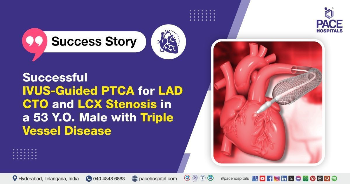
Successful IVUS-Guided PTCA for LAD CTO and LCX Stenosis in a 53-Y.O. Male with Triple Vessel Disease
PACE Hospitals
PACE Hospitals’ expert cardiology team successfully performed a
Percutaneous Transluminal Coronary Angioplasty (PTCA) of the Proximal Left Anterior Descending (LAD) artery and Left Circumflex (LCX) artery using Intravascular Ultrasound (IVUS) guidance in a 53-year-old male diagnosed with Coronary Artery Disease (CAD). The aim of the procedure was to restore proper blood flow to the heart muscle by opening the narrowed arteries, thereby relieving symptoms, improving overall heart function, and reducing the risk of future cardiac events.
Chief Complaints
A 53-year-old male patient with a
body mass index (BMI) of 22 presented to the Cardiology Department at
PACE Hospitals, Hitech City, Hyderabad, with chief complaints of anginal chest pain for the past 3 days, associated with palpitations and shortness of breath (SOB), prompting urgent cardiac evaluation.
Past Medical History
There is no documented history of other chronic conditions, such as diabetes, hypertension, or liver disease, at the time of admission.
On Examination
On examination, the patient was alert, oriented, and clinically stable. There were no signs of pallor, cyanosis, icterus, or pedal edema. Vital signs were within normal limits. Cardiovascular examination revealed normal heart sounds with no murmurs. Respiratory examination indicated clear bilateral air entry. The neurological and abdominal examinations were unremarkable.
Diagnosis
Upon admission to PACE Hospitals, the patient was evaluated by the cardiology team, which included a detailed review of his medical history and a comprehensive clinical examination. The patient underwent a comprehensive diagnostic evaluation to assess cardiac status.
Coronary angiography (CAG) confirmed Triple Vessel Disease (TVD) with 100% proximal occlusion of the left anterior descending (LAD) artery (Chronic Total Occlusion – CTO), 80% stenosis in the non-dominant left circumflex (LCX) artery, and 40–50% long-segment stenosis in the dominant right coronary artery (RCA). Echocardiography findings were consistent with the patient’s clinical condition.
Based on these confirmed findings, the patient was advised to undergo
Coronary Artery Disease Treatment in Hyderabad, India, under the expert care of the Cardiology Department.
Medical Decision Making
After a detailed consultation with Dr. Seshi Vardhan Janjirala, Consultant Cardiologist, a comprehensive evaluation was conducted focusing on the patient’s advanced coronary artery disease (CAD) with triple vessel involvement, including chronic total occlusion (CTO) of the left anterior descending (LAD) artery. Clinical and diagnostic findings confirmed significant stenosis in the left circumflex (LCX) and right coronary artery (RCA).
It was determined that Percutaneous Transluminal Coronary Angioplasty (PTCA) to the LAD and LCX arteries under Intravascular Ultrasound (IVUS) guidance was identified as the most appropriate intervention to restore coronary blood flow, relieve ischemic symptoms, and improve cardiac outcomes while minimizing procedural risks.
The patient and his family members were informed about his diagnosis, the planned procedure, associated risks, and expected benefits aimed at preserving heart function and enhancing his overall prognosis.
Surgical Procedure
Following the diagnosis, the patient was scheduled for Percutaneous Transluminal Coronary Angioplasty (PTCA) Procedure in Hyderabad at PACE Hospitals, under the expert supervision of the Cardiology Department, ensuring optimal care and a smooth recovery process.
The procedure involved the following steps:
- Crossing the Chronic Total Occlusion (CTO) of LAD: The procedure began with careful wiring across the 100% occluded proximal segment of the left anterior descending (LAD) artery. Specialized guidewires and catheters were used to successfully cross the CTO segment, ensuring safe access for stent deployment.
- PTCA and Stent Placement in LAD: Following successful crossing of the CTO, a 2.5 × 38 mm Boston Scientific drug-eluting stent was positioned and deployed in the LAD artery. The deployment was performed with precision to restore vessel patency.
- Intravascular Ultrasound (IVUS) Guidance: IVUS imaging was utilized during the intervention to guide optimal stent placement and expansion. This allowed real-time visualization of the vessel lumen and stent apposition, ensuring adequate coverage and minimizing complications.
- Stenting of LCX Artery: Attention was then turned to the left circumflex (LCX) artery, where a 2.75 × 28 mm Boston Scientific stent was deployed. This procedure was similarly guided by IVUS to confirm accurate stent placement and vessel expansion.
- Restoration of Blood Flow and Final Assessment: Post-stenting, angiography confirmed restoration of normal blood flow with TIMI grade III flow in both LAD and LCX arteries. The procedure was completed without any complications, and the patient remained stable throughout.
Postoperative Care
The procedure and post-procedure course were uneventful. The patient was managed with appropriate medications to optimize cardiovascular health and prevent complications. The vascular access site was monitored regularly for any issues, and supportive care was provided throughout the hospital stay. After a stable recovery, the patient was discharged in stable condition with detailed instructions on medication adherence, diet, and follow-up care.
Discharge Medications
Upon discharge, the patient was prescribed antiplatelet therapy to prevent thrombosis and maintain stent patency. A statin was prescribed to manage cholesterol levels and reduce cardiovascular risk. An additional antiplatelet agent was included for further prevention of clot formation, along with a gastroprotective medication to prevent gastric irritation. Antihypertensive therapy was advised as needed to maintain optimal blood pressure control if systolic blood pressure exceeded 140 mm Hg.
Emergency Care
The patient was informed to contact the emergency ward at PACE Hospitals in case of any emergency or development of symptoms such as chest pain, abdominal pain, and fever.
Review and Follow-up Notes
The patient was advised to return for a follow-up visit after 2 weeks with the cardiologist in Hyderabad at PACE Hospitals, to review his condition.
Conclusion
This case highlights the successful treatment of triple vessel coronary artery disease with chronic total occlusion using intravascular imaging-guided intervention. A multidisciplinary team approach ensured precise planning and patient stability throughout the procedure. With timely intervention and attentive postoperative care, the patient achieved a favorable recovery and improved prognosis.
Precision-Guided Cardiac Care in Multivessel Coronary Artery Disease
Timely diagnosis and the use of advanced intravascular imaging techniques are essential in the management of complex coronary artery disease. Chronic total occlusions, though technically demanding, can be successfully treated with precision-guided percutaneous interventions. Intravascular ultrasound (IVUS) enhances the accuracy of stent placement and reduces the risk of complications. A multidisciplinary approach involving a skilled Cardiologist / Heart specialist ensures comprehensive assessment of patient-specific risks and comorbidities.
Integrating imaging, intervention, and collaborative care leads to safer procedures and better clinical outcomes in high-risk coronary artery disease. Continued advancements in interventional cardiology are expanding the boundaries of what is treatable, offering improved survival and quality of life. Early recognition and individualized treatment planning remain key pillars in achieving optimal patient outcomes.
Share on
Request an appointment
Fill in the appointment form or call us instantly to book a confirmed appointment with our super specialist at 04048486868
Appointment request - health articles
Recent Articles