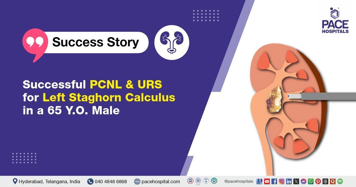
Successful PCNL & URS for Left Staghorn Calculus in a 65-Year-Old Male
PACE Hospitals
The PACE Hospitals expert Urology team successfully performed left percutaneous nephrolithotomy (PCNL), ureteroscopy (URS), and DJ stenting on a 65-year-old male patient who presented with left flank pain. The procedure was done to remove kidney stones, effectively relieving the patient's symptoms and helping restore proper kidney function to support recovery.
Chief Complaints
A 65-year-old male patient with a
Body Mass Index (BMI) of 23, presented to the Urology Department at
PACE Hospitals, Hitech City, Hyderabad, with persistent left flank pain. He reported discomfort that affected his daily activities and had not improved with initial medical management. A detailed evaluation was carried out to determine the underlying cause of his symptoms.
Past Medical History
The patient had a history of small airway disease, preoperative pulmonology evaluation was done to reduce respiratory risks during surgery. Long-acting bronchodilator nebulization was initiated to optimize his lung function and ensure a safer surgical outcome. The absence of
hypertension or
diabetes lowered the risk of other systemic complications.
On Examination
Upon admission, the patient was afebrile with stable vital signs, and his general condition was satisfactory. He was conscious, alert and showed no signs of acute distress. Physical examination revealed mild tenderness over the left flank on deep palpation, consistent with his reported symptoms. However, no palpable mass was detected, and there were no signs of peritonism, such as guarding, rebound tenderness, or abdominal rigidity.
Diagnosis
The patient initially underwent a comprehensive clinical assessment at PACE Hospitals, which included a detailed history and physical examination by the Urology team. During the evaluation, it was noted that the patient had been diagnosed with a left complete staghorn calculus (large, branched kidney stones), which required surgical intervention.
To support the diagnosis and assess the extent of the patient's condition, a series of relevant investigations were conducted.
- CT KUB (Kidney, Ureters, and Bladder): The CT scan of KUB revealed a Large left staghorn calculus and dilation of the upper lateral and middle posterior renal tracts. There was no evidence of ureteral obstruction or additional stones.
- Complete blood count (CBC): The CBC showed normal hemoglobin levels and no neutrophilic predominance, indicating the absence of active systemic infection. This suggested that the patient’s symptoms were likely due to localized inflammation from the renal calculi, rather than a systemic infection.
- Urinalysis: Revealed microscopic hematuria (the presence of red blood cells), a common finding in renal calculi, which supported the patient’s right flank pain, confirming the presence of red blood cells, a common finding in renal calculi, which supported the patient’s left flank pain.
Based on the confirmed findings, the patient was advised to undergo Kidney Stones Treatment in Hyderabad, India, under the expert care of the Urology Department, providing effective stone clearance, alleviating his flank pain, and preventing potential complications including renal function impairment.
Medical Decision-Making (MDM)
Following a detailed consultation with Dr. Abhik Debnath, Consultant Laparoscopic Urologist, a comprehensive evaluation of the patient’s condition was undertaken. Considering his persistent left flank pain and CT scan findings indicating staghorn calculus, the urology team determined that a left percutaneous nephrolithotomy (PCNL), ureteroscopy (URS), with DJ stenting, would be the most suitable and effective intervention.
Surgical Procedure
Following the decision, the patient was scheduled for percutaneous nephrolithotomy (PCNL), ureteroscopy (URS), and DJ stenting Surgery in Hyderabad at PACE Hospitals, under the expert supervision of the Urology Department, ensuring optimal care and a smooth recovery process.
- Anesthesia and Positioning: The patient was administered general anesthesia and positioned appropriately for percutaneous access to the left kidney.
- Initial Evaluation: Preoperative imaging confirmed the presence of a left complete staghorn calculus, occupying all calyces of the kidney. The size and location of the stone necessitated a multi-tract approach.
- Percutaneous Access and Punctures: Under fluoroscopic guidance, four punctures were made to access different parts of the collecting system: Upper lateral calyx, Mid posterior calyx, Lower posterior calyx and Lower anterior calyx
- Tract Dilation: Two tracts upper lateral and middle posterior were selectively dilated to facilitate nephroscope access and allow for effective stone clearance.
- Stone Fragmentation (PCNL): The hard staghorn calculus was fragmented using pneumatic lithotripsy. Careful fragmentation was performed to access and clear all calyceal extensions of the stone.
- Stone Clearance: All visible stone fragments were removed. Complete stone clearance was confirmed intraoperatively via nephroscope inspection and fluoroscopy.
- Ureteroscopy (URS): A ureteroscopy was performed to evaluate the ureter and ensure no residual fragments or obstructions were present.
- DJ Stenting: An antegrade Double J (DJ) stent was placed to ensure adequate drainage of urine from the kidney to the bladder and to support postoperative healing.
- Completion: Hemostasis was achieved. The procedure concluded without complications, and the patient was shifted to the recovery room in stable condition.
Postoperative Care
After surgery, the patient was monitored closely in the surgical intensive care unit to ensure stability. His postoperative course was uneventful. A postoperative chest X-ray showed normal lung fields, and an X-ray KUB revealed no radio-opaque shadows, indicating no residual stones. The nephrostomy tubes were removed on postoperative day 2, and the Foley catheter was removed on postoperative day 3. During his hospital stay, he received intravenous antibiotics to prevent infection, analgesics for pain relief, antiemetics to control nausea, and gastric protectants. The patient was discharged in stable condition.
Discharge Medication
Upon discharge, the patient was prescribed a course of oral medications tailored to support his recovery following the surgical removal of the staghorn calculus. These included oral antibiotics to prevent postoperative infections, analgesics for pain relief, and antipyretics to manage any potential fever. Urinary alkalizers were given to maintain an optimal urinary pH and reduce the risk of stone recurrence. Proton pump inhibitors were prescribed to protect the gastric lining from any medication-related irritation. Additionally, an alpha-blocker was administered to facilitate smooth urinary flow. The patient was also advised to take nutritional supplements to aid in overall recovery and support renal health.
Discharge Advice
The patient was advised to avoid weightlifting, forward bending, straining during bowel movements, and prolonged travel by bus, auto, or two-wheeler. Passage of small stone fragments, mild flank or suprapubic discomfort, occasional blood in urine, cloudy urine, or mild burning were expected and typically resolved within 7–10 days.
He was instructed to maintain good hydration (3–4 L/day), consume citrus fruits and juices, and follow a low-purine, low-salt diet. Physiotherapy, including incentive spirometry, deep breathing exercises, and steam inhalation thrice daily, was recommended to support recovery.
Emergency Care
The patient was informed to contact the Emergency ward at PACE Hospitals in case of any emergency or development of symptoms like fever, abdominal pain, swelling, bleeding, or vomiting.
Review and Follow-up
The patient was advised to return for a follow-up visit with the Urologist in Hyderabad at PACE Hospitals, after 1 month for DJ stent removal, cystoscopy, and URS. This review is necessary to assess the patient's recovery, ensure proper healing, and evaluate the urinary tract for any complications or residual issues that may require further intervention.
Conclusion
This case highlights the effectiveness of percutaneous nephrolithotomy (PCNL), ureteroscopy (URS), with DJ stenting in performing Renal Calculi Treatment in Hyderabad, India, enabling successful removal of kidney stones, thereby facilitating normal urine flow.
Significance of ureteroscopy in interpreting and management of staghorn calculi
Ureteroscopy (URS), also known as ureterorenoscopy, is performed by the urologist/urology doctor. And played a significant role in the interpretation and management of the staghorn calculus, particularly in combination with percutaneous nephrolithotomy (PCNL). It is used to visualize the entire renal pelvis and calyces, guide stone fragmentation, and remove residual fragments. It also allowed for intraoperative assessment of stone composition and identification of any potential complications. The obtained stone fragments through URS for further analysis, helping to determine the stone's nature and its likely response to different treatment options. URS was especially effective in clearing residual stones following PCNL, ensuring a complete and safe surgical outcome.
Share on
Request an appointment
Fill in the appointment form or call us instantly to book a confirmed appointment with our super specialist at 04048486868
Appointment request - health articles
Recent Articles