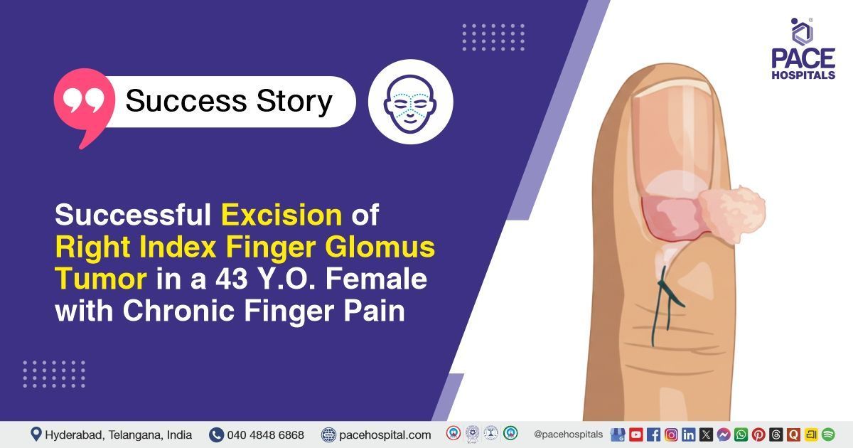
Successful Excision of Right Index Finger Glomus Tumor in a 43 Y.O. Female with Chronic Finger Pain
PACE Hospitals
PACE Hospitals' expert Plastic and Reconstructive Surgery team successfully performed an Excision Biopsy of the Tumor in a 43-year-old female diagnosed with a right index finger glomus tumor, with the aim of complete tumor removal, pain relief, and histopathological confirmation.
Chief Complaints
A 43-year-old female patient with a body mass index (BMI) of 22 presented to the Plastic and Reconstructive Surgery Department at PACE Hospitals, Hitech City, Hyderabad, with complaints of right index finger pain persisting for 12 to 15 years.
Past Medical History
The patient had no known history of chronic medical illnesses such as diabetes mellitus, hypertension, cardiac disease, or respiratory disorders. There was no history of trauma to the right index finger. She had a past surgical history of LSCS (Lower Segment Cesarean Section). No known drug or food allergies were reported.
On Examination
On general examination, the patient was conscious, coherent, and cooperative. She was hemodynamically stable. There was no evidence of pallor, icterus, cyanosis, clubbing, lymphadenopathy, or pedal edema. Vital parameters were within normal limits. Local examination of the right index finger revealed findings consistent with a glomus tumor, with tenderness over the affected area and pain radiating to the rest of the finger. No signs of infection or external trauma were noted.
Diagnosis
Following the clinical examination, the Plastic and Reconstructive Surgery team conducted a thorough assessment, including a detailed review of the patient’s medical and surgical history, with presenting complaints of long-standing pain involving the right index finger. A focused evaluation of the right index finger was performed to assess localized tenderness, nail bed involvement, and functional impairment. Clinical assessment and prior diagnostic evaluation performed outside the hospital were suggestive of a right index finger glomus tumor.
To confirm the diagnosis and determine the extent of the lesion, the right index finger was carefully examined. The assessment revealed a localized glomus tumor involving the right index finger, associated with chronic pain and tenderness. No signs of active infection, trauma, or systemic complications were noted.
Based on the confirmed diagnosis, the patient was advised to undergo Right Index Finger Glomus Tumor Treatment in Hyderabad, India, under the Plastic and Reconstructive Surgery team, for complete tumor excision, symptom relief, and histopathological confirmation.
Medical Decision Making (MDM)
After a detailed consultation with Dr. Kantamneni Lakshmi, Senior Consultant Plastic, Reconstructive & Aesthetic Surgeon, a thorough clinical evaluation was performed focusing on the patient’s presentation of a painful lesion on the right index finger, consistent with a glomus tumor. Imaging studies and clinical examination were reviewed comprehensively to confirm the location, size, and characteristics of the lesion.
It was determined that an excision biopsy of the tumor was identified as the most appropriate intervention to relieve pain, prevent recurrence, and allow for histopathological confirmation of the diagnosis.
The patient and family members were counselled regarding the surgical procedure, potential intraoperative and postoperative risks, and expected outcomes following excision.
Surgical Procedure
Following the decision, the patient was scheduled to undergo Excision Biopsy of the Tumor in Hyderabad at PACE Hospitals under the supervision of the expert Plastic and Reconstructive Surgery Department.
The following steps were carried out during the procedure:
- Anesthesia and Preparation: The patient was administered local anesthesia to the right index finger. After achieving adequate anesthesia, the surgical site was cleaned and prepared under strict aseptic conditions. The finger and surrounding area were painted with antiseptic solution and draped appropriately to maintain a sterile field throughout the procedure.
- Incision and Exposure: A careful incision was made on the lateral ulnar aspect of the right index finger and extended dorsally to provide adequate exposure of the underlying structures. The nail plate and nail bed were gently raised to access the tumor while preserving surrounding tissue integrity.
- Tumor Identification: Upon exposure, the glomus tumor was identified. The lesion was localized in its entirety, and the surrounding tissue was examined to ensure complete delineation of the tumor margins before excision.
- Excision of Tumor: The tumor was excised completely (intoto) with care to avoid damage to adjacent structures. The excised specimen was sent for histopathological examination to confirm the diagnosis and ensure complete removal of the lesion.
- Hemostasis and Closure: Hemostasis was achieved using standard techniques to control bleeding. The wound was then closed in a single layer using 4-0 Ethilon sutures. An aseptic dressing was applied to protect the surgical site and promote healing.
Postoperative Care
The patient’s postoperative period was uneventful. She was managed with pain control medication, antibiotics, acid suppression, and supportive care. The patient remained stable and was discharged with medications and follow-up advice. The biopsy confirmed the tumor was a glomus tumor, showing typical cells with blood vessel involvement. There was no evidence of cancer, and the tumor was completely removed.
Discharge Medications
Upon discharge, the patient was prescribed medications for infection prevention, acid suppression, pain control, and to aid in wound healing and tissue recovery.
Advice on Discharge
The patient was advised to maintain limb elevation to reduce swelling and promote healing. Proper wound care was explained to keep the surgical site clean and dry. A high-protein diet was recommended to support recovery and tissue healing.
Emergency Care
The patient was informed to contact the emergency ward at PACE Hospitals in case of any emergency or development of symptoms such as fever, finger pain and vomiting.
Review and Follow-up Notes
The patient was advised to return for a follow-up consultation with the Consultant Plastic, Reconstructive & Aesthetic Surgeon in Hyderabad at PACE Hospitals 2 days after discharge for wound evaluation and dressing change, with a prior appointment.
Conclusion
This case highlights the successful surgical management of a glomus tumor of the finger. The tumor was excised under local anesthesia without complications, and the patient’s postoperative course was uneventful. Appropriate wound care, pain management, and supportive measures were provided, ensuring a stable recovery. Follow-up was arranged to monitor healing and functional outcome.
Effective Surgical Management of Long-Term Glomus Tumors
Glomus tumors, though rare, can cause chronic finger pain and significantly affect daily activities. Accurate evaluation and imaging are essential to localize the lesion and plan surgery. A skilled Plastic surgeon / Plastic surgery doctor can perform precise excision under local or regional anesthesia, ensuring complete tumor removal while preserving surrounding structures. Histopathology confirms the benign nature of the tumor and ensures proper diagnosis. Postoperative care, including wound management, pain control, and infection prevention, supports smooth recovery and restoration of function. Early recognition, meticulous surgical technique, and careful follow-up are key to achieving optimal outcomes in managing these uncommon hand tumors.
Share on
Request an appointment
Fill in the appointment form or call us instantly to book a confirmed appointment with our super specialist at 04048486868
Appointment request - health articles
Recent Articles