Successful Laparoscopic Left Hepatectomy for Liver Cancer in a 66 Y.O. Woman with Hepatitis C History
PACE Hospitals
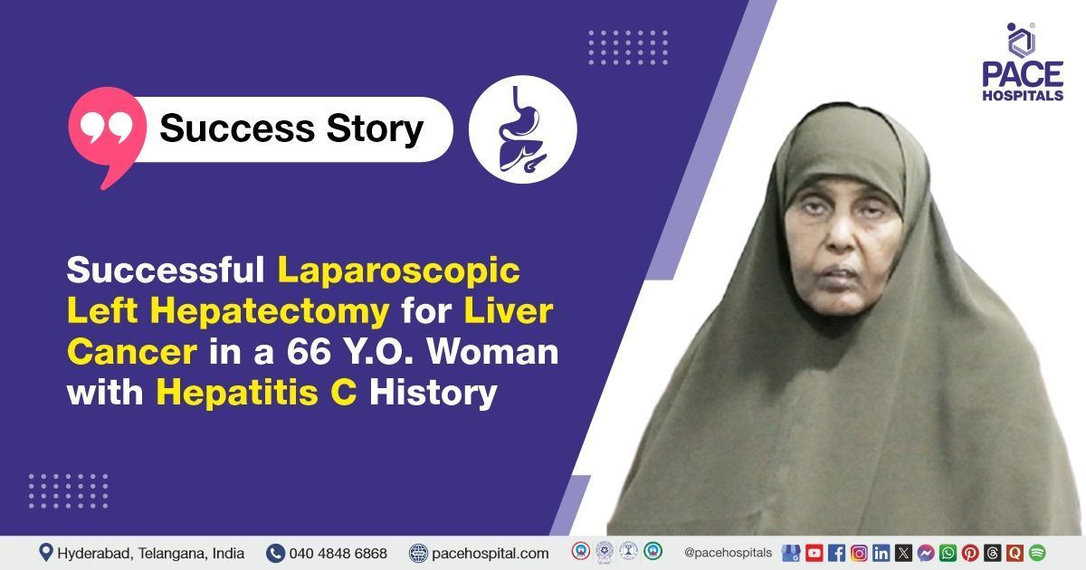
PACE Hospitals’ expert Gastroenterology team successfully performed a Laparoscopic Left Hepatectomy on a 66-year-old female patient diagnosed with a space-occupying lesion in segment IV of the liver, confirmed as Hepatocellular Carcinoma (HCC). The aim of the procedure was to achieve complete surgical removal of the malignant tumor, prevent further progression or metastasis of the disease, and improve the patient's overall prognosis and quality of life.
Chief Complaints
A 66-year-old female patient with a
body mass index (BMI) of 21.2 presented to the Gastroenterology Department at
PACE Hospitals, Hitech City, Hyderabad, with an incidentally detected mass lesion in the liver. She was asymptomatic, reporting no abdominal pain, fever,
jaundice, anorexia, or recent weight loss.
Past Medical History
The patient has a known history of Hepatitis C Virus (HCV) infection, for which she completed a 3-month course of antiviral therapy 17 years ago. She has been diagnosed with hypertension for the past 3 years and is on regular antihypertensive medication. Additionally, she has a 12-year history of hypothyroidism and is maintained on regular thyroid hormone replacement therapy.
On Examination
On examination, the patient’s vital signs were within normal limits. She was conscious, alert, and oriented to time, place, and person. General physical examination revealed no pallor, icterus, cyanosis, clubbing, lymphadenopathy, or pedal edema. The abdomen was soft and non-tender with no evidence of organomegaly or ascites. Cardiovascular, respiratory, and central nervous system examinations were unremarkable.
Diagnosis
Upon admission to PACE Hospitals, the patient was thoroughly evaluated by the Gastroenterology team, which included a detailed review of her medical history and a comprehensive clinical examination. Given her known history of hepatitis and an incidentally detected liver mass, a strong clinical suspicion of a hepatic lesion requiring further investigation was established.
The patient underwent multiple diagnostic tests to evaluate the liver lesion and overall health status. Liver function tests revealed elevated SGPT and SGOT levels, indicating liver injury, along with mildly increased alkaline phosphatase and slightly low serum albumin. Complete blood counts showed neutrophilic leukocytosis suggestive of inflammation or infection.
Histopathological examination of the resected liver tissue identified a cystic lesion within the left lobe consistent with Hepatocellular Carcinoma. Urine culture showed no bacterial growth, and chest X-rays revealed mild pleural effusion on the right side. Additionally, procalcitonin levels were mildly elevated, indicating a low likelihood of bacterial infection.
Based on the confirmed findings, the patient was advised to undergo
Hepatocellular Carcinoma Treatment in Hyderabad, India, under the expert care of the Gastroenterology Department.
Medical Decision Making
After a detailed consultation with the hepatology and surgical teams, including Dr. Govind Verma, interventional Gastroenterologist, Transplant Hepatologist Dr. Padma Priya, Consultant Gastroenterologist and Hepatologist and Dr. Suresh Kumar S, surgical Gastroenterologist, a comprehensive evaluation was undertaken to determine the most appropriate diagnostic and therapeutic strategy for the patient. Considering her history of chronic Hepatitis C infection, hypothyroidism, and the incidentally detected liver mass, the multidisciplinary team collaborated to assess the severity of the hepatic lesion and exclude malignancy or other complications.
Extensive investigations, including liver function tests, CT abdomen imaging, histopathology of resected tissue, and complete blood workup, confirmed the need for surgical intervention. It was determined that a laparoscopic left hepatectomy was identified as the most effective medical approach to remove the cystic lesion and prevent further liver compromise.
The patient and her family were informed about her condition, the procedure, its associated risks, and its potential to alleviate symptoms and enhance her quality of life.
Surgical Procedure
Following the decision, the patient was scheduled to undergo a Laparoscopic Left Hepatectomy Procedure in Hyderabad at PACE Hospitals, under the expert care of the Gastroenterology Department.
The laparoscopic left hepatectomy was performed under general anesthesia as follows:
- Abdominal Exploration and Initial Assessment: Upon laparoscopic entry into the abdominal cavity, the liver appeared grossly normal with no visible mass lesion, no omental or peritoneal deposits, and no evidence of free fluid. The falciform ligament was divided to facilitate access to the left hepatic lobe.
- Hilar Dissection and Vascular Control: The liver was gently lifted to expose the hepatic hilum, where dissection was performed. The left hepatic artery and left portal vein were carefully identified, isolated, and ligated to control vascular inflow to the left liver lobe.
- Parenchymal Transection: Parenchymal transection was initiated adjacent to the falciform ligament using Ligasure and bipolar cautery. This step allowed for the separation of the left hepatic lobe from the remaining liver tissue with controlled hemostasis.
- Outflow Control and Specimen Retrieval: The left hepatic vein (LHV) was identified and divided using a vascular stapler (white load), while the middle hepatic vein (MHV) was preserved. During this stage, 1 unit of packed red blood cells (PRBC) was transfused intraoperatively to compensate for blood loss. The resected liver specimen was retrieved through a midline incision.
- Hemostasis and Closure: A drain was placed at the transected surface of the liver to monitor postoperative bleeding or bile leakage. The procedure was completed uneventfully, and the abdomen was closed in layers.
Postoperative Care
The postoperative period was uneventful. During the hospital stay, the patient was managed with intravenous (IV) antibiotics, analgesics, intravenous proton pump inhibitors (PPIs), laxatives, prokinetics, antihypertensives, and supportive medications. A liver Doppler ultrasound performed showed mild surface nodularity but was otherwise normal. A follow-up CT abdomen revealed post-left hepatectomy status with uniform liver density, normal intrahepatic biliary radicles, and no fluid collections. The patient was discharged in stable condition with appropriate follow-up instructions.
Discharge Medications
Upon discharge, the patient was prescribed an antibiotic to treat infection, a proton pump inhibitor to reduce gastric acid secretion, a mucolytic agent to aid liver function, a hepatoprotective agent for liver support, a multivitamin supplement to improve nutritional status, thyroid hormone replacement for hypothyroidism management, antihypertensive medication to control blood pressure, and a laxative to prevent constipation.
Advice on Discharge
The patient was advised to perform incentive spirometry regularly. She was also recommended to follow a normal diet with high protein content.
Emergency Care
The patient was informed to contact the emergency ward at PACE Hospitals in case of any emergency or development of symptoms like fever, abdominal pain, or vomiting.
Review and Follow-up Notes
The patient was advised to return for a follow-up with the Gastroenterologist in Hyderabad at PACE Hospitals, after 5 days to review her condition, and review with the surgical gastroenterologist after 10days for clip removal.
Conclusion
This case highlights a successful laparoscopic left hepatectomy completed without complications. Comprehensive multidisciplinary care and diligent postoperative management facilitated a smooth recovery. Ongoing follow-up and targeted medical therapy are planned to ensure the best possible long-term outcomes.
Minimally Invasive Surgical Management of Liver Lesions in Hepatocellular Carcinoma
Laparoscopic hepatectomy has emerged as a safe and effective option for managing localized liver lesions, particularly in patients diagnosed with hepatocellular carcinoma (HCC). This case highlights how timely diagnosis, multidisciplinary coordination, and advanced laparoscopic techniques can ensure successful tumor removal with minimal invasiveness. The involvement of a skilled
Gastroenterologist / Gastroenterology doctor plays a pivotal role in diagnosis, treatment planning, and post-operative care. Avoiding large incisions not only minimized recovery time but also reduced the risk of complications. Such patient-centric, precision-based care supports faster recovery and improves overall prognosis.
Share on
Request an appointment
Fill in the appointment form or call us instantly to book a confirmed appointment with our super specialist at 04048486868
Appointment request - health articles
Recent Articles
