Successful Laparoscopic Whipple Procedure for Bile Duct Cancer in a 58-Y.O Female
PACE Hospitals
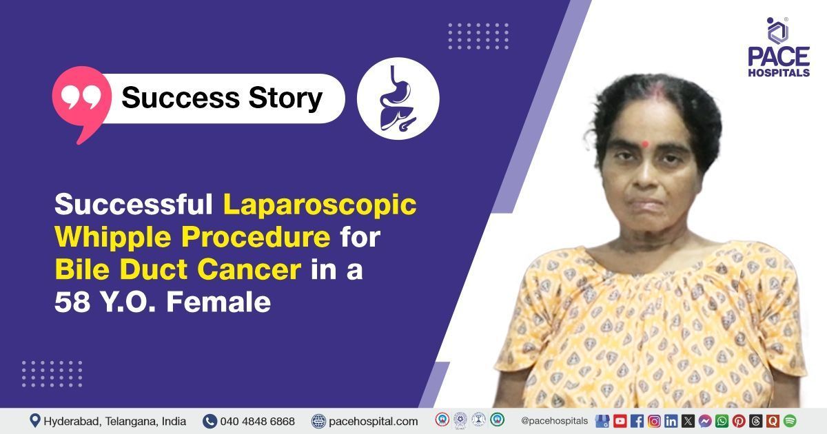
PACE Hospitals’ expert Gastroenterology team successfully performed a Laparoscopic Whipple’s procedure (Pancreaticoduodenectomy) on a 58-year-old female diagnosed with distal cholangiocarcinoma (Bile duct cancer). The procedure aimed to relieve abdominal pain, remove the tumor, relieve biliary obstruction, prevent disease progression and improve the patient's quality of life.
Chief Complaints
A 58-year-old female with a
body mass index (BMI) of 22.4 presented to the Gastroenterology Department at
PACE Hospitals, Hitech City, Hyderabad, with complaints of right upper quadrant abdominal pain and yellowish discolouration of the eyes. These symptoms progressively worsened, prompting further evaluation and management.
Past Medical History
The patient is a known case of systemic hypertension, type 2 diabetes mellitus, and hypothyroidism, all of which were on regular medications. Additionally, she also has a history of Endoscopic Retrograde Cholangiopancreatography (ERCP) with common bile duct (CBD) stenting.
On Examination
The patient was conscious and oriented during the general examination. Vital signs were stable and within normal limits. On systemic examination, the abdomen was soft and non-tender, indicating no acute abdominal findings.
Diagnosis
After admission to PACE Hospitals, the patient underwent a comprehensive clinical evaluation and a thorough review of prior laboratory investigations and imaging studies by the Gastroenterology team.
Initial blood investigations, including complete blood picture (CBP), liver function tests (LFT), and renal function tests (RFT), were within normal limits. However, tumor marker CA 19-9 was found to be elevated. An abdominal ultrasound (USG) showed gallbladder sludge and a left renal cortical cyst, which were noted as incidental findings.
A contrast-enhanced CT (CECT) scan of the abdomen (with oral and IV contrast) revealed a mildly enhancing focal soft tissue density lesion located at the uncinate process of the pancreas. The lesion appeared to cause indentation of the left renal vein and showed evidence of invasion into the terminal common bile duct (CBD), leading to proximal and mid CBD narrowing with minimal intrahepatic biliary radicle dilatation (IHBRD). These radiologic findings were suggestive of a likely neoplastic etiology.
Based on the confirmed diagnosis, the patient was advised to undergo
Bile Duct Cancer Treatment in Hyderabad, India, under the expert care of the Gastroenterology Department.
Medical Decision Making
After a detailed consultation with Dr. Govind Verma, Interventional Gastroenterologist, Dr. M Sudhir, Senior Gastroenterologist, Dr. Padma Priya, Consultant Gastroenterologist, and a cross-consultation with Dr. Suresh Kumar S, Surgical Gastroenterologist, a comprehensive evaluation was conducted to determine the optimal diagnostic and therapeutic approach for the patient diagnosed with distal cholangiocarcinoma (Bile duct cancer) and underlying comorbidities including diabetes, systemic hypertension, and hypothyroidism.
Based on detailed clinical assessment, imaging studies, histopathological evaluation, and blood investigations, the presence of a malignant lesion in the distal common bile duct was confirmed. Surgical intervention was deemed necessary to manage the condition effectively. Following a multidisciplinary discussion, laparoscopic Whipple pancreaticoduodenectomy was identified as the most appropriate treatment option to achieve complete tumor resection and restore biliary drainage.
The patient and her family were informed about the diagnosis, the planned surgical procedure, its associated risks, and its potential to alleviate symptoms and improve her quality of life.
Surgical Procedure
Following the decision, the patient was scheduled to undergo a Whipple procedure (Pancreaticoduodenectomy) in Hyderabad at PACE Hospitals under the expert supervision of the Gastroenterology Department.
The procedure was performed in the following steps:
- Diagnostic Laparoscopy and Exploration: Initial laparoscopic inspection revealed a normal liver without any peritoneal or omental deposits or masses. No evidence of free fluid was seen in the abdominal cavity. The lesser sac was opened, allowing full visualisation of the pancreas along its entire length.
- Mobilisation and Lymphadenectomy: Kocherization was performed to mobilise the duodenum up to the left renal vein. High Dissection Level (HDL) lymphadenectomy was carried out for adequate regional lymph node clearance. Anatomic variations noted included an early-branching right hepatic artery with a retro-common bile duct (CBD) course.
- Vascular and Pancreatic Dissection: The superior mesenteric vein (SMV) was dissected, and the “tunnel of love” was carefully created. Transection of the pancreatic neck was done after freeing the uncinate process by dividing the pancreatic tributaries from the portal vein. The stomach was transected beyond the level of the antrum.
- Common Bile Duct and Specimen Removal: The CBD, containing an indwelling stent, was identified; the stent was removed and sent for culture and sensitivity testing. The resected specimen was placed in a retrieval bag and extracted laparoscopically.
- Reconstruction: Pancreaticojejunostomy was performed using a 4-0 Prolene suture with a modified Blumgart’s technique to ensure secure anastomosis. Hepaticojejunostomy was constructed using 3-0 V-Loc sutures in a continuous running fashion. Gastrojejunostomy was completed using 3-0 silk and Vicryl sutures in four layers. A feeding jejunostomy (FJ) was placed for enteral nutrition support. A surgical drain was placed in the subhepatic space near the pancreaticojejunostomy (PJ) and hepaticojejunostomy (HJ) sites.
- Closure: The abdominal wall was closed meticulously in layers.
Postoperative Care
After the procedure, the patient was closely monitored for stability and postoperative complications. Follow-up ultrasound showed a normal-sized liver with normal echotexture, absent gallbladder (post-cholecystectomy status), and a pancreas obscured by bowel gas. The spleen and both kidneys appeared normal. Mild bilateral pleural effusion was noted, along with an epigastric fluid collection measuring 6.2 x 3.1 cm.
During the hospital stay, the patient was managed with intravenous fluids, intravenous antibiotics, proton pump inhibitors (PPIs), and other supportive medications. The patient showed symptomatic improvement and is being discharged in stable condition with appropriate medications and follow-up care instructions.
Discharge Medications
Upon discharge, the patient was prescribed oral antibiotics to treat the infection and a proton pump inhibitor to protect the stomach lining. Analgesics were provided for pain relief as needed. She was advised to continue thyroid hormone replacement therapy for hypothyroidism, antihypertensive medications to control blood pressure, anticoagulants to prevent blood clots, and antidiabetic treatment to manage blood sugar levels as part of her ongoing care.
Advice on Discharge
The patient was advised to follow a high-protein diet with small, frequent meals to support recovery. Feeding jejunostomy (FJ) feeds are to be continued as per protocol. A strict diabetic diet and a low-salt diet were also recommended to manage blood sugar levels and blood pressure effectively.
Emergency Care
The patient was informed to contact the emergency ward at PACE Hospitals in case of any emergency or development of symptoms like fever, abdominal pain, or vomiting.
Review and Follow-up Notes
The patient was advised to return for a follow-up visit with the Gastroenterologist in Hyderabad at PACE Hospitals after one month and a review with the surgical gastroenterologist after one month, with prior appointments, for comprehensive evaluations to monitor the patient’s progress.
Conclusion
This case highlights the effective multidisciplinary approach in diagnosing and treating a pancreatic neoplasm using laparoscopic Whipple pancreaticoduodenectomy. The patient experienced an uneventful surgery and a smooth postoperative recovery. Close monitoring and continued care have been planned to support long-term health outcomes.
The Importance of Laparoscopic Whipple Pancreaticoduodenectomy in Pancreatic Head Tumors
Laparoscopic Whipple pancreaticoduodenectomy represents a significant advancement in the surgical management of pancreatic head tumors, including neoplastic lesions involving the distal common bile duct. This minimally invasive approach offers precise dissection and tumor removal with reduced postoperative pain, shorter hospital stays, and faster recovery compared to open surgery. The technique requires careful mobilization of surrounding structures such as the duodenum, pancreas, and major vessels, alongside meticulous reconstruction of the pancreatic and biliary drainage.
A
Gastroenterologist / Gastroenterology doctor plays a crucial role in preoperative evaluation and postoperative care, ensuring comprehensive management throughout the patient’s treatment journey. Surgical teams rely on detailed preoperative imaging and intraoperative findings to safely perform this complex procedure. The laparoscopic approach, when feasible, enhances surgical outcomes and quality of life for patients with periampullary neoplasms.
Share on
Request an appointment
Fill in the appointment form or call us instantly to book a confirmed appointment with our super specialist at 04048486868
Appointment request - health articles
Recent Articles


