Successful PCNL, URS & DJ Stent Exchange for Right Staghorn Calculi in a 47-Year-Old Woman
PACE Hospitals
The PACE Hospitals expert Urology team successfully performed a right percutaneous nephrolithotomy (PCNL), ureteroscopy (URS), and DJ stenting on a 47-year-old female patient who presented with right flank pain. The procedure was done to remove kidney stones, effectively relieving the patient's symptoms and helping restore proper kidney function to support her recovery.
Chief Complaints
A 47-year-old woman with a
Body Mass Index (BMI) of 23 presented to the Urology Department at
PACE Hospitals, Hitech City, Hyderabad, with right flank pain. She reported discomfort that affected her daily activities and had not improved with initial medical management. A detailed evaluation was carried out to determine the underlying cause of her symptoms.
Past Medical History
The patient had a history of recurrent nephrolithiasis (kidney stones), specifically a complete staghorn calculi in the right kidney. She previously underwent open nephrolithotomy and percutaneous nephrolithotomy (PCNL) at another hospital for renal calculi. The patient also had several comorbidities, including
diabetes mellitus,
hypertension,
hypothyroidism, and hypovitaminosis vitamin D (deficiency of
vitamin D). The underlying problems likely increased the recurrent formation of stones and complicated the overall management of her nephrolithiasis.
On Examination
Upon admission, the patient was afebrile with stable vital signs, and her general condition was satisfactory. She was conscious, alert, and showed no signs of acute distress. Physical examination revealed mild tenderness over the right flank on deep palpation, consistent with her reported symptoms. However, no palpable mass was detected, and there were no signs of peritonism, such as guarding, rebound tenderness, or abdominal rigidity.
Diagnosis
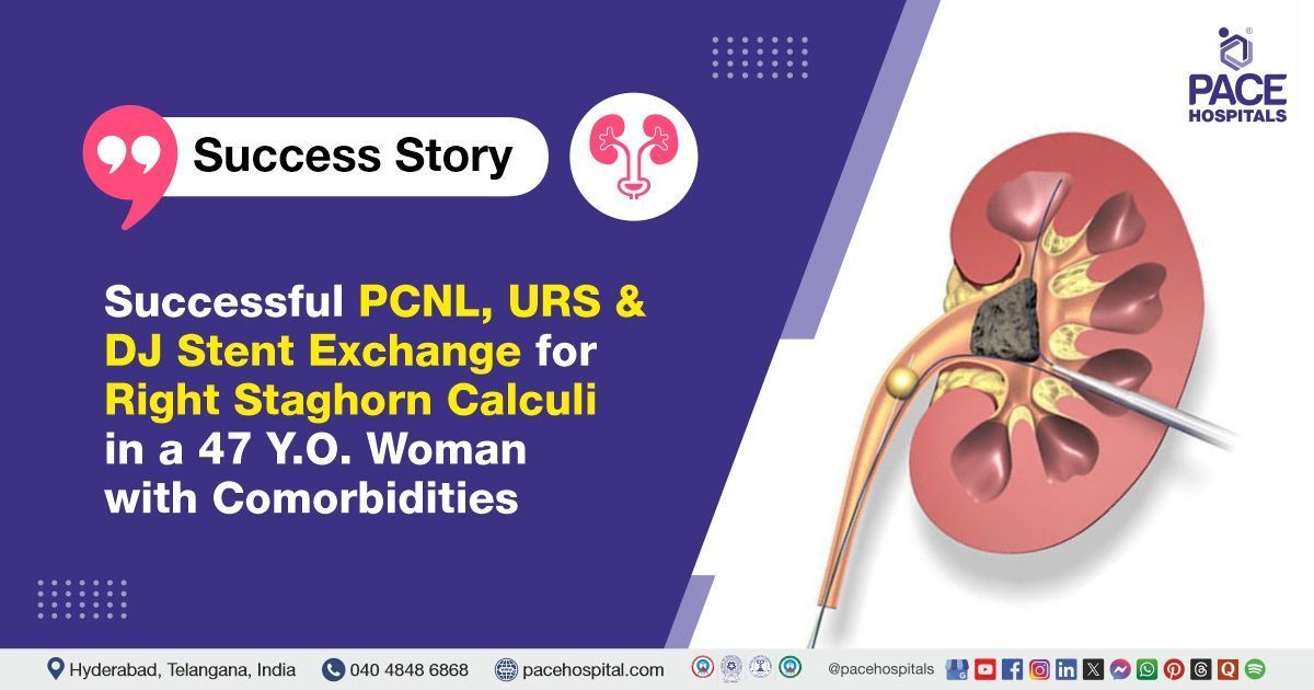
The patient initially underwent a comprehensive clinical assessment at PACE Hospitals, including a detailed history and physical examination by the Urology team. During the evaluation, she was diagnosed with a right complete staghorn calculus (large, branched kidney stones composed mainly of struvite), and identified as a recurrent stone former, which necessitated surgical intervention.
To support the diagnosis and assess the extent of the patient's condition, a series of relevant investigations were conducted.
- Computed Tomography (CT) Scan of the Abdomen and Pelvis: The CT scan demonstrated a safe distance between the colon and the posterior aspect of the right kidney, which was favorable for proceeding with percutaneous intervention. The right kidney appeared scarred and contracted, indicating chronic renal damage, likely due to longstanding calculi and recurrent infections. A complete staghorn calculus was identified within the right kidney, occupying the renal pelvis and calyces. The left kidney was found to be normal in structure and function, providing reassurance regarding overall renal reserve. Additionally, abdominal CT imaging demonstrated bilateral perinephric stranding, further supporting ongoing renal or perinephric inflammation.
- Complete bold count (CBC): The CBC showed normal haemoglobin levels and no neutrophilic predominance, indicating the absence of active systemic infection. This suggested that the patient’s symptoms were likely due to localized inflammation from the renal calculi, rather than a systemic infection.
- Urinalysis: It revealed the presence of abundant pus cells in the urine, consistent with an active urinary tract infection.
- Renogram (renal scan) Findings: The patient’s renogram showed a right renal split function of 45%, indicating the right kidney still contributed a significant portion of total renal function. This was important for surgical and medical planning, despite the kidney’s chronic scarring and staghorn calculus.
Based on the confirmed findings, the patient was advised to undergo Kidney Stones Treatment in Hyderabad, India, under the expert care of the Urology Department, providing effective stone clearance, alleviating her flank pain, and preventing potential complications including renal function impairment.
Medical Decision-Making (MDM)
Following a detailed consultation with Dr. Abhik Debnath, Consultant Laparoscopic Urologist, a comprehensive evaluation of the patient’s condition was undertaken. Considering her right flank pain and CT scan findings indicating a staghorn calculus, the urology team concluded that a right percutaneous nephrolithotomy (PCNL), ureteroscopy (URS), along with DJ stenting, would be the most appropriate and efficacious strategy. Following the treatment, a right DJ stent exchange would be performed to facilitate healing and provide adequate urine outflow during the recovery period.
Surgical Procedure
Following the decision, the patient was scheduled for percutaneous nephrolithotomy (PCNL), ureteroscopy (URS), and DJ stenting Surgery in Hyderabad at PACE Hospitals, under the expert supervision of the Urology Department, ensuring optimal care and a smooth recovery process.
Right PCNL with URS and DJ Stenting
- Anesthesia and Positioning: The patient was administered with general anaesthesia and positioned appropriately for percutaneous access to the right kidney. Access was achieved by making three punctures: one each at the upper, middle, and lower poles of the right kidney, respectively.
- Two tracts were established: one through the upper calyx (21 Fr) and the other through the lower calyx (24 Fr), to optimize stone access and clearance.
Stone Removal: A pneumatic lithotripter was used to fragment the large staghorn calculus after around 90% of the stone bulk had been removed. To ascertain the composition of the stone, portions of the stone were removed and submitted for investigation using Fourier Transform Infrared Spectroscopy (FTIR). It was seen that some of the stone was outside the collecting system and was not visible during nephroscopy, suggesting that it may have extravasated or been displaced.
Stent Placement
Antegrade DJ stent placement was performed to ensure adequate urinary drainage and promote healing.
Completion
Hemostasis was achieved. The procedure concluded without complications, and the patient was shifted to the recovery room in stable condition.
Right DJ Stent Exchange (Performed Two Days Later)
- Stent Exchange: A retrograde DJ stent exchange was performed to maintain urinary drainage and minimize the risk of infection or obstruction. And the position of the stent in the upper pole was confirmed with retrograde pyelography (RGP).
- Nephrostomy Tube Removal: Once adequate urinary drainage was established and the correct positioning of the DJ stent was confirmed, the nephrostomy tubes were removed as part of the postoperative management, ensuring the patient’s safety and readiness for further recovery.
Postoperative Care
After surgery, the patient was monitored closely in the surgical intensive care unit to ensure stability. Her postoperative course was uneventful. Postoperative care involved careful monitoring of urine output and evaluation for signs of infection or haemorrhage (bleeding), The nephrostomy tubes were removed after demonstrating right stent placement, and she was monitored for complications associated with the procedure or pre-existing chronic renal disease. Instructions regarding stent care and subsequent imaging were given to assess for remaining stone fragments and confirm the resolution of infection. During her hospital stay, she received intravenous antibiotics to prevent infection, analgesics for pain relief, antiemetics to control nausea, and gastric protectants. The patient was discharged in stable condition.
Discharge Medication
Upon discharge, the patient was prescribed a course of oral antibiotics for infection prophylaxis, analgesics for pain control, and a proton pump inhibitor for gastric protection. Additional medications included a urinary alkalinizer to help prevent stone recurrence, an alpha-blocker to facilitate the passage of residual stone fragments, and an antioxidant supplement for renal and prostate health. Routine medications for hypertension, diabetes, dyslipidemia, and hypothyroidism were advised to continue. The patient was advised to avoid blood thinners for two months to reduce the risk of postoperative bleeding.
Discharge Advice
The patient was advised to avoid weightlifting, forward bending, straining during bowel movements, and prolonged travel by bus, auto, or two-wheeler. Passage of small stone fragments, mild flank or suprapubic discomfort, occasional blood in urine (Hematuria), cloudy urine, or mild burning were expected and typically will resolve within 7–10 days.
Dietary advice
She was instructed to maintain good hydration (3–4 L/day), consume citrus fruits and juices, and follow a low-purine, low-salt diet, to support recovery.
Emergency Care
The patient was informed to contact the emergency ward at PACE Hospitals in case of any emergency or development of symptoms like fever, abdominal pain, swelling, bleeding, or vomiting.
Review and Follow-up
The patient was advised to return for a follow-up visit with the Urologist in Hyderabad at PACE Hospitals, after 2 months for DJ stent removal, cystoscopy, and URS. She was advised to bring the Fourier Transform Infrared Spectroscopy (FTIR) stone analysis report for assessment to ensure proper healing and identify any residual issues.
Conclusion
This case highlights the effectiveness of percutaneous nephrolithotomy (PCNL), and ureteroscopy (URS), with DJ stenting in performing Renal Calculi Treatment in Hyderabad, India, enabling successful removal of stones in the kidney, thereby facilitating normal urine flow.
Significance of Multitract PCNL in Managing Complex Staghorn Calculi
Multitract percutaneous nephrolithotomy (PCNL) is a specialized technique employed by the urologist/ urology doctor to create multiple access tracts into the kidney, optimizing stone removal and clearance in complex cases. In this case, two tracts (upper and lower calyx) were used to approach different sections of the large staghorn stone. This technique significantly improved stone clearance efficiency, reduced the need for multiple sessions and minimized residual stone burden. Multitract PCNL is particularly valuable in patients with complex, branched calculi, as it allows comprehensive access to all parts of the renal collecting system, enhancing surgical success while maintaining renal safety.
Share on
Request an appointment
Fill in the appointment form or call us instantly to book a confirmed appointment with our super specialist at 04048486868
Appointment request - health articles
Recent Articles