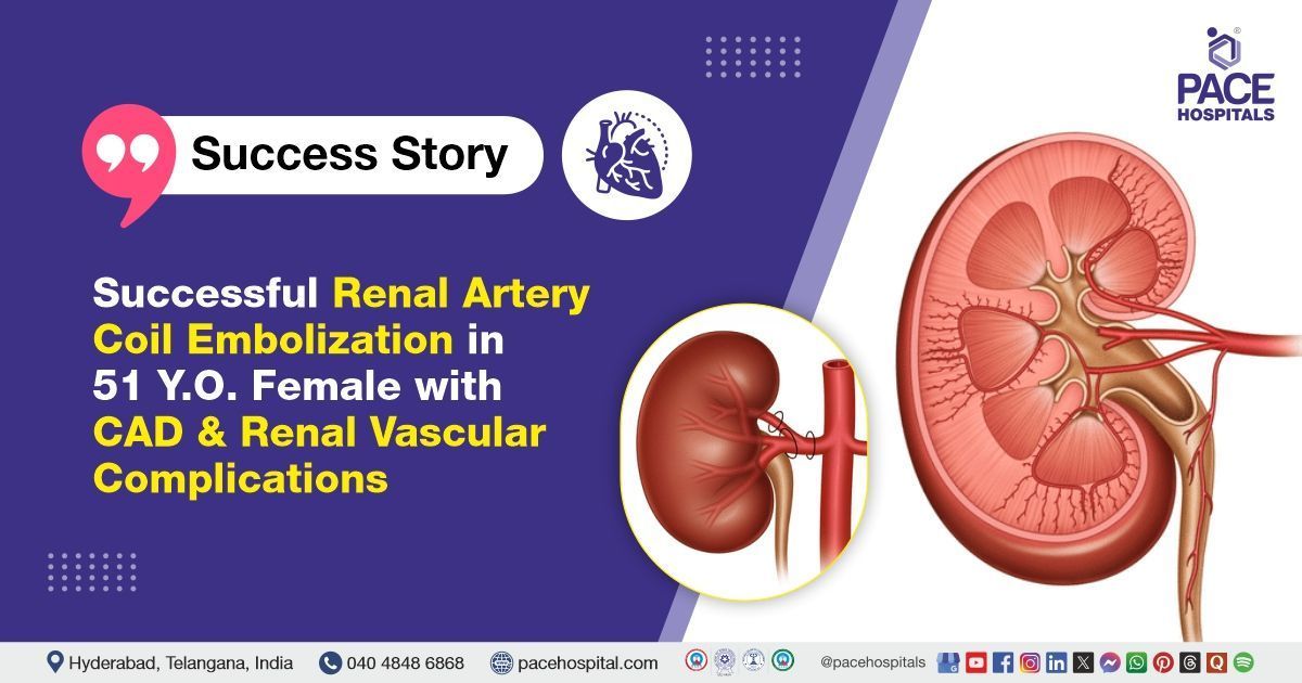
Successful Renal Artery Coil Embolization in 51-Y.O Female with CAD & Renal Vascular Issues
PACE Hospitals
PACE Hospitals’ expert cardiology team successfully performed Renal Artery Coil Embolization on a 51-year-old female patient diagnosed with coronary artery disease (CAD) and double vessel disease (DVD). She presented with left flank pain and shortness of breath (SOB) on exertion. The procedure effectively relieved her flank pain and is expected to improve her overall clinical condition while reducing the risk of further complications, particularly given her underlying CAD.
Chief Complaints
A 51-year-old female patient with a
Body Mass Index (BMI) of 21.3 presented to the Cardiology Department at
PACE Hospitals, Hitech City, Hyderabad, with complaints of left flank pain and shortness of breath on exertion.
Past Medical History
The patient had a known history of type 2 diabetes mellitus and hypertension, for which she was receiving regular treatment. These chronic conditions likely contributed to the development and progression of her coronary artery disease (CAD) and may also have increased her risk of vascular complications, including the renal artery involvement observed in this case.
On Examination
The patient was conscious and coherent on examination. Vital signs were stable. Systemic examination revealed normal heart sounds on cardiovascular assessment. Neurological evaluation showed no focal deficits. Respiratory examination indicated good bilateral air entry, and abdominal examination revealed a soft, non-tender abdomen.
Diagnosis
After the initial examination, the patient underwent a comprehensive evaluation by the cardiology team, which included a detailed clinical assessment, laboratory investigations, and cardiac imaging. These assessments were aimed at determining the severity of her coronary artery disease (CAD) and double vessel disease evaluating her overall cardiac function to guide appropriate management.
The diagnostic workup included a comprehensive evaluation of the patient’s cardiac and renal systems. Cardiac assessment involved ECG, echocardiography, and coronary angiography (CAG), which revealed double vessel disease (DVD. Renal imaging started with Doppler ultrasound, followed by CTA or MRA for detailed vessel visualization. Digital subtraction angiography (DSA) was used during embolization to locate the affected artery. Laboratory tests assessed blood counts, kidney function, coagulation, and diabetes control. Respiratory evaluation with chest X-ray and pulse oximetry helped investigate the shortness of breath and oxygenation status.
Based on the confirmed findings, the patient was advised to undergo
Coronary Artery Disease Treatment in Hyderabad, India, under the expert care of the Cardiology Department.
Medical Decision Making
After thorough consultations with Dr. Seshi Vardhan Janjirala, a cardiologist; Dr. Tripti Sharma, a physician and diabetologist, regarding the patient’s blood sugar levels and hypertension; and Dr. Abhik Debnath, regarding renal vascular complications, a comprehensive evaluation was conducted to develop a tailored diagnostic and therapeutic plan for the patient’s coronary artery disease. Based on her symptoms and cardiac history, the assessment focused on determining the extent and severity of the disease.
It was determined that the patient had a renal artery vascular lesion responsible for her left flank pain. Renal artery coil embolization was identified as the most effective intervention to relieve the vascular obstruction and improve her overall condition.
The patient and her family were thoroughly counseled about her condition, the need for intervention, and the benefits and potential risks of renal artery coil embolization. The primary goal was to ensure timely and effective treatment to relieve the vascular obstruction, improve her condition, and reduce the risk of further complications related to her coronary artery disease.
Surgical Procedure
Following the diagnosis of coronary artery disease (CAD), double vessel disease (DVD) and renal artery vascular lesion, the patient was scheduled for a renal artery coil embolization in Hyderabad at PACE Hospitals, under the expert supervision of the Cardiology Department, ensuring optimal care and a smooth recovery process.
The following are the steps involved in procedure :
- Access and Angiography: Under local anesthesia, vascular access was obtained, usually via the femoral artery. A catheter was guided to the renal artery under fluoroscopic guidance, and angiography was performed to visualize the site of vascular abnormality.
- Identification of Target Vessel: The exact location of the arterial lesion or bleeding was identified through contrast imaging.
- Coil Deployment: Small metallic coils were carefully deployed into the affected segment of the renal artery to occlude blood flow and control bleeding or vascular abnormality.
- Confirmation of Occlusion: Post-deployment angiography was performed to confirm successful occlusion of the target vessel and ensure no further bleeding or abnormal flow.
- Completion and Monitoring: The catheter was withdrawn, hemostasis was achieved at the access site, and the patient was monitored for any immediate post-procedure complications.
Postoperative Care
The patient’s postoperative course was uneventful, with stable vital signs and no complications at the access site. Pain was well controlled, and renal function remained stable with adequate hydration. The patient was discharged in a stable condition with the following medications.
Discharge Medications
Upon discharge, the patient was prescribed a lipid-lowering agent to reduce cardiovascular risk and a nutritional supplement to support recovery. Antihypertensive medication was included to manage blood pressure and improve cardiac function. Thyroid hormone replacement was continued for hypothyroidism. For diabetes, a sulfonylurea and a DPP-4 inhibitor with metformin were prescribed. An antiplatelet agent was given to prevent clot formation, and a nitrate was included for angina control. An analgesic was advised to be taken as needed for pain relief.
Emergency Care
The patient was informed to contact the
emergency ward at PACE Hospitals in case of any emergency or development of symptoms such as chest pain, abdominal pain, and fever.
Review and Follow-up Notes
The patient was advised to return for a follow-up visit with the cardiologist at PACE Hospitals, Hyderabad, after 5 days to assess her recovery. Revascularization for double vessel disease (DVD) was planned as a staged intervention during follow-up.
Conclusion
This case highlights the effective multidisciplinary management of a patient with coronary artery disease, diabetes, hypertension, and renal vascular complications. Timely renal artery coil embolization resulted in symptom relief and clinical stability. Individualized care ensured successful recovery and discharge.
The Role of Renal Artery Coil Embolization in Vascular Complication Management
Renal artery coil embolization is a minimally invasive procedure used to treat renal vascular complications such as bleeding or aneurysms by blocking abnormal blood flow with tiny metallic coils. It is especially beneficial for high-risk patients with conditions like coronary artery disease, diabetes, and hypertension, where surgery poses greater risks. This technique helps preserve kidney function and shortens recovery time. In this case, the
cardiologist/heart specialist played a key role in coordinating care and recommending embolization. The procedure effectively relieved symptoms and prevented further complications. Its precision and safety make it an important option in managing complex vascular conditions.
Share on
Request an appointment
Fill in the appointment form or call us instantly to book a confirmed appointment with our super specialist at 04048486868
Appointment request - health articles
Recent Articles