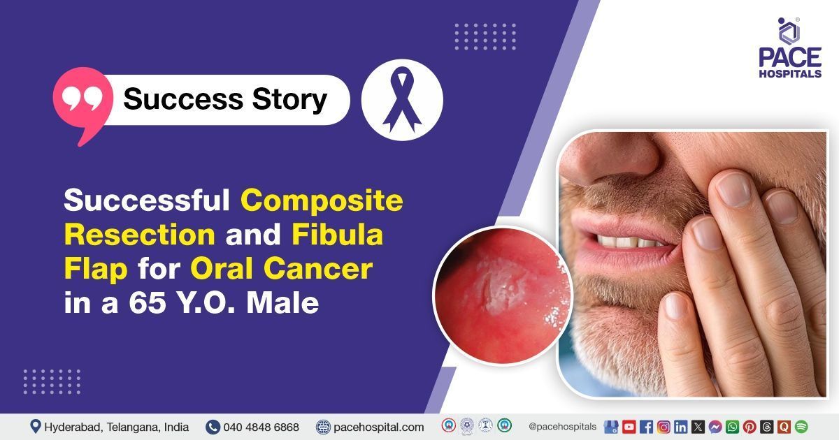
Successful Composite Resection and Fibula Flap for Oral Cancer in a 65 Y.O. Male
PACE Hospitals
PACE Hospitals' expert Oncology team successfully performed a
left composite resection with free fibula flap reconstruction
on a 65-year-old male patient who presented with an ulcer on the left buccal mucosa associated with difficulty in opening his mouth for the past seven months. The surgery was performed to restore jaw function and appearance while ensuring cancer control, relieving symptoms, and improving the patient's overall quality of life.
Chief Complaints
A 65-year-old male patient with a
body mass index (BMI) of 20.2 presented to the Oncology Department at
PACE Hospitals, Hitech City, Hyderabad, with an ulcer on the left buccal mucosa associated with difficulty in opening his mouth for the past seven months, which had begun to interfere with his daily activities.
Past Medical History
The patient is a known case of
Type 2 Diabetes Mellitus and
hypertension for the past eight years, as well as
hypothyroidism, and he is currently on regular medication for all these conditions. Additionally, he had a previous episode of lower motor neuron (LMN) facial palsy, which had fully recovered.
On Examination
Upon admission to PACE Hospitals, the patient was hemodynamically stable with normal vital signs. On cardiovascular examination, S1 and S2 heart sounds were present. Central nervous system examination revealed no focal neurological deficits. Respiratory system examination showed bilateral air entry with normal vesicular breath sounds. The abdomen was soft and non-tender.
On oral examination, there was an ulcer proliferative growth measuring 4 cm × 3 cm over the left buccal mucosa, extending from the first premolar to the retromolar region. Trismus (Jaw Stiffness) was present, with mouth opening restricted to approximately one finger breadth. Palpable, mobile lymph nodes were noted in the left neck at Level 1b and 2b.
Diagnosis
After the initial examination, the oncology team conducted a comprehensive assessment that included clinical evaluation, laboratory investigations, and imaging studies:
Imaging Studies
- Computed tomography (CT) scan with contrast: To acquire detailed images of the tumor and its surrounding areas, a CT scan of the neck was performed. This imaging technique helped pinpoint the precise location of the primary lesion in the left buccal mucosa. It also evaluated possible invasion of nearby tissues, including muscles, neurovascular bundles, and the mandible. The scan also assessed whether any enlarged regional lymph nodes were present, as well as their features. The results were essential for precise staging and surgical planning.
- Chest Imaging: A chest X-ray was performed as part of the staging workup. These investigations help exclude the presence of distant metastasis to the lungs or other thoracic structures. No evidence of distant spread (M0) was found, confirming the disease was localized.
Based on clinical examination and contrast-enhanced CT scan of the neck, a diagnosis of squamous cell carcinoma of the left buccal mucosa (T2N1M0) was established. The imaging was essential for staging and surgical planning, thereby guiding the team toward planning appropriate surgical intervention.
Biopsy and Histopathology
- Incisional biopsy: A tissue sample was taken from the ulcer proliferative lesion on the left buccal mucosa.
- Histopathological examination: The biopsy specimen was analyzed under a microscope, confirming the diagnosis of squamous cell carcinoma. Histopathology identified the type and grade of the tumor and provides information about tumor differentiation and other prognostic factors.
- Fine needle aspiration cytology (FNAC): It was performed to obtain samples of lymph nodes that appeared suspicious on imaging or during a physical examination. This helped confirm or rule out metastatic involvement, thereby improving the staging and treatment strategy
Endoscopy
The patient underwent endoscopy (direct laryngoscopy or pan endoscopy) to visually assess the oral cavity, pharynx, larynx, and upper esophagus. This step was essential to rule out any synchronous (second) primary tumors, which are relatively common in oral squamous cell carcinoma cases.
Based on the comprehensive evaluation, the patient was advised to undergo
oral cavity cancer Treatment in Hyderabad, India, under the expert care of the Oncology Department to ensure complete management of his condition, symptom relief, and prevention of further complications.
Medical Decision-Making (MDM)
After a thorough consultation with Dr Ramesh Parimi, a surgical oncologist, cross-consultants Dr. Kantamneni Lakshmi, Dr. Sashi Vardhan Janjirala, Dr. Pramod Kumar and Dr. Pradeep Kiran Panchadi, a comprehensive evaluation was conducted to determine the most appropriate treatment for the patient.
Based on patient symptoms and the diagnostic findings, it was concluded that a left composite resection with partial mandibulectomy, upper alveolus excision, functional neck dissection and free fibula flap reconstruction would be the most appropriate surgical intervention for treating squamous cell carcinoma of left buccal mucosa (T2 N1 M0). The decision was based on a detailed clinical assessment and aimed at ensuring optimal outcomes through timely and definitive management.
The patient and his family were counselled about the patient's condition, including the associated risks, complications, and benefits. They were also informed about the necessity of surgical intervention to prevent future complications and ensure optimal outcomes.
Surgical Procedure
Following the decision, the patient was scheduled to undergo a left composite resection with partial mandibulectomy, upper alveolus excision, functional neck dissection and free fibula flap reconstruction in Hyderabad at PACE Hospitals under the expert supervision of the Oncology Department.
The following steps were carried out during the procedure:
Preoperative preparation and anaesthesia
The patient received clearance for anaesthesia, positioned in a supine posture with the neck extended, and the head moved to the right, intubated, and the surgical area was prepared and covered in a sterile manner.
Surgical exposure
- Neck Incision and Lymph Node Dissection: A curvilinear incision was made from the mastoid process to the hyoid, providing access to the left neck. Functional block dissection of left neck lymph nodes (L1–L4) was performed, with care taken to preserve vital structures.
- Exposure and Resection of Tumor and Bone: The left mandible and upper alveolus were exposed through combined intra- and extraoral approaches, with release of the masseter muscle and elevation of the periosteum. A left composite resection was then carried out, including partial mandibulectomy, excision of the left temporomandibular joint, and the left upper alveolus. Osteotomies were made with a saw under irrigation to ensure clear tumor margins, and the tumor with involved bone and soft tissue was removed.
- Reconstruction Planning and Fibula Flap obtained: The defect size was measured and prepared for reconstruction. A free vascularized fibula flap was collected from the left leg, preserving the vascular pedicle for subsequent transfer.
- Flap Inset, Anastomosis, and Reconstruction: The fibula flap was transferred to the head and neck region. Microvascular anastomosis was performed to connect the flap’s pedicle to the recipient vessels in the neck. The fibula was shaped and fixed to the residual mandible using plates and screws. Soft tissue from the flap was used as needed to reconstruct the oral lining and external skin
Haemostasis and closure
Haemostasis (bleeding control) was secured, and the wound was closed in layers, with a drain placed, to minimize postoperative complications and promote optimal recovery for the patient.
Postoperative Care
After surgery, the patient's postoperative recovery was uneventful, and he was monitored closely in the surgical intensive care unit to ensure stability. He was transferred to the general ward once his condition was stable. During his hospital stay, he received intravenous antibiotics to prevent infection, analgesics for pain relief, antiemetics to control nausea, and gastric protectants. Intensive spirometry and respiratory exercises were encouraged to support pulmonary function.
Discharge Medications
The patient was discharged in a hemodynamically stable condition, and he was prescribed a medication regimen to support postoperative recovery and minimize complications. This regimen included thyroid hormone replacement for hypothyroidism, combined antihypertensive therapy for blood pressure control, a proton pump inhibitor for stomach acid reduction, and an NSAID for pain and inflammation. A probiotic was given for gut flora, and a potassium supplement to prevent low potassium. Mucolytic agents and inhaled steroids with bronchodilators were prescribed for airway and mucus management. An antibiotic and antiseptic were advised for oral care, while low-dose antiplatelet therapy was continued for cardiovascular protection. An oral antidiabetic agent was maintained for diabetes management.
Advice on Discharge
The patient was instructed to continue daily wound care and maintain regular daily activities as tolerated. Ambulation was permitted with strict non-weight bearing instructions on the left leg (donor site for fibula flap). Intensive spirometry exercises were recommended to support respiratory function during recovery.
Emergency Care
The patient was informed to contact the emergency ward at PACE Hospitals in case of any emergency or development of symptoms such as fever, sudden onset of arm or shoulder pain or swelling, increased redness, swelling, or discharge from the wound.
Review and Follow-up
The patient was advised to schedule a follow-up appointment with the Oncologist in Hyderabad at PACE Hospitals one week after discharge.
Conclusion
This case demonstrates the successful management of oral squamous cell carcinoma (T2 N1 M0) through left composite resection with free fibula flap reconstruction. A multidisciplinary approach ensured complete tumor removal, restoration of jaw function, and improvement in quality of life, with smooth postoperative recovery and ongoing follow-up care.
Rebuilding the Jaw: Successful Composite Resection and Fibula Flap Reconstruction in Oral Cancer
This case study highlights the successful surgical management of oral squamous cell carcinoma (T2N1M0) through a left composite resection followed by mandibular reconstruction using a free fibula flap. The fibula flap, often considered the gold standard for mandibular reconstruction, was chosen for its ample bone length, dependable vascular supply, and adaptability in shaping the graft to match the native jaw contour. This technique allowed for precise restoration of both function and facial symmetry, which is critical for speech, chewing, and aesthetics. An oncologist/cancer specialist approach ensured thorough tumor resection, clear surgical margins, and optimal reconstruction outcomes. Postoperative recovery was uneventful, and the patient benefited from a well-coordinated care plan tailored to his comorbidities. This case reinforces the role of advanced reconstructive techniques in improving the quality of life and long-term prognosis in head and neck cancer patients.
Share on
Request an appointment
Fill in the appointment form or call us instantly to book a confirmed appointment with our super specialist at 04048486868
Appointment request - health articles
Recent Articles