Comprehensive Management of Recurrent Ascites Following Right Hemicolectomy for Abdominal TB
PACE Hospitals
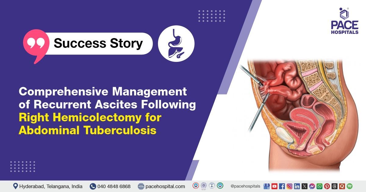
PACE Hospitals’ Surgical Gastroenterology team successfully performed a Exploratory Laparotomy with Adhesiolysis and Drain Placement for a 20-year-old female patient suffering from recurrent loculated ascites following a right hemicolectomy for abdominal tuberculosis (TB).
Chief Complaints
A 20-year-old female patient presented to the Gastroenterology Department at PACE Hospitals, Hitech City, Hyderabad, with complaints of intermittent abdominal pain over the last month. Ten days ago, she developed generalized, non-radiating abdominal pain, along with recurring abdominal distension and discomfort after meals. She also had intermittent bilious vomiting about three days ago.
For the past three years, she has experienced similar symptoms, including reflux, which have been managed with oral antacids for the last year.
Medical History
Delving further, it was understood that the patient underwent a laparotomy for intestinal obstruction in 2014, followed by adhesiolysis with bowel resection. Post-surgery, she received antitubercular treatment (ATT) for four months but later defaulted on the treatment. A CECT scan evaluation revealed peritoneal thickening with loculated ascites and a suspected ovarian mass, possibly a germ cell tumor, along with moderate loculated ascites. Ascitic fluid analysis was negative for AFB, and serum CEA levels were within normal limits. The patient had no other comorbidities.
The previous findings, including peritoneal thickening and a suspected ovarian mass, raised concerns about potential recurrence or related pathology, necessitating further evaluation and management.
Diagnosis
Upon admission to PACE Hospitals, the patient’s vital signs were stable. Following a detailed physical examination, it was noted that she had been suffering from intermittent abdominal pain for the last month.
The diagnostic tests revealed recurrent loculated ascites, indicating a buildup of fluid in the abdomen, which appeared to be a complication following her previous right hemicolectomy for abdominal tuberculosis (TB).
Surgical procedure
Following discussions with Dr. CH Madhusudan, the Consultant Surgical Gastroenterologist, it was concluded that an exploratory laparotomy, including adhesiolysis and drain placement, would be the best course of action for the patient’s treatment.
After being admitted with the aforementioned symptoms, the patient underwent further evaluation and was taken for surgery. An exploratory laparotomy with adhesiolysis and drain placement was successfully performed. This procedure was crucial for addressing bowel obstruction and preventing further adhesion-related complications.
Findings
Abdominal cocoon, a fibrous encapsulation of the small intestine, often presents with small bowel adhesions and enlarged peritoneum, leading to bowel obstruction, pain, and malabsorption, potentially due to inflammation, infection, or previous surgeries.
Loculated collections around the bowel loops are confined pockets of fluid or pus, often seen in infections, abscesses, or inflammation. They can develop due to perforated bowel, inflammatory bowel disease (IBD), or post-surgical complications. These collections can cause pain, fever, and bowel obstruction.
A history of appendectomy surgery is indicated by a tiny ovarian cyst on the right side and a larger 5 x 3 cm cyst on the left. Although ovarian cysts are usually benign but monitoring them is crucial for potential complications or changes in size.
Adhesiolysis is a surgical procedure that removes or releases abnormal fibrous tissue between organs, such as adhesions in the small bowel loops. This procedure alleviates symptoms like bowel obstruction and pain, improves intestinal function, and promotes better recovery and function.
A pelvic collection, which is a confined fluid, pus, or blood accumulation, which is often caused by infection, abscess, or inflammation and can cause pelvic pain, fever, and discomfort. Imaging is crucial for diagnosis, and treatment usually involves drainage or surgical intervention to address the issue.
A peritoneal biopsy is a diagnostic procedure that involves examining the tissue for abnormalities such as infection, inflammation, or malignancy, which can help diagnose conditions like peritonitis, cancer, or other peritoneal diseases, thus guiding treatment and management decisions.
A pelvic drain is a surgical procedure used to remove excess fluid, pus, or blood from the pelvic area, preventing the buildup of a fluid collection or infection. It aids in healing and reduces the risk of further complications by ensuring proper drainage.
Postoperative Care
The recovery following surgery was uneventful, with no complications. The patient was placed under observation during the postoperative period and was managed with a liquid diet on POD-2 and soft diet on POD-3, ultrasound (USG) screening was normal with no significant abnormalities. During the hospital stay, the patient was advised to take medications including intravenous (IV) fluids, antibiotics, intravenous antibiotics and other supportive medications.
Discharge notes
The patient had a satisfactory postoperative recovery and was discharged in a hemodynamically stable condition, with the drain in place, and has received the following discharge instructions.
Discharge Medications
Upon discharge, the patient was prescribed antibiotics, proton pump inhibitors (PPIs), pain relievers, antacids, laxatives and multivitamins, with instructions to continue a low residue (foods high in fiber) diet.
Emergency Care
The patient’s guardians were informed to seek admission for the patient in the Emergency Ward of PACE Hospitals, Hyderabad, if they observe symptoms such as fever, abdominal pain, or vomiting.
Review and follow-up notes
And the guardians were also advised to schedule a follow-up appointment with Dr. CH Madhusudan in the OPD after 5 days in Surgical Gastroenterology Department.
Peritoneal Biopsy in the Management of Recurrent Ascites After Right Hemicolectomy for Abdominal Tuberculosis
Recurrent ascites following abdominal surgery, particularly after a right hemicolectomy for tuberculosis (TB), can be attributed to peritoneal inflammation, adhesions, or fibrosis. A peritoneal biopsy is essential for identifying the underlying cause, as it helps distinguish between active tuberculosis (TB), malignancy, or other inflammatory conditions. Histopathological examination typically reveals granulomatous inflammation in cases of active TB, while chronic TB may show fibrosis and non-specific inflammatory changes. The biopsy results are vital for determining the appropriate treatment plan, with prolonged anti-tuberculosis (TB) therapy often required for confirmed active TB. In cases involving adhesions or peritoneal fibrosis, surgical intervention may be necessary. Early diagnosis and timely treatment are key to preventing complications and enhancing patient outcomes.
Share on
Request an appointment
Fill in the appointment form or call us instantly to book a confirmed appointment with our super specialist at 04048486868
Appointment request - health articles
Recent Articles


