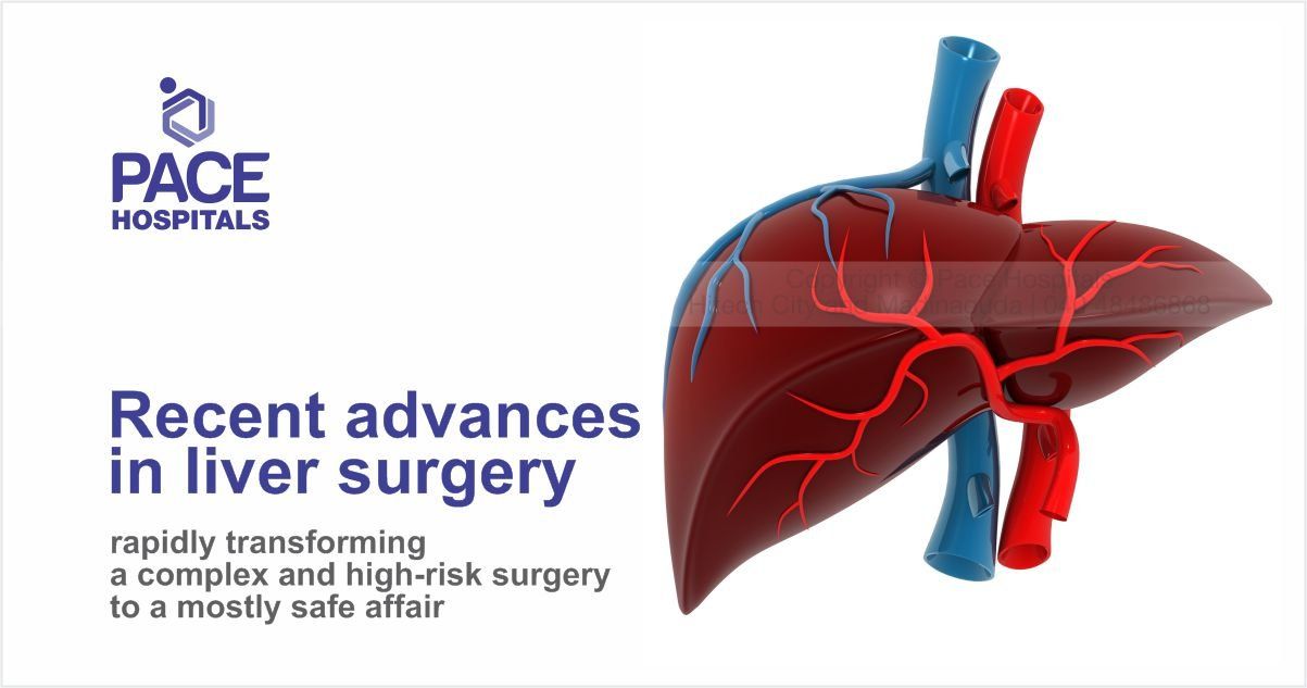
Recent advances in liver surgery: safe and effective for complex and high-risk surgery
Pace Hospitals
Liver surgery has seen great strides have been made over the past 2 decades. Be it diagnostics, imaging, innovative surgical strategies or technology the branch has evolved rapidly transforming a complex and high-risk surgery to a mostly safe affair.
Bloodless liver surgery
Unlike other organs the liver is extremely vascular with half its volume made up of blood in large vascular sinuses. The blood loss in major liver resections was once measured in liters is now in milliliters. This is thanks to concepts like hypotensive-low venous pressure anaesthesia and inflow and total vascular occlusion in which the liver is operated upon after temporarily stopping its blood supply. Devices such as CUSA (ultrasonic aspirator) , Water Jet dissector, and radiofrequency ablator aid in bloodless division of liver while identifying and protecting crucial structures.
Image guided surgery
Beyond the standard Multi slice CT scan and MRI scan, Innovations in functional liver imaging such as Functional MRI, Tc-99 sulfur colloid single photon PET scan and HIDA scan have helped us predict not just the size of the liver remnant but the function. This helps us plan and predict outcomes in patients with diseased and fatty livers where not just the size but the quality of liver matters. Preoperative liver mapping (MEVIS scan), virtual resections with image overlays and navigational systems guide us through liver anatomy in complex liver resections
Innovative surgical strategies
Several innovative surgical strategies have revolutionised liver surgery. The liver has a tremendous capacity to regenerate. By strategies like portal vein embolization and ALLPS we deprive nutrition the tumor half of the liver for a few weeks before surgery. This way the future remnant liver (ie the liver to be left behind after taking out tumor) regenerates even before the surgery. This allows us to do supra-major liver resections with low risk of postoperative liver faiure.
The understanding that the liver anatomy is segmental (like a tree with its branches) has led to parenchyma preserving resections like central hepatectomies, monosegmentectomies taking out only tumor with excellent postoperative function.
Two staged liver resections help us safely treat patients with disease in both halves of the liver. ‘Down staging‘ treatments such as Transarterial chemoembolization , Transarterial radioembolisation are used to preoperatively decrease the size of tumors and convert inoperable tumors to operable.
Robotic and laparoscopic surgery
The 3D visualisation, improved precision and better maneuverability has led to wide application of laparoscopic and robotic surgery in liver resections and transplant. This translates to smaller incisions and faster recovery.
Adjunctive techniques
Adjunctive techniques such as microwave ablation , radiofrequency ablation for image guided destruction of tumours , radiosurgery using transarterialradioembolisation (tumor breakdown by injecting radioactive beads) into it have extended the boundaries and also useful tools in patients unfit for surgery.
Share on
Request an appointment
Fill in the appointment form or call us instantly to book a confirmed appointment with our super specialist at 04048486868
Appointment request - health articles
Recent Articles