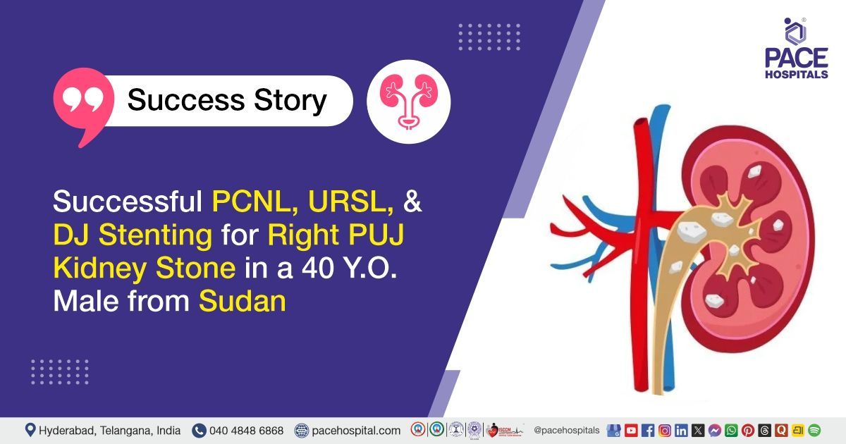
Successful PCNL, URSL, and DJ Stenting for Right PUJ Kidney Stone in a 40 Y.O. Male from Sudan
PACE Hospitals
PACE Hospitals’ Urology team successfully performed Right
Percutaneous Nephrolithotomy (PCNL) with Ureteroscopic Lithotripsy (URSL) and Double-J (DJ) stenting
on a 40-year-old male patient from Sudan diagnosed with a right renal calculus at the pelviureteric junction (PUJ). The procedure aimed to achieve complete stone clearance, relieve urinary obstruction and flank pain, preserve kidney function, and support recovery and improved quality of life.
Chief complaints
A 40-year-old male patient from Sudan with a
body mass index (BMI) of 21 presented to the Urology Department at
PACE Hospitals, Hitech City, Hyderabad, with complaints of right flank pain.
Past Medical History
The patient had no significant past medical history or documented chronic medical conditions. There was no relevant history of previous urological surgery or other major comorbidities.
On Examination
On examination, the patient was conscious, coherent, and oriented, with stable vital signs. General physical examination was satisfactory. Abdominal examination was normal apart from right flank discomfort/tenderness. No significant abdominal distension, palpable mass, or other acute abnormality was noted.
Diagnosis
Upon admission to PACE Hospitals, the patient underwent a comprehensive evaluation by the Urology team, including a detailed clinical assessment and review of his medical history. He presented with right flank pain, raising suspicion of a right renal calculus.
Imaging studies, including ultrasound and computed tomography (CT) evaluation of the abdomen and pelvis, revealed a right renal calculus located at the pelviureteric junction (PUJ) with associated moderate hydronephrosis, indicating obstruction to normal urine drainage from the kidney.
Laboratory investigations and preoperative assessments were performed to evaluate the patient's overall condition and fitness for intervention. The findings supported the diagnosis of an obstructing right renal calculus with hydronephrosis.
Based on the confirmed findings, the patient was advised to undergo
Right Kidney Stone Treatment under the supervision of the Urology Department.
Medical Decision Making (MDM)
After a detailed consultation with Dr. Abhik Debnath (Consultant Urologist), a comprehensive evaluation was conducted to determine the most appropriate therapeutic approach for the patient presenting with right flank pain. Clinical assessment and imaging findings confirmed a right renal calculus at the pelviureteric junction (PUJ) with moderate hydronephrosis.
Based on the stone location, associated urinary obstruction, and the need for complete stone clearance, it was determined that Right Percutaneous Nephrolithotomy (PCNL) with Ureteroscopic Lithotripsy (URSL) and Double-J (DJ) stenting was an appropriate minimally invasive treatment strategy. The planned intervention aimed to fragment and completely remove the renal calculus, relieve the obstruction, improve urinary drainage, and prevent further complications related to hydronephrosis.
The patient and his family members were counselled regarding the diagnosis, planned procedures, potential risks and benefits, expected recovery, postoperative precautions, and the importance of follow-up, including DJ stent removal as advised.
Surgical Procedure
Following the decision, the patient was scheduled for Right Percutaneous Nephrolithotomy (PCNL) with Ureteroscopic Lithotripsy (URSL) and Double-J (DJ) Stenting Surgery in Hyderabad at PACE Hospitals, under the supervision of the Urology Department.
The procedure involved the following steps:
- Preoperative Preparation and Anaesthesia: After completing the required preoperative evaluation and obtaining informed consent, the patient was taken to the operating room and administered general anaesthesia. Standard aseptic precautions were maintained throughout the procedure.
- Renal Access and Tract Dilatation: The patient was positioned appropriately, and a mid-pole posterior calyceal puncture was performed to obtain access to the right kidney. The access tract was gradually dilated up to 21 French (Fr) to facilitate introduction of the nephroscope.
- Stone Identification and Fragmentation: An 18 Fr nephroscope was introduced into the renal collecting system, and the renal calculus was identified. The stone was fragmented using appropriate endourological instrumentation, and the fragments were carefully removed.
- Ureteroscopic Lithotripsy: Ureteroscopic evaluation and lithotripsy were performed as required to facilitate complete stone clearance and ensure unobstructed urinary drainage. Mild mucosal oedema was noted at the site of previous stone impaction.
- DJ Stent Placement: A Double-J (DJ) stent was placed to maintain adequate urine drainage from the kidney to the bladder and support healing following stone removal.
- Completion of Procedure: The renal collecting system was inspected to confirm satisfactory stone clearance. Haemostasis was achieved, and the procedure was completed without complications. The patient was shifted to the recovery area in a stable condition.
Postoperative Care
Following the procedure, the patient was closely monitored during the postoperative period. His recovery was uneventful, and he remained hemodynamically stable. During his hospital stay, he received medicines to prevent infection, relieve pain, control fever or other postoperative symptoms, and protect the stomach from medication-related irritation. Mild flank or suprapubic discomfort, occasional blood or cloudiness in the urine, and mild burning during urination were explained as expected temporary symptoms. The patient showed satisfactory recovery and was discharged in a stable condition.
Discharge Medications
Upon discharge, the patient was prescribed medicines to prevent urinary infection, relieve pain, control fever if required, facilitate smooth urinary flow, maintain an appropriate urinary pH to help reduce the risk of stone recurrence, and protect the stomach from medication-related irritation.
Discharge Advice
The patient was advised to maintain adequate hydration and include citrus fruits in his diet. A low-purine and low-salt diet was recommended to help reduce the risk of recurrent stone formation. He was advised to avoid heavy lifting, excessive forward bending, straining during bowel movements, and prolonged travel by bus, auto, or two-wheeler during the recovery period.
Emergency Care
The patient was advised to contact the emergency ward at PACE Hospitals in case of any emergency or if symptoms such as fever, severe abdominal or flank pain, persistent vomiting, worsening urinary discomfort, or other unusual symptoms developed.
Review and Follow-up Notes
The patient was advised to return for a follow-up visit with the
Urologist
after 2 months.
Conclusion
This case highlights the successful management of a right renal calculus at the pelviureteric junction (PUJ) with moderate hydronephrosis using Right Percutaneous Nephrolithotomy (PCNL), Ureteroscopic Lithotripsy (URSL), and Double-J (DJ) stenting. The procedure achieved complete stone clearance and relief of the urinary obstruction. The patient recovered without significant complications and was discharged in a stable condition with dietary advice, postoperative precautions, and scheduled follow-up for DJ stent removal.
Medical Disclaimer: This information is for general educational purposes only and does not replace consultation with a qualified urologist. Treatment decisions and recovery vary depending on stone size and location, urinary obstruction, kidney function, infection status, anatomy, and other individual clinical factors.
Minimally Invasive Management of PUJ Renal Calculus with Hydronephrosis
Renal calculi located at the pelviureteric junction (PUJ) can obstruct normal urine drainage and lead to hydronephrosis, flank pain, and potential deterioration of kidney function if left untreated. Appropriate imaging helps determine the stone location, size, and degree of obstruction and and guides treatment planning by the
urologist/urology doctor. Minimally invasive procedures such as Percutaneous Nephrolithotomy (PCNL) and Ureteroscopic Lithotripsy (URSL) can be used to fragment and remove stones, while a Double-J (DJ) stent helps maintain urinary drainage during recovery. Adequate hydration, appropriate dietary measures, and follow-up for timely DJ stent removal are important for recovery and reducing the risk of recurrent stones and stent-related complications.
Frequently asked questions (FAQs)
What is a PUJ renal stone?
A PUJ renal stone is a kidney stone located at the pelviureteric junction (PUJ), where the kidney connects to the ureter. If the stone blocks urine flow at this point, it can cause flank pain and swelling of the kidney due to urine buildup.
Can a PUJ stone cause hydronephrosis?
Yes, a PUJ stone is capable of either partly or completely obstructing the normal flow of urine from the kidney through to the ureter, which in turn can cause urine to accumulate within the kidney and lead to hydronephrosis, that is, swelling or enlargement of the kidney's drainage system.
What is a Double-J (DJ) stent and why is it used after stone removal?
A DJ stent is a thin, flexible tube placed between the kidney and bladder. It helps keep the ureter open and allows urine to drain properly from the kidney to the bladder after stone treatment. It may also support healing and help prevent blockage caused by temporary swelling following the procedure.
How long does it take to recover after PCNL and URSL?
Recovery varies depending on the size and location of the stone, the procedure performed, and the patient's overall health. Many patients can gradually return to normal activities within a few weeks, although strenuous activities may need to be avoided for longer. The treating urologist will advise when it is safe to resume specific activities.
Is mild blood in the urine normal after kidney stone surgery?
Mild blood in the urine can occur for a short period after PCNL or URSL because the urinary tract may be temporarily irritated by the procedure or DJ stent. It usually improves with recovery and adequate fluid intake. However, heavy or persistent bleeding, blood clots, or difficulty passing urine should be evaluated promptly.
Can patients experience pain while a DJ stent is in place?
Yes. Some patients may experience mild discomfort in the flank, lower abdomen, or bladder while a DJ stent is in place. Urinary frequency, urgency, or mild burning during urination can also occur. These symptoms generally improve after the stent is removed, but severe or worsening pain should be reported to the urologist.
How much water should be consumed after kidney stone treatment?
Adequate hydration is important after stone treatment, but the exact amount should be individualised according to kidney function, medical conditions, climate, and fluid losses. In general, patients who are able to drink normally are encouraged to consume enough fluids to produce around 2–2.5 litres of urine per day. A urologist may recommend a different amount when medically appropriate.
What happens if a DJ stent is not removed on time?
A DJ stent is generally intended to remain in place only for the period recommended by the treating urologist. If it is left for too long, mineral deposits can build up on the stent, making removal more difficult and increasing the risk of infection, blockage, pain, or urinary complications. Therefore, the scheduled stent-removal appointment should not be missed.
Can a kidney stone cause permanent kidney damage?
A kidney stone does not necessarily cause permanent kidney damage. However, a stone that causes prolonged or severe urinary obstruction, particularly when associated with infection, can potentially damage kidney function. Prompt evaluation and treatment of obstructing stones help reduce this risk.
How can future kidney stones be prevented?
Adequate fluid intake is one of the most important measures for preventing recurrent kidney stones. Depending on the type of stone and individual risk factors, limiting excessive salt intake, maintaining a balanced diet, avoiding excessive intake of certain stone-forming foods, and maintaining an appropriate dietary calcium intake may help. Patients with recurrent or high-risk stones may benefit from stone analysis and metabolic evaluation, including 24-hour urine testing, to develop a personalised prevention plan.
Share on
Request an appointment
Fill in the appointment form or call us instantly to book a confirmed appointment with our super specialist at 04048486868
Appointment request - health articles
Recent Articles