Thromboembolism - Symptoms, Types, Causes, Complications & Prevention
PACE Hospitals
Overview | Epidemiology | Types | Causes | Symptoms | Risk Factors | Complications | Diagnosis | Treatment | Prevention | Venous thromboembolism vs Arterial embolism | Atheroembolism vs Thromboembolism | Thromboembolism vs Embolism | Thromboembolism vs Fat Embolism Syndrome | FAQs | When to Consult a Doctor
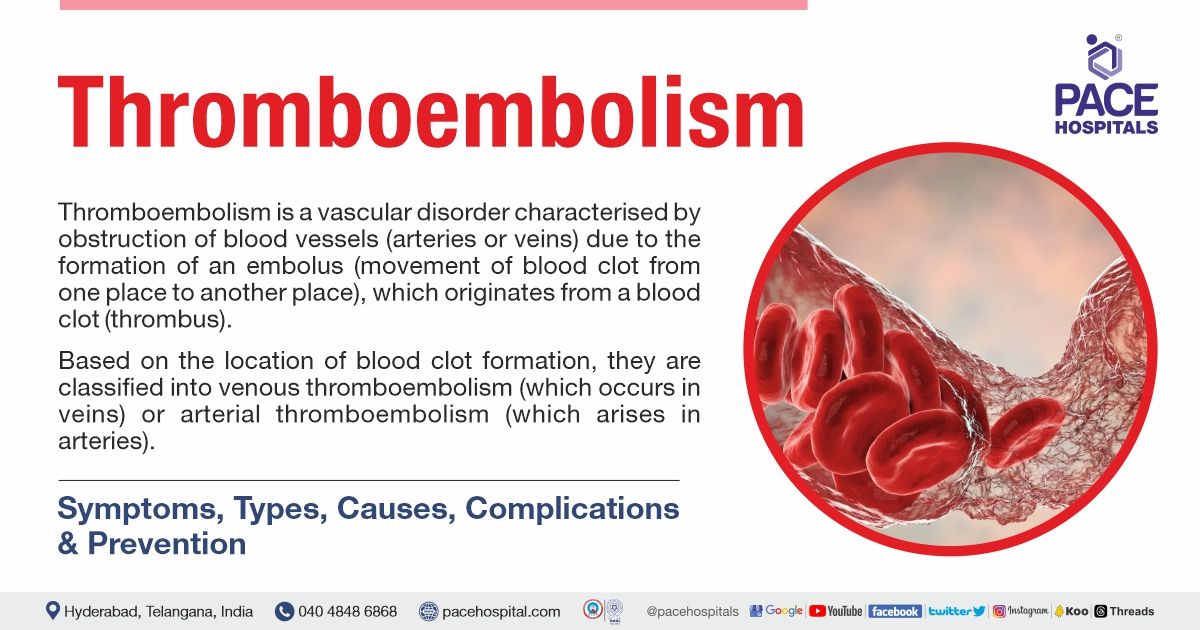
Thromboembolism is a vascular disorder characterised by obstruction of blood vessels (arteries or veins) due to the formation of an embolus (movement of blood clot from one place to another place), which originates from a blood clot (thrombus). Based on the location of blood clot formation, they are classified into venous thromboembolism (which occurs in veins) or arterial thromboembolism (which arises in arteries). Haematologists lead the healthcare team which diagnoses and treats thromboembolic disorders.
While acute myocardial infarction (MI), atherothrombotic stroke and peripheral artery diseases (PAD) constitute thromboembolic arterial diseases, venous thromboembolism (VTE) includes deep venous thrombosis (DVT) and pulmonary embolism (PE).
Thromboembolism meaning
While the thromboembolism definition explains the blood flow obstruction, the word thrombo is derived from the Greek word "thrombos", meaning blood clot, and embolism is derived from the Greek word "embolus", which defines the movement of a blood clot from one place to another place.
Epidemiology of Venous Thromboembolism
Venous thromboembolism (VTE) is the major third cause of death worldwide. The incidence of VTE varies according to different countries, ranging from 1-2 per 1000 person-years in Western Countries and lower in Eastern Countries (<1 per 1000 person-years).
Epidemiology of Arterial Thromboembolism
Globally, the numbers of ischemic stroke cases, deaths, and DALYs increased from 407, 204.9, and 4050 crores, respectively, in 1990 to 786, 315, and 6253 crores, respectively, in 2020.
According to a research study, the total incidence of myocardial infarction is 15%, with a maximum incidence of eight cases in the 51-60-year age group. This is consistent with the study, which found a 12.7% incidence of acute myocardial infarction in cerebrovascular accident patients.
Types of Thromboembolism
While there are various types of thromboembolism derived from the location of a blood clot, the major types of thromboembolic disorders include:
- Venous Thromboembolism
- Deep Venous Thrombosis
- Pulmonary thromboembolism
- Arterial Thromboembolism
- Ischemic Stroke
- Myocardial infarction
- Other thromboembolism
- Systemic thromboembolism
- Cerebral thromboembolism
- Coronary thromboembolism
- Massive thromboembolism
- Thromboembolism in spleen
- Thromboembolism after muscle injury
- Thromboembolic coagulopathy
- Cancer-associated thromboembolism
- Postpartum thromboembolism
- Thromboembolic renal disease
Venous thromboembolism
Venous thromboembolism (VTE) refers to the formation of blood clots in the veins. This is a gradually developing disorder which is often underdiagnosed but can have serious consequences, such as disability and death. Nevertheless, it is a preventable medical condition if diagnosed accurately and early.
Deep vein thrombosis: Deep vein thrombosis (DVT or venous thrombosis) is a medical condition referring to blood clot formation in the veins where the thrombus formation could occur due to either injured veins or the blood's hypercoagulability (increased coagulation affinity). Usually found in the lower extremities, such as the lower leg, thigh, etc. Deep vein thrombosis could cause partial or complete blockage to the blood flow to the respective organs.
Pulmonary thromboembolism: This is also known as pulmonary embolism, a medical condition that disrupts (blocks) the pulmonary arterial blood flow due to the embolus either in the pulmonary artery or its branches. Here, the thrombus is formed elsewhere in the circulatory system but gets detached and passes through the pulmonary artery or its capillaries.
The combination of disrupted pulmonary embolism and deep vein thrombosis is collectively called venous thromboembolism (VTE). Pulmonary emboli originate from deep venous thrombus and are responsible for the most common form of pulmonary thromboembolic disease. Based on the severity of the disease, the pulmonary embolism is of two types:
- Acute pulmonary embolism: In acute embolism, the increased diameter of the pulmonary artery is observed.
- Chronic pulmonary embolism: In chronic embolism, narrowing of distal blood vessels is observed.
Arterial thromboembolism
Arterial embolism is an infarction (tissue death due to inadequate blood supply) of arteries in the blood vessels due to an embolus that adheres (attached) to the arterial wall that obstructs the blood flow. Arterial thromboembolism may lead to cerebral or another vital end-organ infarction.
Ischemic stroke: The deficiency (reduction)of blood supply to the brain is known as an ischaemic stroke. This is a serious condition as it causes the blockage of oxygen and nutrients to the brain, which could cause the death of neurons (brain cells) within minutes. Ischaemic stroke could be due to thrombosis (clot formation) or emboli (movement of blood clot from one place to another).
Myocardial Infarction: Known as a "heart attack," this myocardial infarction (MI) is a cardiac condition in which there is either a complete or a partial obstruction of blood flow to the myocardium (heart wall). Myocardial infarction due to atherosclerotic plaque (fat deposition in arteries) build-up in coronary arteries (blood taken to arteries), leading to a deficient supply of blood to the heart, if it is not treated, leads to sudden death.
Other types of thromboembolism
Systemic thromboembolism: If the thromboembolism condition is observed in systemic arteries, it is considered systemic thromboembolism. Most systemic emboli (80%) are from intra cardiac mural thrombi; two-thirds are associated with left ventricular infarcts (deficient blood supply), and another 25% have dilated left atria (upper heart chamber). The remainder originate from aortic aneurysms (abnormal blood vessels); common arteriolar embolisation sites include the lower extremities (75%) and central nervous system (10%).
Cerebral thromboembolism: If the thromboembolism condition is observed in the cerebral arteries, it is known as cerebral thromboembolism. Cerebral thromboembolism is due to the artery-to-artery embolism. The intracranial arteries include the intracranial internal carotid, basilar, proximal segments of the basilar artery, and anterior cerebral artery, which are affected by cerebral thromboembolism.
Coronary thromboembolism: The thromboembolic forms in the coronary arteries which obstruct the arteries is known as coronary thromboembolism. Coronary embolism mainly affects the anterior descending artery in the regions of the distal epicardial portion and its intramural branches.
Massive thromboembolism: Massive thromboembolism, which is usually observed in pulmonary embolism, defined as obstruction of at least 51% of pulmonary arteries, leads to acute and severe cardiopulmonary failure from right ventricular overload.
Thromboembolism in spleen: Patients who are suffering from splenic vein thrombosis can be symptomatic (shows symptoms) or asymptomatic. These symptoms include abdominal pain, variceal bleeding, splenomegaly (enlarged spleen), and thrombocytopenia (decreased platelets). Splenectomy is a surgical intervention for specific indications, benign and malignant. This procedure could induce venous thromboembolism (VTE).
Thromboembolism after muscle injury: The partial ruptures of muscles in the leg lead to the formation of venous thrombosis. The presence of oedema or haematoma (collection of blood outside the blood vessel), caused by the ruptured muscles, may lead to compression of popliteal or gastrocnemius veins, thus determining the deep vein thrombosis (DVT).
Thromboembolic coagulopathy: Trauma-induced coagulopathy (TIC) (usually >24 hours after injury) is represented by a hypercoagulable state, which may result in thromboembolic events (e.g. deep venous thrombosis (DVT) and pulmonary embolism (PE)).
Cancer-associated thromboembolism: In cancer, coagulation and platelets are activated, which can induce cancer-associated thromboembolism.
Post-partum thromboembolism: The risk for post-partum (post-delivery state) venous thromboembolism (VTE) is higher during the first three weeks after delivery. Women with delivery complications are at a higher risk of post-partum venous thromboembolism, and this risk will be elevated throughout the first 12 weeks after delivery.
Thromboembolic renal disease: The thromboembolic renal disease occurs due to a thrombus in the veins of the renal system. The pathogenesis of thromboembolic renal disease is based on Virchow's triad, which includes vascular endothelial damage, stasis of blood flow, and hypercoagulability.
Thromboembolic Disease Causes
The thromboembolism depends on the formation of thrombus or blood clot in arteries or veins.
- Etiology of venous thromboembolism (venous thromboembolism causes)
- Injury of the deep vein leads to the formation of a blood clot.
- Limited movement in physical activity, so there is a deficient supply of blood circulation in blood vessels, leads to the formation of a blood clot.
- Heavy usage of oestrogen leads to the formation of a blood clot.
Etiology of arterial thromboembolism (arterial thromboembolism causes)
- Increased age
- Smoking
- Increased fibrinogen levels
Thromboembolic Disease Symptoms
Thromboembolic disorder's signs and symptoms may vary depending upon thrombus formation in the venous and arteries.
Venous thromboembolism Symptoms
The following are the venous thromboembolic disease symptoms.
- Tenderness along the veins: Tenderness along the veins, which are sensitive to touch
- Aching pain: An unpleasant sensitised pain and emotional experience associated with potential tissue damage
- Swelling occurs whenever the organs, skin, or other parts of your body enlarge. It leads to inflammation or a buildup of fluid. Swelling can occur internally, or it can affect your outer skin and muscles.
Arterial thromboembolism Symptoms
The following are the arterial thromboembolic disease symptoms.
- Aphasia (slurred speech)
- Agnosia (unable to recognise objects, persons and sounds)
- Apraxia (unable to carry out skilful movements and gestures)
- Monoplegia (paralysis of arm or leg)
- Loss of consciousness
- Chest pain
- Dyspnoea (shortness of breath)
- Palpitation (increased heartbeat)
- Diaphoresis (excessive sweating)
Thromboembolism in Pregnancy and Infants
Thromboembolic disease in pregnancy:
Pregnancy, as such, is a prothrombotic state that enhances venous stasis (cessation of blood flow in veins) mainly due to obstructed venous flow by the enlarging uterus. During 25–29 weeks of gestation, venous flow velocity falls by nearly 50% in the legs. A reduction of venous flow velocity of almost 50% occurs in the legs by weeks 25–29 of gestation. This self-healing condition lasts until approximately 6 weeks post-partum (after delivery). Among pregnant and post-partum women, usually the lower part of the left leg is the most commonly affected site of deep vein thrombosis (DVT)
Risk factors for thromboembolism in pregnancy, specifically for deep vein thrombosis (DVT)
Pregnancy is a prothrombotic state; there are various risk factors which increase the affinity for the development of deep vein thrombosis (DVT)
- Hospitalisation for caesarean delivery
- An inherited thrombophilia (e.g., factor V Leiden mutation, prothrombin gene mutation, or antithrombin III, protein C, or protein S deficiencies) increases the risk of thromboembolism .
Thromboembolic complications in pregnancy:
The risk of developing a thromboembolic disorder is high, during 6 weeks after delivery. Most of the complications due to formation of blood clots results from injuries of the blood vessels during labour(delivery) period. The caesarean deliveries are having higher risk than vaginal delivery.
Thromboembolism infant:
Neonates are at higher risk for development of venous thromboembolism. Central lines (supply of medicine through jugular vein), sepsis (body response improperly to infection), liver dysfunction, fluid fluctuations (imbalance of fluids), and inflammation are the risks for venous thromboembolism (VTE) development in ill neonates.
Risk factors for Thromboembolism
The risk factors for thromboembolism vary, depending on venous and arterial thromboembolism.
Venous thromboembolism risk factors. The following are the risk factors of deep venous thrombosis
- Obesity
- Height
- May–Thurner illness
- Diet
- Tobacco
- Obesity: Obesity increases the risk of venous stasis, higher levels of inflammatory and haemostatic biomarkers, and an increased chance of inducing venous thromboembolism.
- Height: Higher BMI has continuously been linked to an increased risk of venous thromboembolism taller individuals have higher hydrostatic pressure, more venous valves, and a larger venous surface area in the vascular wall of the legs, which leads to an increase in the risk of venous thromboembolism
- May–Thurner illness: May-Thurner syndrome patients are more likely to experience an extensive left iliofemoral (localised blood clot) due to the lumbar spine being compressed by a common iliac artery with a left common iliac vein.
- Diet: Trans fatty acids and red and processed meat increase the risk of thromboembolism
- Tobacco: smoking might enhance coagulability and chronic inflammation; there has been an assumption that smoking increases the risk of venous thromboembolism
Arterial thromboembolism risk factors:
- The following are the risk factors for arterial thromboembolism.
- Smoking
- Diabetes
- High blood pressure
Thromboembolic Complications
Thromboembolic complications are serious medical conditions that occur when a blood clot breaks off from its original location and travels through the bloodstream to another part of the body, where it can block blood flow and cause damage. These complications can be life-threatening and require prompt medical attention.
- Thromboembolic aneurysm: Thromboembolism rarely occurs in large aneurysms where decreased blood flow within the aneurysm leads to blood clots, which are considered thromboembolic aneurysms.
- Pulmonary venous thromboembolism: This is a rare form of venous thromboembolism complications, that is observed in veins of the pulmonary blood vessels. The pulmonary veins are the most common source of arterial thromboembolism. Pulmonary vein thrombosis (PVT) is a rare but potentially lethal disease which could occur due to the direct extension of the tumour into the vein, thus compressing the vein and causing epithelial damage and a hypercoagulable state
- Chronic thromboembolic pulmonary hypertension (CTEPH): Chronic thromboembolic pulmonary hypertension (CTEPH) is one of the complications of venous thromboembolic disease; specifically, CTEPH, due to pulmonary embolism, is a significant cause of chronic pulmonary hypertension, leading to right heart failure and death.
Thromboembolism Diagnosis
Imaging studies are primarily preferred to diagnose any form of thromboembolism. The overall diagnoses for thromboembolism include:
Diagnosis of arterial thromboembolism
The following are the diagnostic procedures to detect (find out) arterial thromboembolism.
- Computed Tomography (CT) scan
- Magnetic Resonance Imaging (MRI) Scan
- MRI Contrast
- Ultrasound
- D dimer
- Transthoracic or transoesophageal echocardiography
- Colour-coded Doppler ultrasonography
- Conventional grey-scale real-time ultrasonography.
- Compression ultrasonography (CUS)
- Computed tomographic pulmonary angiography.
- Complete compression ultrasonography (CUS)
- Venous ultrasound (VUS)
- Compression venous ultrasonography
- Colour Doppler ultrasonography (CDUS)
- Radioisotope scintigraphy
- Plethysmography
- Multidetector CT angiography (MDCTA)
Venous thromboembolism diagnosis:
The diagnostic procedures to detect (find out) venous thromboembolism are similar to arterial thromboembolism; however, the following are the specified tests to detect (find out) venous thromboembolism.
- Pulmonary angiography
- V/Q Scan (lung ventilation perfusion)
- Contrast venography
Thromboembolism Treatment
Venous thromboembolism treatment and arterial thromboembolism treatment have the same pharmacological therapy; however, they might vary in surgical interventions. The standard drug categories which help in managing thromboembolism (both arterial and venous) include:
- Pharmacological treatment
- Conventional anticoagulants
- Direct oral anticoagulants
- Thrombolytic therapy
Surgical Interventions:
The following are the common surgical interventions performed by a vascular surgeon:
Arterial thromboembolism surgical procedures
- Coronary artery bypass graft (CABG)
- Venous thromboembolism surgical procedures
- Venous thrombectomy with a Fogarty catheter
- Pulmonary Embolectomy
Why Choose PACE Hospitals?
Expert Super Specialist Doctors
Advanced Diagnostics & Treatment
Affordable & Transparent Care
24x7 Emergency & ICU Support
Thromboembolism Prevention
The prevention of thromboembolic disease depends on the early diagnosis. The standard measures which prevent thromboembolism include:
Venous thromboembolism prevention
Venous thromboembolism can be prevented early; however, the following measures can reduce the risk of venous thromboembolism.
- Keep moving. Physical activity increases blood circulation and decreases the risk of blood clots.
- Changing positions while sitting, changing sitting positions prevents the risk of blood clot formation.
- Perform simple exercises. Flexing and extending the ankles and knees enhances the contraction of the calf muscles at regular intervals.
- Physical activity after being confined to bed, such as surgery, illness, or injury decreases the risk of blood clot.
- When sitting for long periods, such as when travelling for a long time, standing and walking around every 1 to 2 hours.
Arterial thromboembolism prevention
Arterial thromboembolism cannot be prevented; however, the following measures can reduce the risk of arterial thromboembolism.
- Being active increases blood circulation and decreases the risk of blood clot formation.
- Returning to regular physical activity after surgery decreases the risk of blood clot formation.
- Stretching of the legs while exercising decreases the risk of blood clot formation.
- A reduced in weight enhances the blood circulation, so it reduces the risk of blood clot due to loss of heavyweight.
Prevention of Venous Thromboembolism in Surgical Patients
If the pharmacological prophylaxis is adequately maintained, the risk of bleeding complications will be low. Prophylaxis with mechanical methods is preferred for patients at high risk of bleeding complications. The common prophylactic measures in surgical patients are the graduated compression stockings, intermittent pneumatic compression, and pre-discharge surveillance with blood vessels or venous ultrasonography of the legs.
Thromboembolic stockings:
The following are the various types of stockings used after surgery or as a part of the prevention of thromboembolism.
- Graduated compression stockings
- Elastic compression stockings
- Thromboembolic deterrent stockings
- Graduated compression stockings: The graduated compression thromboembolic stockings are used for the prevention of DVT. Graduated compression stockings are one of the most common mechanical forms of venous thromboembolism (VTE) prophylaxis. They work by displacing blood from superficial veins to deep leg veins, thus increasing the flow through deep veins. A 2010 Cochrane study demonstrated that there is a significant risk reduction for deep vein thrombosis (DVT) after analysing 18 randomised controlled trials with 887 patients.
- Elastic compression stockings: Elastic compression stockings are used for deep vein thrombosis (DVT) patients undergoing ankle surgery but not in patients undergoing lumbar, knee or spinal surgery. Along with elastic compression stockings, pharmacological therapy such as anticoagulant drugs and other physiotherapy methods prevent deep vein thrombosis (DVT).
- Thromboembolic deterrent (TED) stockings: Thromboembolic deterrent (TED) stockings are effective in the reduction of thromboembolic events in post-operative patients. These manufactured stockings create graduated compression from ankle to calf, thus preventing deep vein thrombosis (DVT).
Difference between Venous Thromboembolism and Arterial Embolism
While venous thromboembolism (VTE) and arterial embolism are conditions that involve the formation of blood clots, they occur in different blood vessels and have distinct characteristics.
| Parameters | Venous Thromboembolism | Arterial Embolism |
|---|---|---|
| Shear rate | Low | High |
| Composition | Red blood cells and fibrin | Platelets |
| Diseases | Deep vein thrombosis and pulmonary embolism | Myocardial infarction and Stroke |
Difference between Atheroembolism and Thromboembolism
Atheroembolism vs thromboembolism
Both atheroembolism and thromboembolism are types of embolism that obstruct blood vessels with a foreign substance that travels through the bloodstream. However, there are some critical differences between the two:
| Parameter | Atheroembolism | Thromboembolism |
|---|---|---|
| Composition | Cholesterol crystal emboli | Fragmented pieces of thrombus |
| Origin | Lipid core of arterial plaque | The surface of ulcerated plaque |
| Immediate treatment | Supportive care | Thrombolysis |
| Long term treatment | Risk factor modification | Anticoagulant, antiplatelet |
Difference between Thromboembolism and Embolism
Thromboembolism vs Embolism
Both thromboembolism and embolism block blood vessels. While thromboembolism refers to a deficient supply of blood flow that's caused explicitly by an embolism resulting from a blood clot, embolism, on the other hand, refers to the occlusion of blood vessels caused by an embolus (including air bubbles, blood clot, lipid globules or the tip of an indwelling catheter).
Difference between Thromboembolism and Fat Embolism Syndrome
Thromboembolism vs fat embolism syndrome
Both thromboembolism and fat embolism block blood vessels. Meanwhile, thromboembolism is a disorder that obstructs blood vessels (arteries or veins) caused by an embolism from a blood clot. On the other hand, fat embolism (FE) and fat embolism syndrome (FES) are clinically characterised by the systemic dissemination of fat emboli within the systemic circulation.
- If thromboembolism occurs in arteries, then it's known as thromboarterial disorder. If the same thrombus formation is seen in veins, it's known as venous thromboembolism.
- Fat embolism resembles the presence of fat globules in the microcirculation of the blood vessels. In contrast, fat embolism syndrome is a systemic manifestation of the dissemination of fat molecules or globules in microcirculation.
- A fat embolism (FE) is an intravascular fat that occurs within a blood vessel and causes an obstruction of blood flow. Fat emboli commonly occur due to fractures of the long bones of the lower body, particularly in the femur (thighbone), pelvis, and tibia.
- While fat emboli are common and resolve within themselves, in some cases, they don't fix themselves, leading to a severe condition called fat embolism syndrome (FES).
FAQs - Frequently asked questions on thromboembolism
What are the two types of thromboembolism?
The two types of thromboembolism include venous thromboembolism (VTE) and thromboembolic arterial disease (TAD).
Venous thromboembolism (VTE) can be further classified into two other disorders: deep venous thrombosis (DVT) and pulmonary embolism (PE). Similarly, thromboembolic arterial disease (TAD) can be further classified into three diseases: acute myocardial infarction (AM), atherothrombotic stroke and peripheral artery disease (PAD).
While Venous thromboembolism (VTE) and arterial embolism are conditions that involve the formation of blood clots, they occur in different blood vessels and have distinct characteristics.
How is thromboembolism treated?
Both pharmacological treatment (treatment with medicines) as well as surgical therapies have been developed to treat thromboembolism. The standard drug categories which help in managing thromboembolism include:
- Conventional Anticoagulants (old anticoagulants)
- Direct oral Anticoagulants (new anticoagulants
- Thrombolytic Therapy
What is the term thromboembolism?
The word thrombo is derived from the Greek word "thrombosis", meaning a blood clot; embolism is derived from the Greek word "embolus", which defines obstruction of blood vessels.
Thromboembolism refers to blood flow reduction caused explicitly by an embolism from a blood clot .
What is a high D-dimer level?
A normal D-dimer is considered if less than 0.50. A positive D-dimer is 0.50 or greater. There is not necessarily a critical level for a D-dimer.
At moderate risk of pulmonary embolism and moderate to high risk of deep vein thrombosis if there is a positive D-dimer.
What foods make your blood thinner?
The foods containing antioxidants help in making the blood thinner. They could include fruits, vegetables, olive oil, nuts, seeds, fish, cocoa, etc. A 2020 review found that a diet consisting of antioxidants, such as anti-inflammatory, antithrombotic, and antiplatelet effects, may reduce cholesterol deposition and plaque formation in the blood vessels.
Is a blood clot in the leg curable?
Yes, blood clots in the legs are curable with the following treatment:
- Conventional anticoagulants and direct oral anticoagulants are used to dissolve blood clots.
- Thrombolytics are medications that dissolve blood clots.
- Thrombolytics, which lyses the thrombin and decreases the blood clots.
- Surgical thrombectomy
What foods should you avoid if you have blood clots?
The consumption of ultra-processed foods, which are high in calories, fat, sugar, and salt, leads to the development of ischemic heart diseases such as myocardial infarction due to increased HDL cholesterol and elevated levels of sodium and weight gain.
How does cancer increase the risk of thromboembolism?
The diseased stem cells from the hypercoagulable state enhance the increasing production of substances like tissue factors with increased coagulant activity, which enhances coagulation and leads to thromboembolism formation.
When to Consult a Doctor for Thromboembolism?
Consult a doctor if swelling, leg pain, sudden breathlessness, or chest discomfort is noticed, as these symptoms may sometimes be associated with Thromboembolism. While some blood clots may resolve with timely treatment, others can become serious if left untreated.
Warning Signs:
- Swelling in one leg or arm
- Pain or tenderness in the calf
- Redness or warmth over the skin
- Sudden shortness of breath
- Chest pain while breathing & Rapid heartbeat
- Unexplained dizziness or weakness
- Coughing with blood
- Sudden severe leg pain
If these symptoms are present, it is advisable to consult an Interventional radiologist, Cardiologist, or General Physician. The doctor may recommend a Doppler ultrasound, blood tests, a CT scan, or angiography to confirm the diagnosis and plan appropriate treatment.
Share on
Request an appointment
Fill in the appointment form or call us instantly to book a confirmed appointment with our super specialist at 04048486868
Appointment request - health articles
Recent Articles