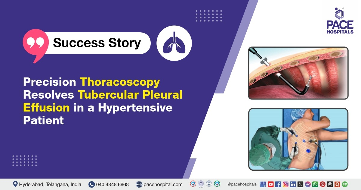
The Breath Restored: Navigating Complex Pleural Tuberculosis with Thoracoscopic Precision
PACE Hospitals
The pulmonology team at PACE Hospitals successfully performed a medical thoracoscopy under general anaesthesia for a middle-aged male patient with persistent tubercular pleural effusion. The procedure enabled precise diagnosis, effective drainage, and adhesiolysis, resolving the chronic pleural inflammation and setting the patient on a clear path to recovery while preventing further pulmonary complications.
Chief Complaints
A middle-aged male presented to the Pulmonology Department at
PACE Hospitals, Hitech City, Hyderabad, with a two-week history of persistent high-grade fever, accompanied by chills, rigors, and noticeable shortness of breath during exertion. The symptoms had progressively worsened, limiting the patient’s daily functioning and prompting evaluation beyond empirical outpatient treatment.
Medical History
The patient had a history of systemic hypertension that was well-managed with daily antihypertensive medication. He had recently undergone ophthalmologic surgery and had no prior diagnoses of
diabetes mellitus,
thyroid dysfunction,
tuberculosis, or
COVID-19. His long-standing occupation in the coal mining sector, with over three decades of exposure to fine coal dust, was a notable risk factor for chronic pulmonary disorders. Additionally, he was a chronic smoker. He reported no known allergies and had completed the
COVID-19 vaccination.
On Examination
At the time of admission, the patient was alert, oriented, and febrile, with a recorded temperature consistent with active infection. Oxygen saturation was within acceptable limits. On auscultation, there was a significant reduction in breath sounds on the right side, particularly in the infra-axillary and infra-scapular regions, suggesting unilateral pleural involvement. Cardiovascular and abdominal examinations did not reveal any abnormalities, and there were no signs of peripheral lymphadenopathy or upper respiratory tract inflammation.
Diagnosis
Radiological evaluation, including chest ultrasound, confirmed the presence of a moderate to large right-sided pleural effusion with basal lung collapse. Laboratory investigations revealed a lymphocyte-predominant exudative effusion. The patient had been recently diagnosed with tubercular pleural effusion at an external facility and had been started on empirical anti-tubercular therapy, but symptoms persisted despite initial management.
Medical thoracoscopy was planned for direct visual inspection, therapeutic drainage, and targeted biopsy. This intervention confirmed multiple loculations within the pleural cavity and an organizing empyema pattern. Histopathological examination of pleural biopsies revealed granulomatous inflammation consistent with tuberculosis. Molecular testing confirmed the presence of Mycobacterium tuberculosis, and pleural fluid culture identified a secondary infection with Acinetobacter baumannii, a gram-negative bacterium known for hospital-acquired resistance. There was no resistance to first-line antitubercular agents.
Medical Decision Making (MDM)
In light of the persistent symptoms, radiological evidence of complex effusion, and the need for diagnostic tissue sampling, the decision was made to proceed with Medical Thoracoscopy in Hyderabad at PACE hospitals. This procedure was selected for its dual benefit offering visual access to the pleural space and enabling effective adhesiolysis and fluid evacuation. It also allowed for comprehensive microbiological and histopathological sampling to refine the diagnosis and direct therapy.
The intervention was led by Dr. Pradeep Kiran Panchadi, Consultant Interventional Pulmonologist. Given the complexity of the case, a multidisciplinary approach was adopted to ensure holistic and safe management. A cross-consultation with Dr. S. Pramod Kumar, Consultant Neurophysician, was sought to evaluate the patient's complaints of tingling and numbness in the thigh, suggestive of a neurological component. Additionally, Dr. Suresh Kumar Sepuri, Consultant Surgical Gastroenterologist & Bariatric Surgeon, was involved intraoperatively to assist with anticipated adhesions and provide surgical support during thoracoscopic intervention.
Surgical Procedure
Medical thoracoscopy was performed under general anaesthesia in a sterile operating room setting. Two ports were placed strategically on the right thoracic wall, facilitating access to the pleural space. Upon entry, the thoracoscopic view revealed dense adhesions and multiple loculated pockets of fluid.
Adhesiolysis was meticulously carried out, and approximately one litre of straw-coloured pleural fluid was drained. Several biopsy samples were collected from the posterior parietal pleura for diagnostic confirmation. Hemostasis was secured, and a large-bore intercostal drainage tube was positioned to allow continuous fluid evacuation during the postoperative period. The patient tolerated the procedure well and was extubated in the operating room.
Postoperative Care
The patient was monitored closely in the inpatient ward following the procedure. Over the subsequent days, his fever subsided, respiratory symptoms improved, and the output from the pleural drain decreased significantly. The drain was removed once the fluid collection had stabilized.
During his hospital stay, the patient received intravenous antibiotics tailored to the sensitivity of the cultured bacteria, systemic anti-inflammatory agents, nebulized bronchodilators, and supportive therapy. Neurological evaluation was performed for symptoms of localized tingling and numbness in the thigh region, which were diagnosed as peripheral neuropathy and managed conservatively with neuropathic agents and vitamin support.
Discharge Medications
At discharge, the patient was advised to continue a structured pharmacological regimen. This included a standard multi-drug anti-tubercular therapy regimen, an oral broad-spectrum antibiotic for bacterial coverage, a neuropathic pain modulator, and dietary supplements including vitamin B complex and protein supplements. A proton pump inhibitor was prescribed for gastric protection, and his regular antihypertensive medication was continued without interruption. The selection and duration of each medication class were based on current clinical guidelines and individualized risk assessment.
Dietary Advice
Nutritional guidance emphasized a high-protein, low-sodium diet to support immune function, tissue repair, and cardiovascular health. Maintaining hydration was equally emphasized to facilitate metabolic clearance and reduce the risk of drug toxicity, particularly hepatotoxicity associated with antitubercular medications.
Advice on Discharge
The patient was discharged in a stable condition with comprehensive home care instructions. These included adherence to medication timing and dosage, adequate rest for the initial recovery period, and attention to signs of possible complications such as jaundice, persistent vomiting, rashes, joint swelling, or visual disturbances.
Suture removal was scheduled for a few days post-discharge, and the patient was advised to avoid strenuous physical activity for two weeks. He was also instructed to monitor for adverse effects of antitubercular therapy, and routine liver function testing along with uric acid levels was recommended every two weeks during the intensive phase of treatment.
Follow-Up
The patient was advised to return for a follow-up appointment with the Interventional Pulmonologist in Hyderabad at PACE Hospitals after 7 days, to be reviewed in the outpatient pulmonology department (OPD).
Conclusion
This case highlights the importance of early thoracoscopic intervention in diagnosing and managing complex pleural tuberculosis. It demonstrates how minimally invasive techniques, supported by a multidisciplinary team, can lead to accurate diagnosis and effective treatment. The outcome reinforces the value of advanced Lung Disease Treatment in Hyderabad, India at PACE Hospitals.
Bridging Diagnosis and Recovery Through Thoracoscopic Intervention
In this case, medical thoracoscopy played a pivotal role in shifting the course of care from uncertainty to recovery. The patient had ongoing fever and breathlessness despite initial fluid drainage and empiric treatment. Imaging suggested persistent pleural effusion with possible loculations, prompting the pulmonologist/pulmonology doctor to make the decision to perform thoracoscopy not as a last resort, but as a timely diagnostic and therapeutic step.
The procedure revealed dense adhesions and multiple loculated pockets of fluid, which were successfully lysed. Targeted biopsies and fluid samples confirmed tuberculosis with a secondary bacterial infection. This enabled the clinical team to tailor therapy precisely, combining anti-tubercular treatment with appropriate antibiotics.
Thoracoscopy served as the turning point resolving symptoms, clarifying diagnosis, and preventing further complications. Its use early in the care pathway exemplified the value of minimally invasive interventions in complex pleural disease, especially when supported by multidisciplinary collaboration.
Share on
Request an appointment
Fill in the appointment form or call us instantly to book a confirmed appointment with our super specialist at 04048486868
Appointment request - health articles
Recent Articles