Successful Percutaneous Cystolithotripsy for Bladder Stone in an Elderly Male, Enhancing Urinary Function
PACE Hospitals
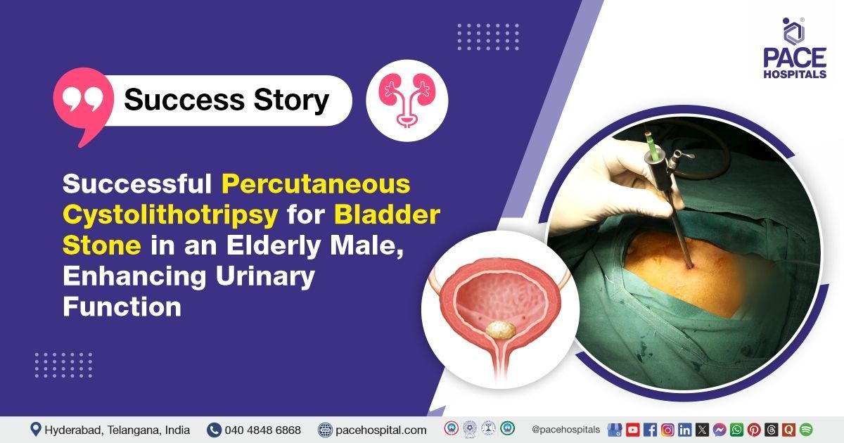
The expert Urology team at PACE Hospitals successfully performed a percutaneous cystolithotripsy (PCCLT) with suprapubic catheter placement on a 71-year-old male patient who had presented with left flank pain. He was referred from a rehabilitation centre and was on a tracheostomy tube, receiving oxygen therapy, and supported with low-dose inotropic medication. The procedure was done to remove a bladder stone, which helped ease his symptoms and improve his urine flow, supporting his overall recovery.
Chief Complaints
A 71-year-old male patient was referred from a rehabilitation center to the Urology Department at
PACE Hospitals, Hitech City, Hyderabad, with complaints of localized and persistent left flank pain. The pain was not associated with any urinary symptoms. Given the nature and duration of his symptoms, a comprehensive urological evaluation was undertaken to investigate a possible underlying renal pathology.
Past Medical History
The patient had a history of recurrent urinary tract infections (UTIs), some of which were complicated by episodes of urosepsis, reflecting a severe and recurring pattern of infection. He was dependent on a tracheostomy tube, receiving supplemental oxygen was supported by mild inotropic management and had a known history of hypertension and a prior cerebrovascular accident (CVA), which resulted in significant neurological deficits and left him chronically bedridden.
His prolonged immobility, combined with impaired neurological function, likely contributed to poor bladder emptying and limited personal hygiene, both recognized risk factors for recurrent UTIs (urinary tract infections). These factors created an environment conducive to bacterial colonization and infection, leading to repeated episodes of sepsis and further worsening of his overall health.
On Examination
Upon admission, the patient was on a tracheostomy tube, receiving supplemental oxygen, and was maintained on mild inotropic support. He was afebrile with stable vital signs and was noted to be chronically bedridden due to a previous cerebrovascular accident. The patient was conscious, alert, and showed no signs of acute distress. Physical examination revealed mild tenderness over the left flank, consistent with his reported pain. No palpable abdominal mass was detected, and there were no signs of peritonism, such as guarding, rebound tenderness, or rigidity, on abdominal examination.
The findings indicated a localized renal or urological issue, consistent with the patient's history of recurrent UTIs and left flank pain. In the absence of systemic or abdominal signs, the medical team suspected vesical calculi as the likely underlying cause.
Diagnosis
Following the initial examination, the patient underwent a thorough review of his medical history and a detailed clinical examination by the urology team. Based on his presenting symptoms and initial examination, there was a strong clinical suspicion of a vesical calculus. These findings warranted further diagnostic investigations, which confirmed the diagnosis and led to the decision to proceed with the percutaneous cystolithotripsy (PCCLT) with suprapubic catheter (SPC) placement.
To support the diagnosis and evaluate the extent of any underlying complications, several investigations were performed, including:
A CT scan of the kidneys, ureters, and bladder (CT KUB) played a crucial role in identifying the source of the patient's symptoms. In this case, the imaging revealed the presence of vesical calculi, which correlated with the patient's history of recurrent urinary tract infections and chronic lower urinary tract obstruction. Additionally, no evidence of hydronephrosis or ureteric obstruction was noted, further confirming that the bladder stones were likely contributing to his symptoms. The CT findings provided critical insight into the urological pathology and guided further management decisions.
Hematological investigations showed anisocytosis with microcytic hypochromic RBCs and pencil cells, indicating iron deficiency anemia. WBCs showed neutrophil predominance, suggesting infection. Mild thrombocytopenia with giant platelets was also noted, likely to reflect a reactive process.
Based on the confirmed diagnosis, the patient was advised to undergo Vesical Calculus Treatment in Hyderabad, India, under the expert care of the Urology Department. This comprehensive management plan aimed to address his condition, minimize the risk of future complications, and improve his overall quality of life through timely surgical intervention and thorough post-operative care.
Medical Decision-Making (MDM)
After a thorough consultation with Dr. Kataradu Ravichandra, Consultant Laparoscopic Urologist, Andrologist & Kidney Transplant Surgeon, a comprehensive evaluation was conducted to determine the most appropriate diagnostic and therapeutic approach for the patient.
Considering his persistent left flank pain, history of recurrent urinary tract infections, and his existing medical condition—including a tracheostomy tube and dependence on supplemental oxygen, the medical team concluded that percutaneous cystolithotripsy (PCCLT) with suprapubic catheter (SPC) placement would be the most suitable and effective intervention.
This approach aimed to relieve his symptoms and prevent further complications. The patient and his family were thoroughly counselled regarding his condition, its benefits, potential risks, and the necessity of surgical intervention to avoid further complications and preserve renal function.
Surgical Procedure
Following the decision, the patient was scheduled for Percutaneous Cystolithotripsy Surgery in Hyderabad at PACE Hospitals with suprapubic catheter (SPC) placement under the expert supervision of the Urology Department, ensuring optimal care and a smooth recovery process. The following steps were carried out during the procedure:
- Anesthesia and Preparation: The patient was taken to the operating room and administered general anesthesia, considering his existing medical condition. Standard aseptic precautions were followed throughout the procedure.
- Diagnostic Cystoscopy: An initial cystoscopy was performed to evaluate the bladder and confirm the presence of calculi. Multiple vesical stones were visualized within the bladder. The prostate was also assessed and noted to be Grade 1 and non-obstructive, with no evidence of contributing to urinary outflow obstruction.
- Suprapubic Access: Following cystoscopy, a suprapubic puncture was made to gain direct access to the bladder. A small skin incision was made above the pubic symphysis, and a tract was established using a guidewire and dilators under cystoscopic guidance.
- Stone Fragmentation (Lithotripsy): A nephroscope (a tubular medical instrument used to examine a kidney through a small skin incision) was introduced through the suprapubic tract into the bladder. Vesical calculi were fragmented using a lithotripter (a medical device uses shock wave to break down kidney stones) and all visible stone fragments were evacuated using forceps and irrigation.
- SPC (Suprapubic Catheter) Placement: A suprapubic catheter was inserted through the established tract to ensure continuous bladder drainage and prevent urinary retention during the recovery period.
- Hemostasis and Wound Closure: Bleeding was controlled, and the suprapubic wound was dressed appropriately.
Postoperative Care
The procedure was uneventful, and the patient had a smooth postoperative recovery, with no complications observed during the post-surgical period. During hospitalization, he was closely monitored and administered intravenous fluids to maintain adequate hydration and support overall recovery.
Intravenous antibiotics were given as a prophylactic measure to prevent postoperative infections, considering his history of recurrent UTIs (urinary tract infections) and compromised general condition. Additional supportive medications, including analgesics and antispasmodics, were also provided to manage discomfort and promote healing.
Discharge Medications
At the time of discharge, the patient was advised to continue alpha-blockers as part of his ongoing urological management, particularly to support bladder emptying and reduce lower urinary tract symptoms. In addition, he was instructed to resume his previous medications for chronic conditions, including antihypertensives and other supportive therapies. This comprehensive discharge plan was tailored to his medical history and current recovery status, aiming to prevent recurrence of urinary complications and ensure overall stability of his health.
The patient was advised to continue bladder irrigation for two days or until hematuria resolved, to prevent clot formation, maintain catheter patency, and support bladder healing post-procedure.
Emergency Care
The patient was informed to contact the Emergency ward at PACE Hospitals in case of any emergency or development of symptoms such as fever, hematuria, or dysuria.
Review and Follow-Up
The patient was advised to follow up with the Urologist in Hyderabad at PACE Hospitals, after 4 days for per-urethral catheter removal and to monitor signs of infection or discomfort while maintaining adequate hydration.
Conclusion
The case demonstrates the successful management of vesical calculi in a high-risk patient through percutaneous cystolithotripsy (PCCLT) with suprapubic catheter (SPC) placement. The procedure was completed without complications, leading to symptom relief and improved recovery, highlighting the effectiveness of timely surgical intervention and coordinated care.
Interconnected Pathways: Understanding and Managing Ureteric and Vesical Calculi
Ureteric and vesical calculi, though located in different parts of the urinary tract, are often interconnected due to shared risk factors such as dehydration, recurrent infections, and urinary stasis. A ureteric stone may originate in the kidney and travel to the bladder, where it can develop into a vesical calculus if not expelled. Bladder outlet obstruction or neurogenic bladder can contribute to stone formation in both locations. Under the guidance of a urologist/urology doctor, diagnosis is typically confirmed through imaging such as CT KUB, followed by appropriate surgical interventions—ureteroscopy with laser lithotripsy for ureteric stones, and percutaneous cystolithotripsy (PCCLT) for vesical stones. In some cases, combined or staged procedures may be necessary, along with catheter placement for urinary drainage.
Share on
Request an appointment
Fill in the appointment form or call us instantly to book a confirmed appointment with our super specialist at 04048486868


