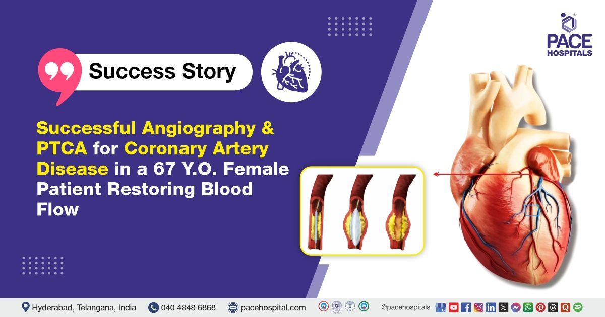
Successful Angiography & PTCA for Coronary Artery Disease in a 67-Year-Old
PACE Hospitals
The expert cardiology team at PACE Hospitals successfully performed coronary angiography (CAG), followed by percutaneous transluminal coronary angioplasty (PTCA) to the left circumflex artery (LCX) and right coronary artery (RCA), on a 67-year-old female patient who presented with complaints of chest pain, profuse sweating, and shortness of breath. The procedure aimed to restore proper blood flow to the heart muscle, relieve symptoms, and lower the risk of future cardiac events.
Chief Complaints
A 67-year-old female patient presented with
BMI of 23.2 presented to the cardiology Department at
PACE Hospitals, Hitech City, Hyderabad, with sudden-onset chest pain that had been ongoing for the past two days, accompanied by sweating, shortness of breath, and palpitations. She denied any history of orthopnea (shortness of breath that occurs when a person is lying flat and is relieved by sitting or standing up). The nature and persistence of her symptoms raised concern for possible cardiac involvement, prompting further clinical evaluation.
Past Medical History
The patient's medical history was unremarkable, with no prior history of
diabetes,
hypertension, or renal disease. She denied any previous surgical interventions or significant chronic illnesses. This was her first episode of cardiac-related symptoms, which warranted a thorough diagnostic workup to determine the cause of her chest pain and associated complaints.
On Examination
Upon admission to PACE Hospitals, the patient's vital signs were stable. General examination revealed normal findings. Cardiovascular examination showed normal heart sounds (S1 and S2). The central nervous system was non-focal, with no neurological deficits. Respiratory examination demonstrated bilateral air entry with normal vesicular breath sounds. Abdominal examination revealed a soft, non-tender abdomen with no evidence of organomegaly. These findings supported her initial clinical stability despite the presenting cardiac symptoms.
Diagnosis
After the initial examination, the patient underwent a detailed evaluation by the cardiology team, including clinical assessment, lab tests, and cardiac imaging to determine the extent of cardiac involvement.
Following a comprehensive physical examination, the patient was found to have experienced sudden-onset chest pain that had persisted for the past two days, accompanied by profuse sweating, shortness of breath, and palpitations. Diagnostic investigations were conducted to evaluate a possible cardiac cause. An electrocardiogram (ECG) revealed ischemic changes suggestive of myocardial involvement. A two-dimensional echocardiogram (2D ECHO) showed regional wall motion abnormalities, indicating impaired cardiac function. Coronary angiography (CAG) confirmed coronary artery disease (CAD) involving both the left circumflex (LCX) and right coronary artery (RCA), diagnosed as double vessel disease (DVD), establishing the diagnosis and guiding further management.
Based on these confirmed findings, the patient was advised to undergo Coronary Artery Disease Treatment in Hyderabad, India, under the expert care of the Cardiology Department. Timely intervention was recommended to restore adequate blood flow to the heart, reduce cardiac workload, and prevent potential complications such as myocardial infarction or heart failure, thereby improving overall clinical outcomes.
Medical Decision Making (MDM)
After a thorough consultation with Dr. Sashi Vardhan Janjirala, a Cardiologist, a comprehensive evaluation was undertaken to formulate an appropriate diagnostic and therapeutic plan tailored to the patient’s cardiac condition. Given her symptoms, along with initial findings suggestive of coronary artery disease, the evaluation aimed to accurately assess the extent and severity of the condition. The goal was to initiate timely and effective treatment to prevent further cardiac complications and improve the patient’s overall prognosis.
The patient and her family were thoroughly counselled regarding her condition, the recommended coronary angiogram (CAG) and percutaneous transluminal coronary angioplasty (PTCA) to the left circumflex artery (LCX) and right coronary artery (RCA), including its benefits, potential risks, and the necessity of surgical intervention to avoid further complications and preserve cardiac function.
Surgical Procedure
Following the diagnosis of double vessel disease involving the LCX and RCA, the patient was scheduled for an Angioplasty Procedure in Hyderabad at PACE Hospitals, under the expert supervision of the Cardiology Department, ensuring optimal care and a smooth recovery process.
Percutaneous transluminal coronary angioplasty (PTCA) was performed as a primary intervention for both affected vessels. The procedure involved the following steps:
- Preparation: The patient was stabilized, and informed consent was obtained.
- Catheterization: A catheter was inserted via the radial artery to access the coronary circulation.
- Angiographic Imaging: The coronary vessels were visualized, confirming the LCX and RCA blockages.
- Balloon Angioplasty: A balloon catheter was advanced to the sites of stenosis in both arteries and inflated to widen the narrowed areas.
- Stenting: Drug-eluting stents were deployed in both the left circumflex artery (LCX) and right coronary artery (RCA) to ensure long-term patency and prevent restenosis.
- Post-Procedure Assessment: The angiography confirmed successful reopening of both vessels with good flow, and no immediate complications were noted.
Postoperative Care
The patient’s postoperative recovery was smooth and uneventful, with no complications observed during the immediate postoperative period. After the procedure, she was closely monitored in the intensive care unit (ICU) for six hours to ensure stable hemodynamic status and early detection of any potential complications.
During this period, her vital signs, including heart rate, blood pressure, and oxygen saturation, were carefully observed, and any necessary adjustments to medications or interventions were promptly made. The patient responded well to postoperative care, with no signs of adverse events, and was later transferred to a regular ward for continued recovery.
Discharge Medications
Upon discharge, the patient was prescribed a combination of medications to aid recovery and prevent complications. Oral antiplatelets were given to prevent blood clots and ensure proper blood flow. Analgesics were provided for pain relief. Proton pump inhibitors were prescribed to protect the stomach from irritation when used with antiplatelet medications and NSAIDs.
Benzodiazepines were prescribed to manage anxiety, while NSAIDs were used to relieve mild pain and inflammation, with careful monitoring for side effects. Additional supportive treatments were prescribed to manage blood pressure and cholesterol, reducing the risk of future cardiovascular events.
Emergency Care
The patient was informed to contact the Emergency ward at PACE Hospitals in case of any emergency or development of symptoms such as chest pain, abdominal pain, and fever.
Review and Follow-Up
The patient was advised to return for a follow-up visit with the Cardiologist in Hyderabad at PACE Hospitals after 10 days to assess her recovery from the angioplasty and stenting procedure. The visit helped evaluate the effectiveness of the treatment, monitor for any complications, and adjust her care plan as needed for optimal recovery and long-term heart health.
Conclusion
This case highlights the effectiveness of PTCA in Heart Disease Treatment in Hyderabad, India, emphasizing its ability to improve blood flow and restore heart function.
The Role of Angiography in the Diagnosis and Management of CAD and Double Vessel Disease (DVD)
Angiography played a crucial role in managing coronary artery disease (CAD) and double vessel disease (DVD) by providing a detailed visual representation of the coronary blood vessels. This allowed for accurate diagnosis, risk stratification, and guidance for treatment. The cardiologist/heart specialist reviews the angiographic images and confirms the presence of heart disease by clearly identifying any narrowing or blockages in the coronary arteries. By assessing the extent and location of atherosclerosis, angiography helped determine the severity of the disease, which was critical in evaluating the patient’s risk for future heart attacks and other cardiac events. This information guided the decision to proceed with percutaneous coronary intervention (PCI) to restore blood flow and prevent further complications.
Share on
Request an appointment
Fill in the appointment form or call us instantly to book a confirmed appointment with our super specialist at 04048486868
Appointment request - health articles
Recent Articles