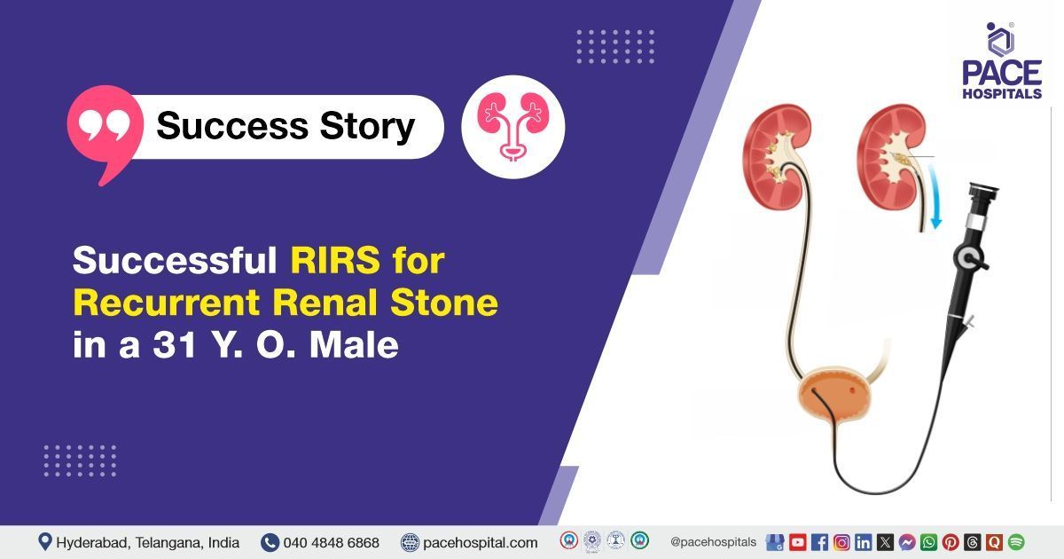
Successful RIRS for Recurrent Kidney Stone in a 31-Year-Old Male
PACE Hospitals
The Urology team at PACE Hospitals successfully performed Right Retrograde Intrarenal Surgery (RIRS) with DJ stenting on a 31-year-old Male who presented with the complaint of right flank pain. The procedure removed the renal stone, alleviated symptoms, and promoted optimal renal function and recovery.
Chief Complaints
A 31-year-old male with a
BMI of 23.4 presented to the Urology Department at
PACE Hospitals, Hitech City, Hyderabad, with complaints of persistent right flank pain that had been ongoing for several days. He did not report any associated urinary symptoms such as dysuria, haematuria, nausea, or vomiting. In view of the localized and persistent nature of his symptoms, an urological evaluation was initiated, leading to the diagnosis of a right renal calculus.
Past Surgery
The patient’s medical history was unremarkable, with no known comorbidities or prior urological interventions. Notably, a non-obstructing renal calculus had previously been identified in the left kidney, however, as it was not causing any urinary obstruction or related symptoms, no intervention was deemed necessary at the time.
This episode represented his first presentation with flank pain, prompting a comprehensive diagnostic evaluation.
On Examination
On admission, the patient was afebrile with stable vital signs, and his general condition was satisfactory. Physical examination revealed mild right flank tenderness on deep palpation, correlating with his primary complaint. There were no palpable masses or signs of peritoneal irritation, and the systemic examination was otherwise unremarkable, suggesting a localized urological pathology.
Diagnosis
The patient initially underwent a comprehensive clinical assessment at PACE Hospitals, which included a detailed history and physical examination by the Urology team. During the evaluation, it was noted that the patient had a previously diagnosed non-obstructing renal calculus on the left side, which had been managed medically at the time and did not require surgical intervention.
Recently, however, the patient developed right-sided flank pain, raising clinical suspicion for residual or recurrent renal calculi or related complications on the right side. To investigate further, the patient underwent right-sided ureteroscopy (URS) with DJ stent placement. During the procedure, the presence of right renal calculi was confirmed, which was determined to be the source of the patient's ongoing symptoms.
Other diagnostic investigations suggestive of renal calculi include:
- Urinalysis: The urinalysis showed significant abnormalities, including plenty of red blood cells (RBCs/HPF) and leukocytes (125/µL), along with positive blood (+++). These findings are consistent with microscopic haematuria and suggest possible mucosal irritation or mild urinary tract inflammation. The presence of RBCs and leukocytes in the urine indicates an ongoing irritative or inflammatory process in the urinary tract, which could be related to stone-related irritation or post-procedural inflammation from the prior DJ stenting.
- Urine Culture and Sensitivity: A urine culture revealed no bacterial growth, confirming that the urine was sterile at the time of testing. This is a crucial finding as it indicates that there is no active infection, making surgical interventions such as RIRS safer to perform.
- Complete Blood Count (CBC): The CBC showed a neutrophil predominance, which could reflect a localized inflammatory response, likely related to stone disease or prior interventions. However, the total WBC count was within normal limits, suggesting no systemic infection but indicating localized inflammation.
- C-Reactive Protein (CRP): The CRP was significantly elevated, indicating the presence of an ongoing inflammatory process, which could be secondary to stone irritation or post-procedural inflammation from the DJ stenting.
- Renal Function Tests: The serum creatinine (0.9 mg/dL) and electrolytes were within normal limits, confirming that the patient's renal function was well-preserved, which is a positive finding when planning for surgical interventions such as RIRS.
- Coagulation Profile: Prothrombin Time (PT) and International Normalized Ratio (INR) were within normal limits, indicating no significant bleeding risk and confirming the patient’s suitability for RIRS.
Collectively, the diagnostic findings confirmed the presence of a moderate-sized calculus in the right renal pelvis, which was responsible for the patient’s persistent flank pain, despite the absence of overt obstruction or signs of infection. This supported the decision to proceed with definitive surgical management through RIRS.
Based on the confirmed findings, the patient was advised to undergo Kidney Stone Treatment in Hyderabad, India, under the expert care of the Urology Department, providing effective stone clearance, alleviating his flank pain, and preventing potential complications such as obstruction, infection, or renal function impairment.
Medical Decision-Making (MDM)
Following a detailed consultation with Dr. Abhik Debnath, Consultant Laparoscopic Urologist, a thorough evaluation of the patient's condition was carried out. Given the persistence of right flank pain and ureteroscopic findings revealing a moderate-sized calculus in the right renal pelvis, the urology team concluded that Right Retrograde Intrarenal Surgery (RIRS) with DJ stenting represented the most appropriate course of treatment.
This intervention was selected to ensure complete removal of residual or recurrent stone fragments and to address any anatomical or inflammatory changes resulting from prior procedures. Additionally, it aimed to provide symptomatic relief and reduce the risk of future complications such as infection, obstruction, or decline in renal function. The treatment plan was tailored to the patient's clinical profile and prior urological history, ensuring a targeted and effective approach.
Surgical Procedure
Given the patient's tight ureter, a primary stenting procedure followed Right Retrograde Intrarenal Surgery in Hyderabad at PACE Hospitals was scheduled under the expert care of the Urology Department to ensure optimal treatment and facilitate a smooth recovery.
Following a detailed pre-anaesthetic evaluation and after obtaining informed consent, the patient underwent Right Retrograde Intrarenal Surgery (RIRS) with DJ stenting under general anaesthesia.
During the procedure, mild swelling was found in the right ureter where the stone had initially lodged. A stone in the lower part of the kidney was difficult to break apart, but the doctors used a Holmium YAG laser to successfully fragment it. Afterward, all stone fragments were removed, and the ureter was checked to ensure no stones were left behind. The procedure went smoothly, and the patient was monitored for recovery.
Postoperative Care
The Patient underwent the procedure as planned, with his intraoperative and postoperative course being largely uneventful. On Postoperative Day 1 (POD 1), he experienced a mild episode of fever, which resolved without further complications. Following this, the patient’s condition stabilized, and he became asymptomatic. He was discharged in stable condition with specific instructions for follow-up care and management of the stent.
Discharge Medications
At the time of discharge, the patient was prescribed medications aimed at supporting recovery and preventing potential complications. Antibiotics were prescribed to prevent or treat any infections following the surgery, while analgesics were provided to manage pain. To protect the stomach from irritation caused by the medications, a proton pump inhibitor was included in the treatment plan. Additionally, alkalinizing agents were recommended to maintain urinary alkalinity and ease discomfort during urination.
To facilitate proper urine flow and bladder function, alpha blockers and beta-3 agonists were prescribed, addressing the patient's urinary symptoms. For local care, a topical anti-inflammatory ointment was applied to the vein cannulation sites to alleviate swelling and discomfort. A follow-up review has been scheduled for stent removal and cystoscopy to monitor progress and ensure optimal healing.
Discharge Notes
In addition to pharmacological therapy, the patient received detailed dietary and lifestyle guidance. She was advised to maintain adequate hydration with an intake of 3–4 liters of fluids per day and to follow a low-purine, low-sodium diet with an emphasis on citrus fruits and juices to help prevent recurrence of renal calculi.
Activity restrictions included avoiding weight lifting, forward bending, and long-distance travel via bus, auto-rickshaw, or two-wheeler for a defined period. She was also informed that mild flank discomfort, occasional haematuria, or cloudy urine may persist for up to 7–10 days after discharge, which are expected postoperative symptoms.
Emergency Care
The patient was informed to contact the Emergency ward at PACE Hospitals in case of any emergency or development of symptoms like fever, abdominal pain, swelling, bleeding or vomiting.
Review and Follow-up Notes
The patient was advised to return for a follow-up visit with the Urologist in Hyderabad at PACE Hospitals, after 1.5 months for the removal of the DJ stent and to undergo cystoscopy. This review is necessary to assess the patient's recovery, ensure proper healing, and evaluate the urinary tract for any complications or residual issues that may require further intervention.
Conclusion
This case highlights the effectiveness of Retrograde Intrarenal Surgery (RIRS) in performing Renal Calculi Treatment in Hyderabad, India, enabling successful removal of stones in the kidney, thereby facilitating normal urine flow.
The Diagnostic Value of Ureteroscopy (URS) in Managing Renal Calculi
Ureteroscopy (URS) plays a vital role in the diagnosis of renal calculi by allowing direct visualization of the urinary tract, including the ureters and kidneys. This enables precise identification of the stone’s size, location, number, and composition, which may not be clearly detected through imaging alone. URS is particularly useful when radiological findings are inconclusive, providing real-time assessment of the urinary tract. It also helps in identifying associated abnormalities such as ureteral strictures, mucosal inflammation, or anatomical variations that may contribute to stone formation. By offering detailed visual confirmation, URS enhances diagnostic accuracy and supports informed clinical decision-making by a qualified urologist/urology doctor.
Share on
Request an appointment
Fill in the appointment form or call us instantly to book a confirmed appointment with our super specialist at 04048486868
Appointment request - health articles
Recent Articles