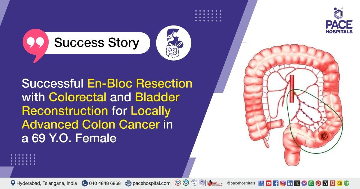
Successful En-Bloc Resection with Colorectal and Bladder Reconstruction for Locally Advanced Colon Cancer in a 69 Y.O. Female
PACE Hospitals
PACE Hospitals’ expert Surgical Gastroenterology and multidisciplinary team successfully managed a 69-year-old female patient diagnosed with well-differentiated adenocarcinoma of the sigmoid colon with bladder involvement. The patient presented with stool and occasional bleeding through the urethra, urinary burning, white discharge, constipation, and significant weight loss. The treatment involved extensive
surgical resection with bladder reconstruction,
urinary tract reconstruction, vascular repair, colorectal anastomosis, and protective ileostomy, followed by structured postoperative care and monitoring.
Chief complaints
A 69-year-old female patient with a
body mass index (BMI) of 15 presented to the Surgical Gastroenterology Department at
PACE Hospitals, Hitech City, Hyderabad with complaints of passing stool through the urethra for approximately 8 months, associated with occasional bleeding through the urethra. She also reported white urinary discharge, burning sensation while passing urine, and constipation for the same duration. During this period, she had significant weight loss of approximately 20 kg.
Past Medical history
The patient had a history of previous hysterectomy. There were no known significant comorbidities or previous history of malignancy or oncological treatment.
On Examination
On examination, the patient was evaluated for her urinary, gastrointestinal, and general symptoms. Clinical assessment was suggestive of significant weight loss with features of abnormal communication between the bowel and urinary tract. The abdominal and pelvic examination was performed to assess for any palpable mass, tenderness, or other abnormalities. Further evaluation was carried out to determine the extent of the colorectal tumour and its involvement with the surrounding pelvic structures.
Diagnosis
Upon admission to PACE Hospitals, following a detailed clinical assessment and review of the patient's medical history, colonoscopy, histopathological findings, and imaging studies, the Surgical Gastroenterology team evaluated her for urinary and gastrointestinal symptoms associated with an underlying colorectal malignancy.
Colonoscopy revealed a circumferential ulcerated growth at the sigmoid colon with critical narrowing, and multiple biopsy samples were obtained. Histopathological examination confirmed well-differentiated adenocarcinoma with prominent villoglandular morphology. PET-CT demonstrated long-segment asymmetric circumferential thickening at the rectosigmoid junction abutting the bladder base, along with positive lymph nodes in the sigmoid and perilesional regions.
Based on the clinical evaluation and diagnostic findings, the patient was diagnosed with well-differentiated adenocarcinoma of the sigmoid colon with rectosigmoid junction and bladder base involvement, indicating locally advanced disease requiring extensive surgical management.
Following confirmation of the diagnosis, the patient was advised to undergo
Sigmoid
Colon Cancer Treatment in Hyderabad, India, along with management of bladder involvement, under the expert care of the multidisciplinary Surgical Gastroenterology and Urology teams.
Medical Decision Making (MDM)
After a detailed consultation with the Dr. Suresh Kumar S, Surgical Gastroenterologist, Bariatric and Metabolic Surgeon, GI and HPB Oncologist, and Liver Transplant Surgeon a comprehensive evaluation was performed to determine the most appropriate surgical approach.
Considering the patient’s history of passing stool and occasional bleeding through the urethra, urinary symptoms, constipation, significant weight loss, and clinical and radiological findings suggestive of well-differentiated adenocarcinoma of the sigmoid colon with bladder involvement, further evaluation confirmed a locally advanced colorectal malignancy involving the adjacent pelvic structures. Colonoscopy, histopathological examination, and PET-CT findings helped establish the diagnosis and determine the extent of tumour involvement.
Based on the clinical assessment, biopsy findings, and imaging studies, it was determined that radical en-bloc resection of the sigmoid colon growth along with subtotal cystectomy and bladder reconstruction was the most appropriate surgical management strategy. The planned approach aimed to achieve complete removal of the tumour along with the involved adjacent structures, while restoring bowel continuity and urinary tract function through colorectal anastomosis, ileostomy creation, bladder reconstruction, bilateral ureteric reimplantation, and other required reconstructive procedures.
The patient and her family members were counselled regarding the diagnosis, extent of disease, planned surgical procedure, need for bowel and urinary reconstruction, expected benefits, potential risks, postoperative catheter and wound care, nutritional requirements, and the importance of regular surgical and urological follow-up.
Surgical Procedure
Following the decision, the patient was scheduled for radical en-bloc resection of the sigmoid colon growth along with subtotal cystectomy and bladder reconstruction in Hyderabad at PACE Hospitals, under the expert care of the Surgical Gastroenterology Department.
The following steps were carried out during the procedure:
- Tumour Assessment and Staging: Cystoscopy and staging laparoscopy were performed to assess the extent of the sigmoid tumour and its involvement with the urinary bladder and surrounding pelvic structures.
- En-bloc Tumour Resection: The sigmoid colon tumour was radically resected along with the involved portions of the adjacent structures, including the affected bladder wall and surrounding tissues, to achieve complete tumour removal.
- Bladder and Urinary Tract Reconstruction: Subtotal cystectomy was performed, followed by augmentation ileocystoplasty, bladder neck repair, and bilateral ureteric reimplantation to reconstruct the urinary tract. A suprapubic catheter was also placed for urinary drainage.
- Vascular and Bowel Reconstruction: The involved right external iliac vein was repaired using patch reconstruction. Colorectal anastomosis was then performed to restore bowel continuity, along with creation of a protective ileostomy.
- Omental Patching and Postoperative Drainage: Greater omental patching was performed as part of the reconstruction. Appropriate urinary drainage was maintained with the suprapubic and Foley catheters, followed by close postoperative monitoring and supportive care.
Postoperative Care
Following surgery, the patient was closely monitored for recovery and any early postoperative complications. Measures were taken to prevent infection, control postoperative pain, reduce the risk of blood clots, protect the stomach, and provide adequate nutritional support for healing. Regular bladder irrigation was performed through the suprapubic and Foley catheters to maintain urinary drainage and bladder hygiene. Urine output, catheter function, bowel activity, surgical wounds, and overall clinical condition were monitored regularly. Blood transfusion was administered when clinically indicated to manage blood loss or maintain adequate haemoglobin levels.
Discharge Medications
At discharge, the patient was advised to continue the prescribed medications to prevent infection, control postoperative pain, reduce the risk of blood clots, provide gastric protection, and support recovery following the extensive colorectal and bladder surgery. She was also advised to continue the prescribed supportive measures for bowel function and overall nutritional recovery, along with the required catheter and bladder care as instructed by the treating team.
Emergency Care
The patient was advised to contact the
emergency ward at PACE Hospitals in case of any emergency or development of symptoms such as fever, severe or worsening abdominal pain, persistent vomiting, abdominal distension, reduced or absent urine output, blood in urine, abnormal catheter drainage, bleeding, wound discharge, or any sudden deterioration in her condition.
Review and Follow-up Notes
The patient was advised to return for a follow-up visit with the Surgical Gastroenterologist in Hyderabad at PACE Hospitals after one week to assess postoperative recovery and wound healing.
Conclusion
This case highlights the successful surgical management of well-differentiated sigmoid colon adenocarcinoma with extensive bladder and adjacent pelvic structure involvement. The patient underwent en-bloc tumour resection with bladder reconstruction and bowel and urinary tract reconstruction, followed by satisfactory postoperative recovery and discharge with detailed care instructions.
Management of Colorectal Cancer with Bladder Involvement
When colorectal cancer extends into the urinary bladder, treatment requires careful assessment of the tumour’s local spread and involvement of nearby structures. An en-bloc resection may be considered in selected patients to remove the primary tumour and directly involved organs together. A Surgical gastroenterologist / Surgical gastroenterology doctor may coordinate with urologists, oncologists, and other specialists to plan appropriate treatment.
Depending on the extent of bladder and urinary tract involvement, reconstruction may be required to restore urinary drainage. Bowel reconstruction or temporary diversion may also be performed when necessary to support healing. Such complex procedures require detailed planning, multidisciplinary coordination, and close postoperative monitoring, followed by regular follow-up and further cancer management when indicated.
Frequently Asked Question
Why was the colon tumour removed together with part of the bladder?
When a colon cancer grows into a nearby organ such as the bladder, separating the tumour from that organ may leave cancer cells behind. In such situations, the surgeon may remove the affected structures together in one operation, known as en-bloc resection, to achieve complete tumour removal.
Can the bladder function normally after part of it is removed?
In many suitable patients, the bladder can be reconstructed after removing the involved portion. Depending on the extent and location of the tumour, a segment of the small intestine may be used to increase or reconstruct the bladder. Urinary function is monitored closely during recovery.
Why were both ureters reimplanted into the reconstructed bladder?
The ureters carry urine from the kidneys to the bladder. If the tumour or surgery involves the area where the ureters normally enter the bladder, they may need to be reattached to the reconstructed bladder. This allows urine to continue flowing from the kidneys into the bladder.
Why was an ileostomy created after the bowel was joined?
After the diseased part of the colon is removed and the bowel ends are joined, the new connection needs time to heal. A temporary ileostomy diverts stool through an opening in the abdomen, reducing the amount of stool passing through the healing bowel connection.
Can the ileostomy be closed later?
In many cases, a protective ileostomy is temporary. Once the bowel connection has healed and the patient has recovered sufficiently, the surgical team may consider closing the stoma. The timing depends on healing, overall health, and the patient's treatment plan.
How long will the urinary catheter remain after bladder reconstruction?
The catheter is usually kept in place until the reconstructed bladder and urinary connections have had adequate time to heal. The exact duration differs from patient to patient and is decided by the urology team based on recovery and follow-up findings.
Will chemotherapy be required after surgery?
Not every patient requires chemotherapy after surgery. The need for additional treatment depends mainly on the final biopsy report, lymph-node involvement, tumour stage, surgical margins, and other features of the cancer. The oncology team decides whether further treatment is appropriate after reviewing the complete pathology report.
What follow-up is needed after this type of surgery?
Regular follow-up is important to check healing and monitor bowel, bladder, urinary, and stoma function. The treating team may also recommend periodic blood tests, imaging, colonoscopy, and cancer surveillance based on the final stage and pathology findings.
Which symptoms should be reported after discharge?
The patient should contact the treating team if she develops fever, increasing abdominal or pelvic pain, repeated vomiting, abdominal swelling, reduced urine output, blood in the urine, catheter problems, wound discharge, or any unusual changes around the stoma.
How long does recovery take after bowel and bladder reconstruction?
Recovery time differs from one patient to another and depends on the extent of surgery and overall health. Bowel and urinary functions may take some time to return to normal, while catheter and stoma care may be needed during the early recovery period.
Share on
Request an appointment
Fill in the appointment form or call us instantly to book a confirmed appointment with our super specialist at 04048486868
Appointment request - health articles
Recent Articles