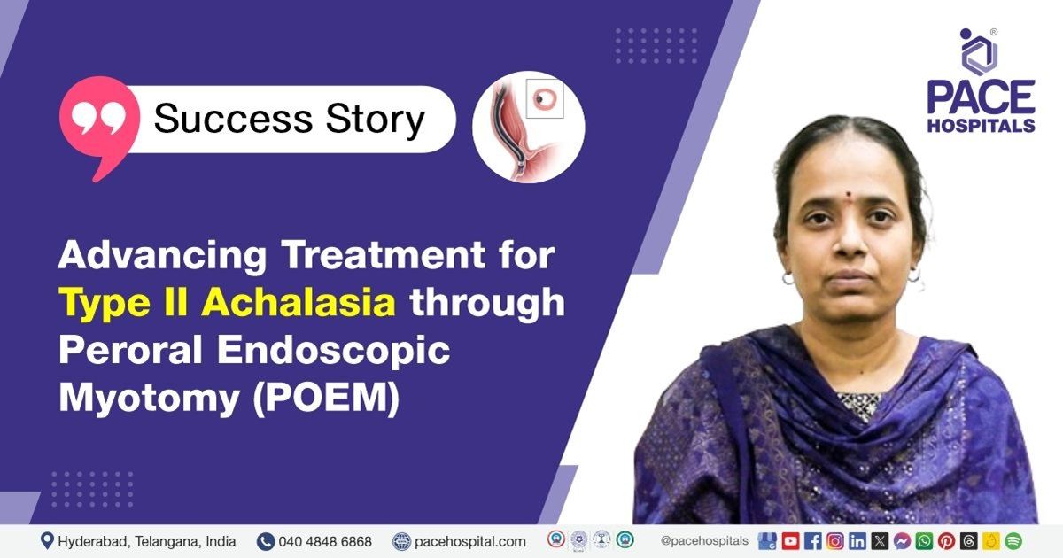
Advancing Treatment for Type II Achalasia through Peroral Endoscopic Myotomy
PACE Hospitals
PACE Hospitals' Gastroenterology team successfully performed a POEM (Peroral Endoscopic Myotomy) procedure on a 41-year-old female with achalasia cardia (Eckardt’s score: 9), improving her ability to eat and swallow comfortably.
Medical History
A 41-year-old female patient approached the gastroenterology department of PACE Hospitals, Hyderabad, with chief complaints of difficulty swallowing and chest tightness for 3 years and was diagnosed outside as Achalasia cardia type II. The patient also appeared to have lost weight and had a history of regurgitation.
The patient’s physical and systemic examination was normal. Upon evaluating the patient’s medical history, no other existing comorbidities were observed. She was conscious and coherent when she approached the hospital with no pallor or icterus.
Diagnosis
Considering the severity of the patient’s swallowing difficulty, she was admitted to PACE Hospitals, Hyderabad, for further treatment and management of her medical condition. She underwent an endoscopy and a barium swallow test. Endoscopy helps widen the oesophageal lumen and check for food residue in the oesophagus, whereas a barium swallow test assesses the oesophageal motility.
The diagnostic procedures performed, and an oesophageal manometry procedure was done thereafter, which again revealed Achalasia Cardia type 2 with an Eckardt’s score of 9 and median integrated relaxation pressure (IRP) of 19.5 (<15mmHg considered normal).
Treatment
As the patient was diagnosed with achalasia cardia type II with severe difficulty in swallowing, Chief gastroenterologists, hepatologists and therapeutic endoscopists Dr. Govind Verma, affirmed that Peroral Endoscopic Myotomy (POEM) procedure would help the patient recover from dysphagia and provide relief from associated symptoms.
On the day of the procedure, the patient was explained about the disease condition and treatment modality, and consent was obtained from her. Short-acting general anaesthesia was administered as premedication.
An
Upper Gastrointestinal (UGI) endoscopy was done initially to examine the fundus. It appeared normal, but moderate resistance was felt at the gastroesophageal junction at 37 cm. To prevent infection, she was given antibiotics before the myotomy procedure. CO2 monitoring was done continuously throughout the procedure. The mucosa was infiltrated with diluted methylene blue 9 cm above the gastroesophageal junction. Submucosal tunneling was done by an incision in the mucosa with a T- knife. Vessels were coagulated with a coag grasper, and a hybrid knife was used to incise the circular muscle. After the procedure, the tunnel was closed with medorah clips, and the endoscope was passed across the gastroesophageal junction much easier.
Post-procedure
The procedure was successfully done with no complications. The patient was managed with intravenous fluids, antibiotics, proton pump inhibitors and other supportive care during hospitalization. She was strictly kept on ‘nil by mouth’ (NBM) for two days post-procedure. The oral gastrograffin test revealed no evidence of contrast extravasation, and transient retention of contrast was seen in the lower oesophagus. Later patient was discharged with medical advice.
Discharge notes
The patient was stable and well tolerant to liquid diet while she was discharged. She was advised to consume only liquid diet followed by soft diet for the next 5 days. To preserve better motility of the oesophagus, she was objected to consume cool liquids.
With a prior appointment, the patient was asked to get review of the condition by Dr. Phani Krishna Ravula and Dr. Govind Verma in the OPD after a week. However, she was instructed to be admit anytime in the medical emergency ward in case of adverse symptoms like fever, abdominal pain or vomiting.
Oesophageal Manometry and Barium Swallow Comparison
Oesophageal manometry and barium swallow studies are very important for analysing the factors that lead to swallowing disorders. A barium swallow is an X-ray examination in which the patient swallows a thick liquid containing barium, that is used as a contrast medium. Oesophageal manometry measures the pressure and constriction of the muscles located within the oesophagus when swallowing occurs. This test helps in determining specific patterns of muscle activity within the oesophageal region including constriction which is either weak or constrictions that are too strong. On the contrary, catheters can sample various muscle groups and indirectly measure the degree of barium incorporation, providing insight into the contraction level.
Share on
Request an appointment
Fill in the appointment form or call us instantly to book a confirmed appointment with our super specialist at 04048486868
Appointment request - health articles
Recent Articles