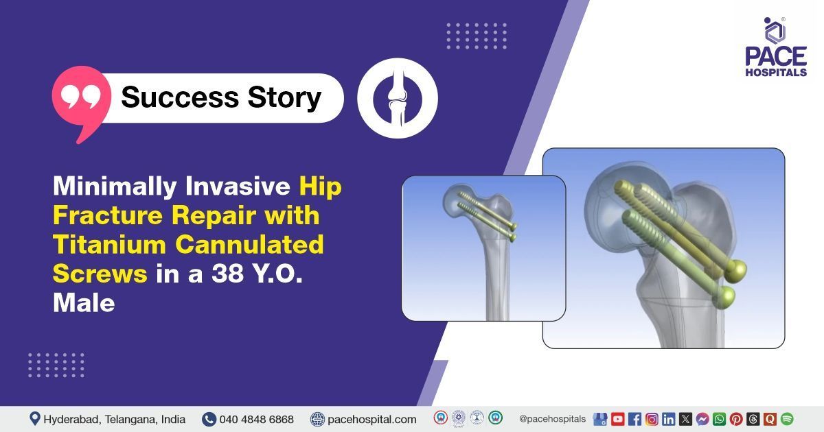
Minimally Invasive Hip Fracture Repair with Titanium Cannulated Screws in a 38 Y.O. Male
PACE Hospitals
The Orthopaedic team at PACE Hospitals successfully performed a percutaneous cannulated cancellous (CC) screw fixation under spinal anaesthesia on a 38-year-old male patient who presented with complaints of right hip pain and inability to bear weight on the right lower limb, thereby aiming to restore joint stability, relieve pain, and enhance his mobility and weight-bearing capacity for improved functional recovery.
Chief Complaints
A 38-year-old male patient with a
body mass index (BMI) of 26.8, presented to the Orthopaedic Department at
PACE Hospitals, Hitech city, Hyderabad, with complaints of pain over the right hip joint, associated with an inability to bear weight on the right lower limb. The symptoms were reported to have developed following an alleged history of trauma.
Past History
The patient had no significant history of known drug allergies or chronic illnesses. The absence of such comorbidities is favourable, as it reduces the risk of perioperative complications and supports a smoother postoperative recovery.
General Examination
Upon admission to PACE Hospitals, the patient's vital signs were stable. On general examination, no abnormalities were noted. Local examination revealed that the right foot was externally rotated. There was tenderness over the right hip joint, and the bitrochanteric compression test was positive. The range of motion at the right hip was restricted and painful, suggesting intra-articular involvement or fracture. No distal neurovascular deficit was noted, indicating preserved blood supply and nerve function to the affected limb.
Diagnosis
Following admission to PACE Hospitals, the patient underwent a comprehensive evaluation, including a detailed medical history review and clinical examination by the Orthopaedic team. Clinical findings raised a strong suspicion of a post-traumatic fracture of the right femoral neck. To confirm the diagnosis, further imaging was performed, including an X-ray and CT scan, which conclusively revealed a post-traumatic fracture of the right femoral neck (a break in the upper part of the right thigh bone near the hip caused by an injury or trauma).
After a thorough clinical and radiological assessment, the patient was advised to undergo Femoral Fracture Treatment in Hyderabad, India, under the expert care of the Orthopaedic Department to ensure comprehensive management.
Medical Decision-Making (MDM)
In view of the patient’s symptoms, functional limitations in performing daily activities, and the confirmed diagnosis of a post-traumatic fracture of the right femoral neck, surgical intervention was deemed necessary. Following a detailed discussion with the patient’s guardians and after obtaining informed consent, Dr. Anand V. Agroya, Consultant Orthopaedic Surgeon, recommended percutaneous cannulated cancellous (CC) screw fixation as the most appropriate and effective treatment option for optimal stabilization and recovery.
Surgical Procedure
Following the decision, the patient was scheduled to undergo Percutaneous Cannulated Cancellous (CC) Screw Fixation Surgery in Hyderabad at PACE Hospitals, under the expert supervision of the Orthopaedic Department to ensure optimal stabilization and promote a successful recovery.
Before surgery, the patient was diagnosed with HBsAg and underwent further tests to assess fitness for the planned procedure. A thorough pre-operative workup was completed, including a pre-anaesthesia checkup and counselling about potential complications and the possibility of requiring hip replacement. Following this, the patient was scheduled for surgery.
As planned, the patient underwent percutaneous cannulated cancellous (CC) screw fixation surgery under spinal anaesthesia. During the procedure, 6.5 mm titanium screws were inserted through small incisions, guided by imaging techniques such as fluoroscopy. The cannulated design of the screws allowed for precise placement, minimizing soft tissue damage, promoting quicker recovery, and reducing the risk of complications compared to traditional open surgery. This method was particularly beneficial for treating fractures of the femoral neck, where early mobilization is essential for optimal recovery.
Postoperative Care
The postoperative period was uneventful. Following the surgery, the patient was transferred to a room where intravenous antibiotics, analgesics, and other supportive treatments were initiated. On postoperative day (POD) 1, the patient developed a fever, which was promptly managed with a paracetamol injection, leading to its resolution. On POD 2, the dressing was changed, and the wound appeared healthy with no signs of infection. An X-ray of the pelvis with both hips was conducted, which confirmed that the implant was in place and aligned appropriately.
Discharge Notes
The patient was discharged on postoperative day (POD) 2 in a hemodynamically stable condition, with prescribed medications and instructions for home care, including recommendations for rest, wound care, and follow-up appointments.
Discharge Medications
Upon discharge, the patient was prescribed antibiotics to prevent infection, analgesics to manage postoperative pain, non-steroidal anti-inflammatory drugs (NSAIDS) to reduce inflammation and discomfort, and a proton pump inhibitor (PPI) to protect the gastric lining from potential irritation caused by the NSAIDS.
Advice on Discharge
The patient was advised to refrain from walking, standing, or bending the right knee until further instructions, to prevent undue stress on the healing femoral neck and to ensure proper fixation of the implant. Physiotherapy was to be continued as recommended, with a focus on chest, calf, and ankle exercises, to maintain overall mobility, prevent muscle atrophy, and promote circulation while avoiding strain on the affected hip.
Emergency Care
The patient was informed to contact the Emergency ward at PACE Hospitals in case of any emergency or development of symptoms like fever, abdominal pain, or vomiting.
Review and Follow-Up Notes
The patient was advised to return to the Orthopaedic Department at PACE Hospitals for a follow-up appointment after one week, for suture removal and further evaluation of the surgical site and overall progress.
Conclusion
This case underscores the effectiveness of advanced orthopaedic procedures and comprehensive care in managing complex fractures, particularly fracture of the neck of the femur. The availability of specialised facilities and experienced surgeons ensures optimal outcomes, with cutting-edge techniques and personalised care playing a key role in successful recovery.
Fluoroscopy-Enhanced Precision in Femoral Neck Fracture Fixation
Fluoroscopy plays a vital role in minimally invasive orthopaedic procedures performed by orthopedic doctor / orthopaedic surgeon, particularly in the management of femoral neck fractures. It provides real-time X-ray imaging, allowing for accurate guidance during the placement of cannulated cancellous screws. This technique enhances surgical precision, minimises soft tissue disruption, and reduces the risk of intraoperative complications. Additionally, the use of fluoroscopy supports better bone alignment, accelerates recovery, and contributes to improved overall patient outcomes. Its integration into CC screw fixation has significantly advanced orthopaedic surgical practices by enabling safer and more effective interventions with smaller incisions.
Share on
Request an appointment
Fill in the appointment form or call us instantly to book a confirmed appointment with our super specialist at 04048486868
Appointment request - health articles
Recent Articles