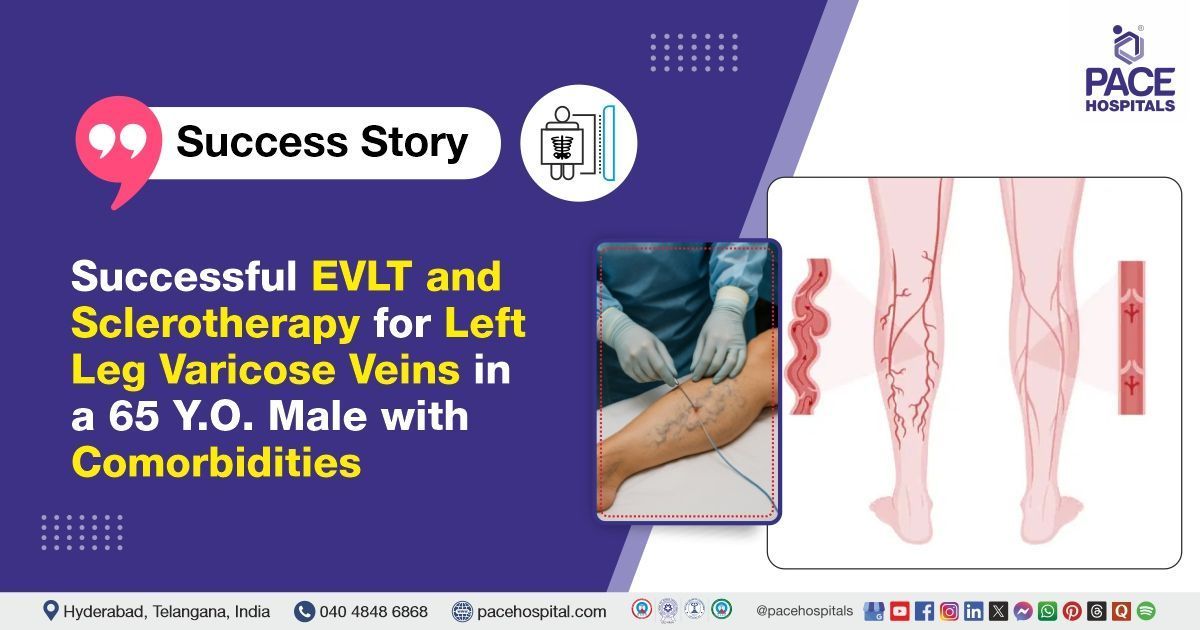
Successful EVLT & Sclerotherapy for Left Leg Varicose Veins in a 65-Year-Old Male
PACE Hospitals
PACE Hospitals Expert Interventional Radiology Team successfully performed Endovenous Laser Therapy (EVLT) and Sclerotherapy on a 65-year-old male patient with Varicose Veins in the Left Lower Limb who presented with bilateral dilated veins (left greater than right leg), pigmentation, itching, and recurrent wounds on the left leg. These minimally invasive procedures were carried out to relieve symptoms, prevent further complications, and improve the patient’s mobility and quality of life.
Chief Complaints
A 65-year-old male with a
Body Mass Index (BMI) of 22.4 presented to the Interventional Radiology Department at
PACE Hospitals, Hitech City, Hyderabad, with complaints of dilated (enlarged) and prominent veins in both lower limbs, which were more pronounced on the left side. He also reported pigmentation and persistent itching in the left leg, along with a history of recurrent wounds in the same area.
Past Medical History
The patient is a known case of
diabetes mellitus and has a history of cerebrovascular accident (CVA), both of which are well-controlled with regular medication. He had no recent episodes of exacerbation or complications. He remained stable with the prescribed medication.
On Examination
Upon admission to PACE Hospitals, the patient was hemodynamically stable. Cardiovascular and respiratory examinations were normal, with normal heart sounds (S1, S2) and adequate bilateral air entry. Abdominal examination revealed a soft and non-tender abdomen. Examination of the lower limbs demonstrated enlarged superficial veins, varicose veins bilaterally, with the left leg exhibiting pigmentation, persistent itching, and a history of recurrent wounds.
Diagnosis
Following the clinical examination, the interventional radiology team conducted a comprehensive assessment that included a detailed review of the patient’s medical history and a focused physical evaluation. A color Doppler ultrasound was performed to evaluate venous reflux, valve competence, and the extent of venous dilation.
The imaging demonstrated evidence of bilateral varicosities in the lower limbs, with incompetent perforator veins more predominant on the left side compared to the right. These findings corroborated the clinical suspicion of chronic venous insufficiency and highlighted a predominant left-sided venous pathology with associated superficial venous incompetence. The results were integrated with the clinical assessment to support the overall evaluation of the patient’s venous status.
Based on the confirmed diagnosis, the patient was advised to undergo
Varicose Veins Treatment in Hyderabad, India, under the care of the Interventional Radiology Department, to ensure comprehensive management of his condition.
Medical Decision Making
After consultation with Dr. Lakshmi Kumar C.H., Interventional Radiologist, a thorough evaluation was undertaken to determine the most suitable diagnostic and therapeutic strategy for the patient's venous condition. Following a detailed assessment, including clinical correlation and imaging findings, it was concluded that conservative management alone would be insufficient.
Based on their expert assessment, it was concluded that Varicose vein surgery would be the most effective procedure to address the patient’s condition.
The patient and his family were thoroughly counselled about the severity of the condition, the planned surgical procedure, potential risks, and the necessity for surgery.
Surgical Procedure
Following the decision, the patient was scheduled to undergo Endovenous laser therapy (EVLT) and Sclerotherapy Surgery in Hyderabad at PACE Hospitals under the supervision of the expert Interventional Radiology Department. The following steps were carried out during the procedure:
The procedure was performed under spinal and tumescent anaesthesia. After ensuring adequate anaesthesia, the following steps were carried out:
1. Ablation of Left Great Saphenous Vein (GSV): The left GSV was targeted for ablation, starting from just above the ankle and extending up to 6 cm proximal to the saphenofemoral junction (SFJ). A laser radial fibre was used for the ablation, with continuous ultrasound guidance to confirm correct placement and effective treatment.
2. Ablation of Left Anterior Accessory Saphenous Vein (AASV): The left AASV, located in the upper thigh, was similarly accessed and ablated using the laser radial fibre, following standard endovenous techniques.
3. Ablation of Left Small Saphenous Vein (SSV): The left SSV was ablated from the apex of the calf up to 4 cm proximal to the popliteal fossa, again utilizing the laser radial fibre and ultrasound guidance for precision.
4. Foam Sclerotherapy: Residual varicosities in the left leg were treated using foam sclerotherapy with anionic surfactant. The foam was injected directly into the affected superficial varicosities under ultrasound guidance to ensure accuracy.
5. Haemostasis and Dressing: Haemostasis (stopping of blood flow) was carefully secured at all access points. The treated limb was then wrapped with compression dressings using crepe bandages to support post-procedure venous return and reduce the risk of complications. The surgery was completed successfully without intraoperative complications.
Postoperative Care
The postoperative period was uneventful. The patient was monitored in the ICU and remained hemodynamically stable throughout. He received intravenous antibiotics, analgesics, anticoagulants, proton pump inhibitors, and supportive care. Symptomatic improvement was noted with no complications. The patient was discharged in stable condition, with post-discharge care instructions and follow-up advice.
Discharge Medications
Upon discharge, the patient was prescribed oral antibiotics to prevent infection and proton pump inhibitors for gastric protection. Analgesics were given for pain relief, and antipyretics were provided to control fever. Anticoagulants were prescribed to lower the risk of blood clots following the vein treatment. Additional supportive care measures were recommended to promote healing and prevent complications. The patient was advised to follow the medication plan and attend scheduled follow-up visits.
Advice on Discharge
The patient was advised to avoid crossing the legs and to limit prolonged standing or sitting to no more than 30 minutes at a time. He was also advised to refrain from lifting weights heavier than 5 kilograms for one week. Additionally, the patient was instructed to wear above-knee Class II compression stockings during the daytime for a period of two months and to continue other medications as advised by other physicians.
Emergency Care
The patient was informed to contact the Emergency ward at PACE Hospitals in case of any emergency or development of symptoms like fever, leg swelling or severe leg pain or chest pain or breathlessness.
Review and Follow-Up Notes
The patient was advised to return for a follow-up visit with the Interventional Radiologist in Hyderabad at PACE Hospitals, after 14 days. During this visit, his recovery would be assessed to evaluate the treatment outcome and determine if any further management was necessary.
Conclusion
This case highlights the effective management of a patient with bilateral varicose veins more prominent on the left side, particularly in a patient with diabetes and a history of cerebrovascular disease. Through Endovenous laser ablation combined with foam sclerotherapy. It emphasizes the importance of accurate diagnosis and individualized treatment planning in achieving optimal outcomes for patients with chronic venous insufficiency.
Comprehensive Care of Varicose Veins in a Diabetic, Post-Stroke Patient by Integrating Advanced Minimally Invasive Techniques
The presence of diabetes and a prior cerebrovascular accident (CVA) required heightened vigilance throughout perioperative care, wound management, and post-procedure monitoring, as these comorbidities increase the risk of delayed healing, infection, and thrombotic events. Close interdisciplinary collaboration and individualized follow-up were essential to address these risks.
In this context, the expertise of the
interventional radiologist/ interventional radiology doctor was crucial not only in selecting and tailoring minimally invasive techniques to minimize complications related to the patient’s underlying conditions, but also in coordinating care with other specialists, ensuring optimal glycemic and vascular control, providing detailed patient counselling, and maintaining vigilant post-procedure surveillance. This comprehensive approach contributed to a safe and effective outcome, despite the added complexity of significant comorbidities.
Share on
Request an appointment
Fill in the appointment form or call us instantly to book a confirmed appointment with our super specialist at 04048486868
Appointment request - health articles
Recent Articles