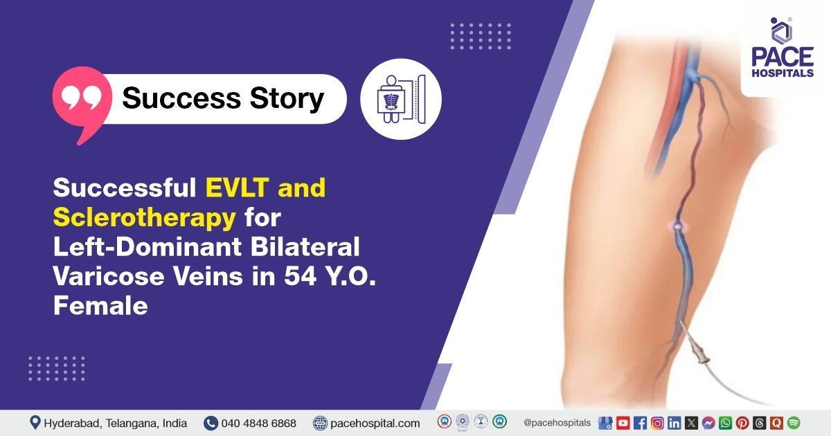
Successful EVLT and Sclerotherapy for Left-Dominant Bilateral Varicose Veins in 54-Y.O. Female
PACE Hospitals
The Radiology team at PACE Hospitals successfully performed Endovenous Laser Therapy (EVLT) and sclerotherapy on a 54-year-old female patient who had been experiencing varicose veins in both legs, with the left side being more severely affected, relieving her of long-standing pain, swelling, and discomfort in her left leg.
Chief Complaints
A 54-year-old female patient with a
BMI of 27.8 presented to the Interventional Radiology Department at
PACE Hospitals, Hitech City, Hyderabad, with complaints of dilated veins and swelling, along with eczema affecting both legs.
Past History
The patient had a known medical history of chronic inflammatory conditions, including
Crohn's disease and
rheumatoid arthritis, both of which have contributed to ongoing systemic inflammation and joint discomfort. Additionally, she has been dealing with eczema, a persistent skin condition characterized by dryness, itching, and irritation, further complicating the clinical picture and affecting the skin integrity of both legs.
Diagnosis
After admission to PACE Hospitals, the patient underwent a thorough medical history review followed by a detailed clinical examination conducted by the Interventional Radiology team. Based on the initial assessment, there was a strong clinical suspicion of varicose veins affecting both lower limbs, more prominently on the left side. This prompted further diagnostic evaluation to confirm the condition and determine the appropriate course of treatment.
To support the diagnosis and guide treatment planning, the Interventional Radiologist carried out a series of investigations:
Colour Doppler Scan: This imaging revealed the presence of varicosities in both legs, along with incompetent perforators, which were more pronounced on the left side compared to the right.
Duplex Ultrasound: This non-invasive imaging test was performed to visualize the veins, assess blood flow, and detect venous reflux. It revealed significant venous insufficiency in the left lower limb, particularly involving the great saphenous vein (GSV), anterior accessory saphenous vein (AASV), and small saphenous vein (SSV), confirming the need for targeted interventions such as endovenous laser therapy and sclerotherapy.
Ankle-Brachial Index (ABI): This test was conducted to evaluate arterial circulation in the lower limbs and to rule out underlying peripheral arterial disease. The ABI results were within the normal range, indicating adequate arterial supply and confirming that the patient was suitable for compression therapy and endovenous procedures.
CEAP Classification: The CEAP system was used to classify the severity and stage of the patient's chronic venous disease. Based on the findings, the patient was categorized as having advanced venous disease, with more severe involvement in the left leg. This classification supported the decision to prioritize intervention on the left side.
Based on the confirmed diagnosis, the patient was advised to undergo Varicose Veins Treatment in Hyderabad, India, under the expert care of the Interventional Radiology Department to minimize the risk of future complications.
Medical Decision-Making (MDM)
After a thorough consultation with the consultant Interventional radiologist, Dr. Lakshmi Kumar CH, a comprehensive evaluation was conducted to determine the most suitable diagnostic and therapeutic approach for the patient.
Based on their expert assessment, it was concluded that Endovenous Laser Therapy and Sclerotherapy would be the most effective procedures to treat the varicose veins in the left lower limb and address the patient's condition.
Surgical Procedure
Following the clinical decision, the patient was scheduled for Endovenous Laser Therapy and Sclerotherapy at PACE Hospitals, Hitech City, Hyderabad, under the expert supervision of the Interventional Radiology Department.
Endovenous laser therapy (EVLT) and sclerotherapy are minimally invasive treatments used to manage varicose veins, especially in the left lower limb. EVLT uses laser energy to heat and seal veins, causing them to collapse and absorb into the body. This procedure offers quick recovery and minimal discomfort. Sclerotherapy injects a solution directly into veins, causing them to scar and close off over time. Both treatments reduce symptoms, improve leg appearance, and prevent complications from venous insufficiency.
Upon admission, the procedure was performed under spinal and tumescent anaesthesia. The treatment involved laser ablation of multiple affected veins in the left lower limb. Specifically, the left great saphenous vein (GSV) extending from above the ankle to 6 cm proximal to the saphenofemoral junction (SFJ), the left anterior accessory saphenous vein (AASV) in the upper thigh, and the left small saphenous vein (SSV) extending from the apex of the calf to 4 cm proximal to the popliteal fossa, were successfully ablated using a radial laser fiber.
In addition, prominent varicosities in the left leg were treated using foam sclerotherapy with 1% sodium tetradecyl sulphate (STS). The procedure was completed with successful haemostasis, and a compression dressing was applied using crepe bandages to aid recovery and support venous return.
Postoperative Care
The patient had a smooth postoperative recovery, with no complications observed during the post-surgical period. During hospitalization, she was administered intravenous fluids to maintain hydration and support recovery, along with antibiotics to prevent postoperative infections. Analgesics were prescribed to manage pain and ensure patient comfort, while proton pump inhibitors (PPIs) were given to protect the gastric lining from potential irritation due to the use of medications.
Additionally, anticoagulants were administered as a precautionary measure to reduce the risk of deep vein thrombosis (DVT), considering her limited mobility during the initial recovery phase and underlying venous insufficiency.
Discharge Notes
The patient was discharged in a hemodynamically stable condition.
Discharge Medications
Upon discharge, the patient was prescribed antibiotics, analgesics, proton pump inhibitors (PPIs), and oral anticoagulants.
Advice on Discharge
The patient was advised to avoid crossing her legs and to limit prolonged standing or sitting beyond 30 minutes to promote healthy blood circulation and prevent venous stasis. Lifting weights over 5 kilograms was restricted for one week to avoid strain on the treated veins. She was instructed to wear above-knee Class II compression stockings during the daytime for two months to support vein function and reduce the risk of recurrence. Additionally, continuation of her regular medications was recommended to maintain stability of her underlying conditions, as prescribed by her respective physicians.
Emergency Care
The patient’s guardians were advised to seek immediate admission to the Emergency ward at PACE Hospitals, Hyderabad, if the patient develops fever, leg swelling, severe leg pain, chest pain, or breathlessness, as these symptoms may indicate potential post-procedural complications such as infection, deep vein thrombosis, or pulmonary embolism, requiring urgent medical evaluation.
Review and Follow-Up Notes
The patient was advised to return for a follow-up visit with the Interventional Radiologist in Hyderabad at PACE Hospitals,14 days after discharge, to monitor the healing of the treated veins, assess the effectiveness of the procedure, and promptly manage any emerging concerns or complications specific to her condition.
Conclusion
This case highlights the successful use of Endovenous Laser Therapy and sclerotherapy in treating varicose veins in the left lower limb, where injections helped close and shrink the enlarged veins. The procedure significantly improved the patient’s symptoms, demonstrating the effectiveness of minimally invasive methods in managing chronic venous disease.
Role of Duplex Ultrasound in the Diagnosis and Management of Bilateral Varicose Veins
Duplex ultrasound is a crucial diagnostic tool in the evaluation of bilateral varicose veins, offering a comprehensive assessment of both the superficial and deep venous systems. This non-invasive imaging modality, typically conducted by a interventional radiologist/interventional radiology doctor, integrates conventional ultrasound with Doppler flow analysis to assess venous anatomy and blood flow direction. It plays a key role in detecting valvular incompetence, venous reflux, and incompetent perforators—major factors contributing to the development of varicose veins. In cases with asymmetrical involvement, such as greater severity in the left leg, duplex ultrasound facilitates accurate mapping of the affected veins, thereby enabling the radiologist to plan and perform targeted, minimally invasive interventions like endovenous laser therapy or sclerotherapy with precision.
Share on
Request an appointment
Fill in the appointment form or call us instantly to book a confirmed appointment with our super specialist at 04048486868
Appointment request - health articles
Recent Articles