Successful Local Excision for Breast Lumps to Rule Out Cancer in 41-Year-Old Female
PACE Hospitals
The PACE Hospitals’ expert oncology team successfully performed a
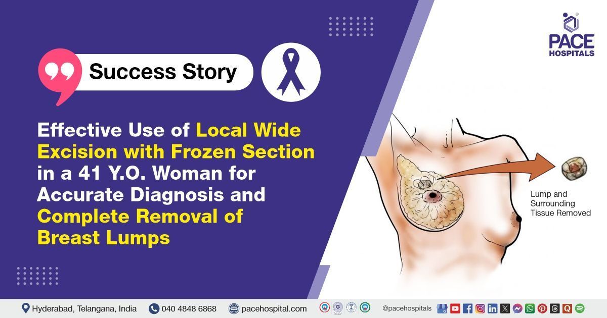
local wide excision with frozen section on a 41-year-old female patient who presented with multiple lumps in both breasts. The procedure was specifically carried out to remove the lumps in the left breast, effectively relieving the patient's symptoms and promoting optimal breast function and recovery.
Chief complaints
A 41-year-old female patient with a
BMI of 22 presented to the Oncology Department at
PACE Hospitals, Hitech City, Hyderabad, with complaints of multiple lumps in both breasts, seeking further diagnosis and management. There was no associated history of fever or pain, indicating a non-inflammatory and likely non-infectious presentation of the disease.
Past Medical History
The patient had no known allergies or significant comorbidities. She has no history of chronic medical conditions such as
diabetes,
hypertension, or
cardiovascular disease, and is not currently on any long-term medications. This indicates that her overall health was stable, without any underlying conditions that could complicate her treatment or recovery.
Menstrual and Obstetric History
The patient’s obstetric history is noted as G1P1A0L1: G1 (Gravida 1) indicates she was pregnant once; P1 (Para 1) means she had one delivery beyond the age of viability (typically over 20 weeks); A0 (Abortus 0) signifies no history of abortions or miscarriages; and L1 (Living 1) confirms she has one living child. A hysterectomy was performed following the delivery, resulting in amenorrhea. Furthermore, she has no history of gynaecological conditions that might affect her overall health or recovery.
General Examination
Upon admission to PACE Hospitals, the patient was hemodynamically stable, with stable vital signs. A general examination revealed that she was conscious, coherent, and cooperative. Cardiovascular and respiratory assessments were unremarkable, with normal heart sounds (S1, S2) and clear bilateral lung fields on auscultation, indicating no signs of cardiovascular or respiratory distress. The abdominal examination was also normal, with a soft, non-tender abdomen and no signs of distension or guarding, suggesting no acute abdominal concerns.
During the breast examination, palpable lumps were noted in both breasts, warranting further diagnostic evaluation, such as imaging, to determine their nature and clinical significance.
Diagnosis
After the initial examination, the patient underwent a comprehensive assessment by the oncology team, which included detailed clinical evaluation, laboratory investigations, and imaging studies, including:
- Ultrasound (USG): Revealed benign lumps in both breasts, with two lumps in the left breast located in the upper outer quadrant near the areola.
- Mammography: Mammography raised mild suspicion due to irregularities and a denser appearance of the lumps, prompting further evaluation to rule out malignancy.
- MRI (magnetic resonance imaging): It showed suspicious characteristics, with irregularities in the lumps that required additional investigation to confirm their benign nature.
- FNAC (Fine Needle Aspiration Cytology) Test: The left breast lump showed scattered atypical cells, raising concern for potential malignancy, though not definitive.
Given the radiological and cytological findings, a biopsy was recommended for a more definitive diagnosis.
Based on the comprehensive evaluation, the patient was advised to undergo
Breast Tumor Treatment in Hyderabad, India, under the expert care of the Oncology Department to ensure complete management of her condition, symptom relief, and prevention of further complications.
Medical Decision-Making (MDM)
Following a detailed consultation with Dr. Ramesh Parimi, a surgical oncologist, and a thorough evaluation of the patient's condition, the oncology team concluded that a local wide excision with a frozen section was the most appropriate treatment plan. This procedure is used to determine if a lump is cancerous or to serve as a treatment for breast cancer.
Based on the patient’s symptoms and the findings from the diagnostic investigations, this approach was chosen to remove the breast lumps while ensuring clear margins to minimize the risk of recurrence. The frozen section technique allowed for real-time examination of the tissue during surgery, providing immediate feedback on the nature of the lumps and ensuring that any potentially malignant tissue was adequately addressed. The comprehensive treatment plan was designed to maximize the patient’s chances of complete recovery while minimizing the risk of future complications.
The patient and the family were counseled about the patient's condition, including the associated risks, complications, and benefits. They were also informed about the necessity of surgical intervention to prevent further complications and ensure optimal outcomes.
Surgical Procedure
Following the decision, the patient was scheduled for Breast Lump Excision Surgery in Hyderabad at PACE Hospitals, with frozen section under the expert supervision of the Oncology Department, ensuring optimal care and a smooth recovery process.
Before proceeding with the surgery, the medical team ensured the patient's medical stability and obtained informed consent to facilitate proper alignment and recovery. The following steps were carried out during the procedure:
- Anesthesia and Positioning: The patient was given local anesthesia and positioned for optimal access to the left breast.
- Circumareolar Incision: A circumareolar incision (a surgical cut made in a semicircular shape around the outer edge of the areola) was made to access the lumps in the upper outer quadrant.
- Wide Excision: A local wide excision was performed, removing the lumps along with a surrounding margin of healthy tissue to ensure clear margins.
- Frozen Section (a rapid microscopic analysis of tissue specimens during surgery): A frozen section technique was used for real-time and immediate pathological assessment of the excised tissue to confirm its nature.
- Closure: After confirming clear margins, the incision was closed with fine sutures for optimal cosmetic results.
Both lumps were sent for frozen section analysis, which showed no malignancy but revealed atypical cells, indicating the need for further evaluation and monitoring.
Postoperative Care
After surgery, the patient was monitored closely in the surgical intensive care unit to ensure stability. She was then transferred to a private ward once her condition was stable. During her hospital stay, she received intravenous antibiotics to prevent infection, analgesics for pain relief, antiemetics to control nausea, and gastric protectants.
Once she was stable and exhibited no signs of distress, the patient was discharged with appropriate post-discharge care instructions. As per the postoperative plan, the drain was removed, and she was advised to follow up as scheduled. Her condition remained hemodynamically stable throughout the recovery period.
Discharge Medications
Upon discharge, the patient was prescribed a combination of oral medications, including antibiotics to prevent infection, analgesics for pain relief, antipyretics to manage any fever, and antifibrinolytics to reduce the risk of excessive bleeding.
In addition, other supportive treatments were provided to aid recovery and ensure proper healing post-surgery. These medications were tailored to the patient’s condition, addressing both the surgical recovery and the need for infection prevention and symptom management.
Emergency Care
The patient was informed to contact the Emergency ward at PACE Hospitals in case of any emergency or development of symptoms such as pain in the abdomen, fever, and vomiting.
Review and Follow-Up
The patient was advised to schedule a follow-up appointment with the Oncologist in Hyderabad at PACE Hospitals, one week after discharge. During this visit, the patient’s recovery was to be evaluated to determine the need for any further management.
Conclusion
This case highlights the effectiveness of local wide excision with frozen section in performing Breast Lump Treatment in Hyderabad, India, enabling successful removal of breast lumps, thereby facilitating normal function.
Significance of FNAC in the interpretation and management of breast lumps
Fine needle aspiration cytology technique, also called FNAC biopsy, is a less invasive procedure performed to obtain a cell sample from a lesion using a thin-bore needle to confirm a diagnosis or to choose the treatment. It is a rapid, cost-effective procedure that serves as an ideal first-line diagnostic tool. FNAC involved the use of a thin needle to aspirate cells from the breast lump, thereby minimizing the need for more invasive surgical biopsies.
This technique was particularly valuable in identifying the presence of malignant cells within the breast tissue, enabling early detection and facilitating timely intervention. Additionally, FNAC provided essential cytological information that guided further diagnostic and therapeutic decisions, improving patient outcomes while reducing procedural risks. Oncologist/cancer specialist often rely on FNAC due to its high accuracy, sensitivity, and specificity in diagnosing breast lesions, especially in identifying malignant cells.
Share on
Request an appointment
Fill in the appointment form or call us instantly to book a confirmed appointment with our super specialist at 04048486868
Appointment request - health articles
Recent Articles