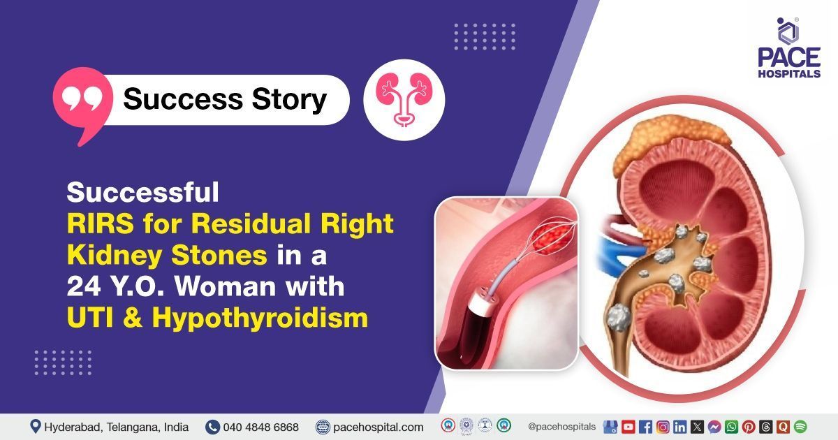
Successful RIRS for Residual Right Kidney Stones in a 24 Y.O. Woman
PACE Hospitals
The PACE Hospitals expert Urology team successfully performed Right
Retrograde Intrarenal Surgery (RIRS) with DJ Stenting on a 24-year-old female patient who presented with
right flank pain, on and off fever and dysuria (painful or uncomfortable urination). The procedure was performed to
remove kidney stones, effectively relieving the patient's symptoms and helping to restore proper kidney function to support recovery.
Chief Complaints
A 24-year-old -female with a
Body Mass Index (BMI) of 24 presented to the Urology Department at
PACE Hospitals, Hitech City, Hyderabad, with right flank pain, on and off fever and dysuria (painful or uncomfortable urination). Because of the localised nature of her symptoms, a detailed evaluation was carried out to determine the underlying cause of her symptoms.
Past Medical History
The patient had a medical history of bilateral percutaneous nephrolithotomy (PCNL) performed at another centre for staghorn calculi. She had
urinary tract infections (UTIs) and urosepsis in the past; residual stone fragments increased her risk of infection and obstruction, and she also had
hypothyroidism. Due to these factors, close follow-up was essential to manage infections, prevent recurrence, and address comorbidities.
On Examination
Upon admission to PACE hospitals, the patient was hemodynamically stable with mild fever. Cardiovascular examination revealed normal S1 and S2 heart sounds. And there were no neurological deficits. Respiratory assessment revealed normal bilateral air entry and vesicular breath sounds.
Diagnosis
The patient initially underwent a comprehensive clinical assessment at PACE Hospitals, which included a detailed history and physical examination by the Urology team. Based on her presenting symptoms, there was a strong clinical suspicion of residual calculi on the right side of the kidney.
To support the diagnosis and assess the extent of the condition, a series of relevant investigations were performed.
- A non-contrast Computed Tomography (NCCT) scan: It revealed the presence of right renal calculi localized to the upper and mid-poles of the kidney. These stones were consistent with nephrolithiasis, likely contributing to the patient’s history of recurrent urinary tract infections and urosepsis.
- Complete blood count (CBC): The CBC showed normal haemoglobin levels, with no neutrophilic predominance, indicating the absence of active systemic infection. This suggested that the patient's symptoms were likely due to localized inflammation from the renal calculi rather than a systemic disease.
- Urine Culture: The urine culture was positive, confirming a urinary tract infection.
- Parathyroid Hormone (PTH) Test: The PTH level was borderline high, indicating possible parathyroid dysfunction or secondary effects related to her renal condition.
- Serum Calcium Test: The calcium level was mildly elevated.
- Vitamin D Test: The vitamin D level was within normal range.
Based on the confirmed findings, the patient was advised to undergo kidney stone treatment in Hyderabad, India, under the expert care of the Urology Department, which provides effective stone clearance, reduces her flank pain, and prevents potential complications such as obstruction and infection or renal function impairment.
Medical Decision Making
Following a detailed consultation with
Dr. Abhik Debnath, Consultant Laparoscopic Urologist, an evaluation of the patient's condition was undertaken. Considering her right flank pain and diagnostic findings indicating calculus in the upper and mid-pole of the right kidney, the urology team concluded that Right Retrograde Intrarenal Surgery (RIRS) with DJ stenting would be the most appropriate course of treatment for the
Kidney Stone.
Surgical Procedure
Following the decision, the patient was scheduled for Right Retrograde Intrarenal Surgery (RIRS) with DJ stenting surgery in Hyderabad at PACE Hospitals, under the expert supervision of the urology department, ensuring optimal care and a smooth recovery process.
- Anaesthesia and Positioning: The patient was administered general anaesthesia and positioned appropriately for retrograde intrarenal surgery.
- Ureteral Access and Sheath Placement: The urethra and bladder were navigated, the right ureteric orifice was cannulated, and an access sheath was inserted to facilitate repeated instrument exchanges and reduce ureteral trauma.
- Introduction of Digital Ureterorenoscope: A digital ureterorenoscopy was advanced through the access sheath into the right renal collecting system, allowing clear visualization of the calyces.
- Stone Identification: Multiple calculi were identified in the upper and mid-pole calyces. A lower pole calculus was noted on retrograde pyelogram (RGP) but was not visualized during renoscopy, as it was located in a cut-off calyx outside the accessible collecting system.
- Laser Fragmentation and Dusting: The upper and mid-pole stones were targeted and completely fragmented and dusted using a solid-state laser, ensuring thorough clearance of visible stones.
- Stone Fragment Retrieval: Stone dust and fragments were carefully collected using a basket device, and representative samples were collected for Fourier Transform Infrared (FTIR) stone analysis.
- Management of Lower Pole Calculus: The lower pole stones, being outside the collecting system in a cut-off calyx and not accessible endoscopically, were left in situ. Given its location and inaccessibility, no intervention was attempted, in line with prudent clinical practice.
- Stent Placement: A double-J (DJ) stent was placed on strings to ensure proper drainage and facilitate easy removal during follow-up.
- Hemostasis: It is the process of stopping bleeding, and it was achieved. The procedure concluded without complications, and the patient was shifted to the recovery room in stable condition.
Postoperative Care
After surgery, the patient was monitored closely in the surgical intensive care unit to ensure stability. Her postoperative course was uneventful. The Foley catheter had been removed on postoperative day 2. During her hospital stay, she received intravenous antibiotics to prevent infection, analgesics for pain relief, antiemetics to control nausea, and gastric protectants. The patient was discharged in stable condition.
Discharge Medications
Upon discharge, the patient was prescribed a tailored course of oral medications to support recovery following surgical removal of the residual renal calculus. The regimen included oral antibiotics to prevent postoperative infections, analgesics for pain relief, and antipyretics to manage any potential fever. Urinary alkalizers were given to provide optimal urinary pH and minimise the risk of stone recurrence. Proton pump inhibitors were given to protect the gastric lining from potential medication-induced irritation. Thyroid hormone replacement was initiated to address hypothyroidism, and thiazide diuretics were prescribed to reduce urinary calcium excretion. An alpha-blocker was administered to facilitate smooth urinary flow. Additionally, the patient was advised to take nutritional supplements to support overall recovery and renal health.
Advice on Discharge
The patient was advised to follow activity restrictions including avoiding weightlifting, forward bending, and long-distance travel via bus, auto-rickshaw, or two-wheeler for a specified period. She was also informed that mild flank discomfort, occasional haematuria, or cloudy urine may persist for up to 7–10 days after discharge, which are expected postoperative symptoms.
Emergency Care
The patient was informed to contact the Emergency ward at PACE Hospitals in case of any emergency or development of symptoms like fever, abdominal pain, swelling, bleeding, or vomiting.
Review and Follow-Up
The patient was advised to return for a follow-up visit with the Urologist in Hyderabad at PACE Hospitals, one month after the surgery for the removal of the DJ stent. Additionally, a further review was scheduled for 3 months, which include repeat assessments of serum calcium, parathyroid hormone, serum albumin, and an X-ray of the kidneys, ureters, and bladder (KUB), complete urine examination (CUE) and urine culture. This review is necessary to assess the patient's recovery, ensure proper healing, and evaluate the urinary tract for any complications or residual issues that may require further intervention.
Conclusion
This case highlights the effectiveness of Retrograde Intrarenal Surgery (RIRS) in performing Renal Calculi Treatment in Hyderabad, India, enabling the successful removal of stones in the kidney, thereby facilitating normal urine flow.
The Essential Role Non-contrast-enhanced computed tomography (NCTT) in the diagnosis of Residual Renal Calculi
Non-contrast-enhanced computed tomography (NCCT) is the predominant and gold standard imaging test used by the urologist/urology doctor to diagnose residual right renal calculi after intervention. NCCT offers high sensitivity and specificity, making it superior to older modalities, such as intravenous pyelogram (IVP) and ultrasound, for detecting even tiny residual stone fragments. It provides detailed information about stone size, location, density, and volume, which is crucial for planning further management or intervention.
NCCT can also help differentiate between various stone types, including radiolucent stones that may not be visible on plain X-rays. Low-dose NCCT protocols are often used to minimize radiation exposure while maintaining diagnostic accuracy, especially in non-obese patients. The test is fast, non-invasive, and widely available, making it the preferred choice in clinical practice for evaluating residual stones. Additionally, NCCT can identify other urinary tract abnormalities or complications, ensuring comprehensive assessment after stone treatment.
Share on
Request an appointment
Fill in the appointment form or call us instantly to book a confirmed appointment with our super specialist at 04048486868
Appointment request - health articles
Recent Articles