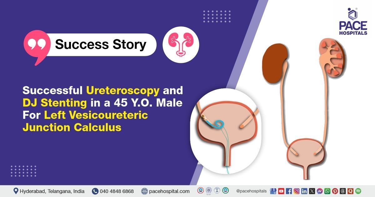
Successful Ureteroscopy and DJ Stenting in a 45-Y.O Male for Left Vesicoureteric Junction Calculus
PACE Hospitals
The PACE Hospital's expert Urology team successfully performed a
Left Ureteroscopy (URS) with Double J (DJ) stenting on a 45-year-old male patient who presented with left flank pain and had not responded to medical expulsion therapy. The procedure was performed to remove the stone located at the left vesicoureteric junction (VUJ), effectively relieving the patient's symptoms and restoring proper kidney function to facilitate recovery.
Chief Complaints
A 45-year-old male patient with a
body mass index (BMI) of 20, presented to the Urology Department at
PACE Hospitals, Hitech City, Hyderabad, with left flank pain that did not improve following initial medical expulsion therapy (unsuccessful treatment using medications without surgery). A detailed evaluation was carried out to determine the underlying cause of his symptoms.
Past Medical History
The patient had no history of diabetes, hypertension, trauma, chronic illnesses, or known drug allergies.
On Examination
On general examination, the patient was conscious, coherent, and oriented with stable vital signs. A systemic examination, including an abdominal assessment, revealed mild tenderness in the left flank region with no palpable masses, and respiratory examination findings were normal. Although his ongoing symptoms suggested an underlying issue that required further investigation.
Diagnosis
Upon admission to PACE Hospitals, the patient was thoroughly evaluated by the Urology team, which included a detailed review of his medical history and a comprehensive clinical examination. Given his history of left flank pain and failed medical expulsion therapy, there was a strong clinical suspicion of a urinary tract obstruction due to calculus.
The patient underwent imaging studies, including ultrasound and non-contrast CT of the kidneys, ureters, and bladder (CT KUB), which revealed a 4mm calculus at the left vesicoureteric junction (VUJ), causing mild hydroureteronephrosis (swelling of the kidney due to urine backup). Laboratory investigations showed normal renal function and no evidence of active infection. The patient was diagnosed with left VUJ calculus resistant to medical management.
Based on the confirmed findings, the patient was advised to undergo
Ureteric Stone Treatment in Hyderabad, India, under the expert care of the Urology Department.
Medical Decision Making
After a detailed consultation with Dr. Abhik Debnath, Consultant Laparoscopic Urologist, a comprehensive evaluation was conducted to determine the optimal diagnostic and therapeutic approach for the patient presenting with left flank pain and a confirmed left vesicoureteric junction (VUJ) calculus. Based on detailed clinical evaluation and imaging studies, including ultrasound and CT scan of the abdomen, surgical intervention was deemed necessary.
It was determined that the patient had a left vesicoureteric junction (VUJ) calculus, which was causing left flank pain and mild hydronephrosis. Ureteroscopy (URS) with Double J (DJ) stenting was identified as the most effective intervention to relieve the obstruction, alleviate symptoms, and restore normal urinary flow.
The patient and his family were informed about his condition, the procedure, its associated risks, and its potential to alleviate symptoms and enhance his quality of life.
Surgical Procedure
Following the decision, the patient was scheduled for Left Ureteroscopy (URS) and DJ Stenting Procedure in Hyderabad at PACE Hospitals, under the expert supervision of the Urology Department, ensuring optimal care and a smooth recovery process.
- Preoperative Preparation and Spinal Anaesthesia: The patient was put on the surgical bed in the lithotomy position after informed consent and a careful preoperative evaluation. Throughout the treatment, spinal anaesthesia was used to ensure sufficient pain alleviation and muscle relaxation. Preoperative IV antibiotics were administered to prevent infection.
- Cystoscopic Evaluation: A diagnostic cystoscopy was first performed to assess the bladder and locate the ureteric orifices. The bladder mucosa appeared normal, with no evidence of wall thickening or lesions. The left ureteric orifice was identified for further intervention.
- Ureteroscopy (URS): A semirigid ureteroscope was introduced into the left ureter. Upon entry, the distal ureter showed signs of inflammation and edema. A 4 mm calculus was visualised at the left vesicoureteric junction (VUJ). Notably, purulent discharge was observed while inserting the guidewire, indicating infection at the site.
- Stricture Identification: During ureteroscopic navigation, a tight distal ureteric inflammatory stricture was encountered. The scope was advanced carefully through this narrowed segment to minimise ureteral trauma and to allow visualisation of the obstructing stone.
- Stone Fragment Management: The stone at the VUJ was managed using standard endoscopic techniques. The fragments were either retrieved or allowed to pass into the bladder safely, depending on size and location.
- DJ Stent Placement: A Double J (DJ) stent was inserted from the renal pelvis to the urinary bladder to ensure unobstructed urine drainage, allow ureteral healing, and prevent recurrent obstruction. Proper stent placement was confirmed endoscopically.
- Hemostasis: It is the process of stopping bleeding, and it was achieved. The procedure concluded without complications, and the patient was shifted to the recovery room in stable condition.
Intraoperative findings
- Edema was observed at the left vesicoureteric junction (VUJ) along with fragments of calculi
- An inflammatory stricture was noted in the distal ureter.
- Upon insertion of the guidewire, purulent discharge was also encountered, indicating the presence of infection.
Postoperative Care
The postoperative course was uneventful. During the hospital stay patient was managed with intravenous antibiotics, analgesics, antiemetics, and gastric protectants. He experienced no significant complications and was stable at the time of discharge.
Discharge Medications
Upon discharge, the patient was prescribed an oral antibiotic to prevent infection, an analgesic and antipyretic for pain and fever control, and a proton pump inhibitor to protect the stomach lining. An alpha-blocker was given to improve urinary flow, and urinary alkalizers were prescribed to maintain optimal urinary pH and minimize the risk of stone recurrence. A nutritional supplement was recommended to support overall healing.
Advice on Discharge
The patient was advised to avoid weightlifting, forward bending, straining during bowel movements, and prolonged travel by bus, auto, or two-wheeler. Passage of small stone fragments, mild flank or suprapubic discomfort, occasional blood in urine, cloudy urine, or mild burning were expected and typically resolve within 7–10 days.
Emergency Care
The patient was informed to contact the emergency ward at PACE Hospitals in case of any emergency or development of symptoms like fever, abdominal pain, swelling, bleeding, or vomiting.
Review and Follow-up Notes
The patient was advised to return for a follow-up visit after 2-3 months for DJ stent removal, check URS, and cystoscopy with the Urologist in Hyderabad at PACE Hospitals, to review his condition. This review is necessary to assess the patient's recovery, ensure proper healing, and evaluate the urinary tract for any complications or residual issues that may require further intervention.
Conclusion
This case highlights the effectiveness of left ureteroscopy (URS), with DJ stenting in the management of a left vesicoureteric junction (VUJ) calculus. The procedure enabled successful removal of the obstructing ureteral stone, thereby restoring normal urine flow, alleviating symptoms, and improving the patient’s overall health and quality of life.
Comprehensive Infection Management in Endoscopic Ureteric Procedures
The intraoperative findings of edema, inflammatory stricture, and purulent discharge indicate an active infection and inflammation at the ureteric site that require prompt and careful management to prevent serious complications such as urinary tract infection or urosepsis. Addressing these infectious and inflammatory changes during ureteroscopy is crucial to prevent ongoing tissue damage and serious complications such as urinary tract infections or urosepsis. The
urologist / urology doctor ensures effective management through careful stone removal, appropriate use of antibiotics before and after surgery, and adequate urinary drainage via stenting. Additionally, microbiological cultures of urine, stone fragments, or renal pelvis fluid guide targeted antibiotic therapy to reduce infection risk. Close postoperative monitoring and strict adherence to antimicrobial protocols by the urology team are essential to minimise complications and promote safe recovery.
Share on
Request an appointment
Fill in the appointment form or call us instantly to book a confirmed appointment with our super specialist at 04048486868
Appointment request - health articles
Recent Articles