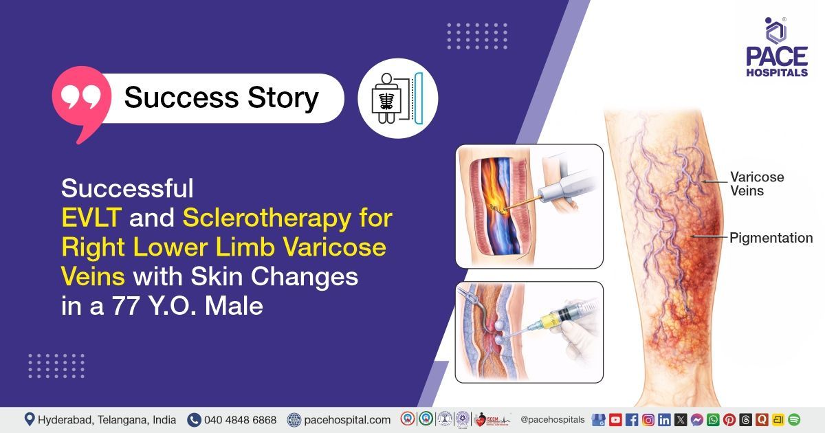
Successful EVLT and Sclerotherapy for Right Lower Limb Varicose Veins in a 77 Y.O. Male
PACE Hospitals
PACE Hospitals’ expert Interventional Radiology team successfully performed Endovenous Laser Therapy (EVLT) along with Sclerotherapy on a 77-year-old male patient with varicose veins in both lower limbs (Right>Left), who presented with complaints of dilated veins, skin pigmentation, hardening of the skin, itching, and swelling in the right leg, with the aim of closing the affected veins, improving blood circulation, relieving symptoms, and preventing further complications associated with varicose veins.
Chief Complaints
A 77-year-old male with a body mass index (BMI) of 24 presented to the Interventional Radiology Department at PACE Hospitals, Hitech City, Hyderabad, with complaints of dilated veins in the right lower limb, along with skin pigmentation, hardening of the skin, itching, and swelling in the right leg.
Past Medical History
The patient had no known history of drug allergies or chronic illnesses. The absence of comorbid conditions was considered clinically favorable, as it minimized the risk of intraoperative and postoperative complications and supported a smoother, more stable recovery.
On Examination
On examination, the patient was conscious, coherent and oriented. General physical examination appeared normal with a stable overall condition. Local examination of the lower limbs revealed abnormally dilated and tortuous superficial veins, more prominent in the right lower limb compared to the left. The skin over the right leg showed abnormal pigmentation and areas of skin hardening. Mild abnormal swelling was present in the right lower limb.
On palpation, the veins were abnormally prominent and compressible with no abnormal local rise in temperature. Peripheral pulses in both lower limbs were normal and palpable. No abnormal ulcers, bleeding, or wounds were observed. The examination findings were consistent with
varicose veins involving both lower limbs, with more on the right side.
Diagnosis
Following the clinical examination, the Interventional Radiology team conducted a comprehensive assessment, which included a detailed review of the patient’s medical history and a focused clinical evaluation. A color Doppler ultrasound was performed to assess venous reflux, valve competence, and the extent of venous dilation in both lower limbs.
Imaging studies revealed varicosities of both lower limb veins with incompetent perforator veins, with findings more pronounced in the right lower limb. Considering the patient's clinical symptoms, physical examination findings, and Doppler ultrasound results, a provisional diagnosis of varicose veins of both lower limbs (right greater than left) was established.
Based on the confirmed diagnosis, the patient was advised to undergo
Varicose Veins Treatment in Hyderabad, India, under the care of the Interventional Radiology Department, to ensure comprehensive management of his condition.
Medical Decision Making (MDM)
After consultation with Dr. Lakshmi Kumar Chalamarla, Interventional Radiologist, a thorough evaluation was undertaken to determine the most suitable diagnostic and therapeutic strategy for the patient's venous condition. Following a detailed assessment, including clinical correlation and colour Doppler findings, it was concluded that conservative management alone would be insufficient.
It was determined that the patient had varicose veins responsible for dilated veins, pigmentation, and skin hardening, itching, and swelling in the right leg. Endovenous Laser Therapy (EVLT) combined with foam sclerotherapy was identified as the most effective intervention to ablate the diseased veins, relieve swelling, and improve his overall condition.
The patient and his family were thoroughly counselled about the severity of the condition, the planned endovenous procedure, potential risks, and the necessity for intervention to ensure comprehensive management of the varicose veins.
Surgical Procedure
Following the decision, the patient was scheduled to undergo Endovenous laser therapy (EVLT) and Sclerotherapy surgery in Hyderabad at PACE Hospitals under the supervision of the expert Interventional Radiology Department.
The following steps were carried out during the procedure:
- Anesthesia and Preparation: The patient was positioned appropriately in the procedure room. Spinal anesthesia was administered to ensure regional numbness. Tumescent anesthesia was infiltrated along the course of the targeted veins to provide local anesthesia, reduce bleeding, and protect surrounding tissues during the laser ablation. The right lower limb was cleaned and draped under sterile conditions.
- Access and Cannulation of Target Veins: The right great saphenous vein (GSV) and right small saphenous vein (SSV) were identified using ultrasound guidance. The GSV was cannulated just below the knee, and the SSV was cannulated at the apex of the calf. Guidewires and introducer sheaths were inserted to establish secure access for the laser fiber.
- Endovenous Laser Ablation (EVLA): A radial laser fiber was advanced through the introducer sheaths into the GSV up to 6 cm proximal to the saphenofemoral junction (SFJ) and into the SSV up to 4 cm proximal to the popliteal fossa. Controlled laser energy was delivered along the length of the veins to thermally ablate the incompetent veins, causing vein closure. Ultrasound monitoring ensured complete closure and prevented damage to surrounding structures.
- Adjunctive Foam Sclerotherapy: Residual varicosities in the right leg that were not suitable for laser ablation were treated with foam sclerotherapy using an anionic surfactant. The sclerosing agent was carefully injected into the superficial varicosities under ultrasound guidance to induce vein closure and eventual resorption.
- Hemostasis, Dressing, and Post-Procedure Care: Hemostasis was secured at all puncture sites. A compression dressing with crepe bandages was applied to reduce swelling and support vein closure. The patient was monitored post-procedure for stability, vital signs, and potential complications before discharge instructions were provided.
Postoperative Care
The postoperative period was uneventful. The patient was monitored and remained hemodynamically stable throughout. He received medications for infection prevention, pain relief, anticoagulation, acid suppression, and supportive care. Symptomatic improvement was noted with no complications. The patient was discharged in stable condition with post-discharge care instructions and follow-up advice.
Discharge Medications
Upon discharge, the patient was prescribed medications for infection prevention, acid suppression, anticoagulation, and pain relief. He was also advised to continue other medications as prescribed by his treating physicians.
Advice on Discharge
The patient was instructed to avoid crossing legs and prolonged standing or sitting for more than 30 minutes, and to refrain from lifting heavy weights for one week. Use of above-knee compression stockings during the daytime was recommended for two months to support vein healing.
Emergency Care
The patient was informed to contact the emergency ward at PACE Hospitals in case of any emergency or development of symptoms like fever, leg swelling, severe leg pain, chest pain or breathlessness.
Review and Follow-up Notes
The patient was advised to return for a follow-up visit with the Interventional Radiologist in Hyderabad at PACE Hospitals, after 14 days.
Conclusion
This case highlights the management of bilateral lower limb varicose veins, more pronounced on the right side, using endovenous laser therapy and foam sclerotherapy. The procedure was performed under regional and tumescent anesthesia and was uneventful. The patient showed good postoperative recovery. He was discharged in stable condition with medications and post-discharge care instructions.
Minimally Invasive Strategies for Complex Lower Limb Varicosities
Minimally invasive approaches, such as Endovenous Laser Therapy combined with foam sclerotherapy, play a pivotal role in the management of varicose veins with incompetent perforators. Under the expertise of an interventional radiologist/interventional radiology doctor, precise ablation of affected veins can be achieved, effectively relieving symptoms and reducing recurrence. These techniques limit procedural complications, minimize postoperative discomfort, and enable faster rehabilitation.
Careful patient selection, proper anesthesia planning, and structured postoperative support further enhance safety and outcomes. By addressing both truncal and residual veins, these strategies provide comprehensive management of the venous system. Overall, minimally invasive interventions guided by an interventional radiologist represent the evolution of vascular care toward efficient, patient-centered treatment that prioritizes both clinical efficacy and quality of life.
Share on
Request an appointment
Fill in the appointment form or call us instantly to book a confirmed appointment with our super specialist at 04048486868
Appointment request - health articles
Recent Articles