Successful Laparoscopic BMG Ureteroplasty for Complex Right Upper Ureteric Stricture
PACE Hospitals
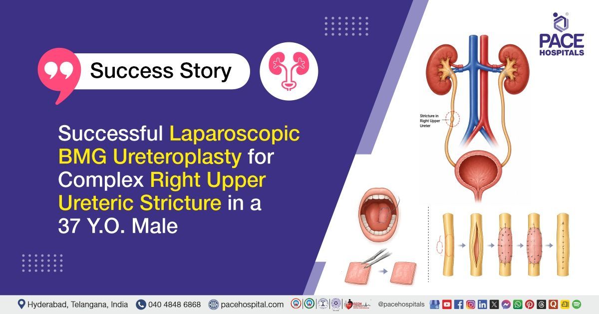
PACE Hospital’s expert Urology team successfully performed a Right Laparoscopic Ureteric Reconstruction with Buccal Mucosal Graft (BMG) Ureteroplasty on a 37-year-old male patient diagnosed with a complex right upper ureteric stricture measuring 6 cm in length with a severely narrowed lumen (3 Fr caliber), associated with right upper ureteric calculi and a poorly functioning right kidney with a differential renal function of 18%. The procedure was undertaken to relieve the ureteric obstruction, restore normal urinary drainage, preserve the remaining kidney function, and improve the patient’s long-term renal and urological outcomes.
Chief Complaints
A 37-year-old male patient with a body mass index (BMI) of 21 presented to the Urology Department at PACE Hospitals, Hitech City, Hyderabad, with complaints of right-sided flank pain.
Past Medical History
The patient had a history of right upper ureteric calculus for which he had previously undergone Ureteroscopic Lithotripsy (URSL) with double-J (DJ) stent placement at another healthcare facility. Subsequent evaluation revealed a long-segment right upper ureteric stricture associated with residual ureteric calculi and reduced function of the right kidney. There was no documented history of significant medical comorbidities.
On examination
On examination, the patient was conscious, coherent, and oriented and hemodynamically stable. Cardiovascular and respiratory system examinations were normal with normal heart sounds and bilateral clear air entry. Abdominal examination revealed right flank tenderness without guarding or rigidity, and no palpable mass was noted. Surgical site examination was normal with no evidence of active bleeding or discharge.
Bowel sounds were normal. No peripheral edema or lymphadenopathy was observed. Overall systemic examination findings were within normal limits except for mild right flank tenderness consistent with the underlying urological condition.
Diagnosis
Upon admission to PACE Hospitals, the patient was thoroughly evaluated by the Urology team, including a detailed review of his medical history and a comprehensive clinical examination. He presented with right-sided flank pain. Based on the initial assessment, a provisional diagnosis of right upper ureteric stricture with calculus was made.
The patient was further evaluated with relevant investigations, including complete blood picture, which showed neutrophilic leukocytosis at initial assessment with subsequent normalization trend, serum electrolytes which were within normal limits, renal function tests including serum creatinine and blood urea which remained within normal range, liver function tests which were within normal limits, coagulation profile including PT, INR, APTT, bleeding time, and clotting time which were within normal limits, urine routine examination which was normal with no hematuria, proteinuria, or evidence of infection.
Urine culture and sensitivity which showed no bacterial growth, blood grouping and Rh typing which revealed B positive blood group, viral screening including HIV, HBsAg, and HCV which were non-reactive or negative, chest X-ray PA view which was normal with clear lung fields, X-ray KUB which demonstrated right-sided DJ stent in situ, 2D echocardiography which showed normal cardiac chamber sizes, good biventricular function, grade I diastolic dysfunction, and no significant structural abnormalities.
Based on these confirmed findings, the patient was advised to undergo Right Upper Ureteric Stricture with Calculus Treatment in Hyderabad, India, causing obstructive uropathy and reduced right renal function under the expert care of the Urology Department.
Medical Decision Making (MDM)
After a detailed consultation with Dr. Vishwambhar Nath, Senior Consultant Urologist & Renal Transplant Surgeon, and Dr. Abhik Debnath, Consultant Laparoscopic Urologist, Endourologist, Andrologist & Kidney Transplant Surgeon, a comprehensive evaluation was conducted focusing on the patient’s presentation of right flank pain and obstructive uropathy. Clinical examination and diagnostic assessment, including complete blood picture, serum electrolytes, renal function tests, liver function tests, coagulation profile, urine routine examination, urine culture and sensitivity, viral markers, blood grouping and Rh typing, X-ray chest PA view, X-ray KUB, and 2D echocardiography, confirmed the presence of right upper ureteric stricture with associated ureteric calculi causing significant urinary obstruction and a reduced functioning right kidney with moiety function of 18 percent, with no evidence of active infection or major cardiopulmonary contraindications for intervention.
It was determined that Right Laparoscopic Ureteric Reconstruction with Buccal Mucosal Graft (BMG) Ureteroplasty was the most appropriate intervention in view of the long segment right upper ureteric stricture with associated ureteric calculi causing obstruction to urine flow and deterioration of renal function, with the aim of relieving obstruction, restoring urinary drainage, and preserving the remaining renal function.
The patient and his family members were informed about the diagnosis of right upper ureteric stricture with calculus causing obstructive uropathy and reduced renal function, the planned management approach, the associated risks, benefits, and expected outcomes, and consent was obtained for proceeding with surgical treatment.
Surgical Procedure
Following the decision, the patient was scheduled to undergo Right Laparoscopic Ureteric Reconstruction with Buccal Mucosal Graft (BMG) Ureteroplasty Surgery in Hyderabad at PACE Hospitals, under the expert care of the urology department.
The procedure involved the following steps:
- Patient Positioning and Anaesthesia: The patient was taken up for surgery under general anaesthesia and positioned appropriately for laparoscopic renal and upper ureteric access. Standard aseptic precautions were followed, and pneumoperitoneum was created for laparoscopic entry.
- Intraoperative Ureteric Assessment: Intraoperative evaluation confirmed a long segment right upper ureteric stricture of approximately 6 cm with 3 Fr calibre. Associated findings included upper ureteric residual calculi measuring 8 mm each, along with small renal calculi measuring 2 to 3 mm. Retrograde pyelography demonstrated a long segment stricture not permitting passage of a 4 Fr ureteric catheter; however, a 0.025-inch guidewire was successfully negotiated across the stricture up to the pelvicalyceal system. Ureteroscopy findings were consistent with RGP, showing a normal and accommodative distal and mid ureter.
- Laparoscopic Exposure and Adhesiolysis: Laparoscopic access to the right upper ureter was obtained. Moderate adhesions were noted around the upper ureter below the pelviureteric junction, which were carefully released to expose the diseased ureteric segment.
- Stricture Management and Graft Preparation: The strictured ureteric segment was identified, and a small portion of the stricture was sent for histopathological biopsy. A lateral stricturotomy was performed to open the narrowed segment. Buccal mucosal graft (BMG) was harvested and tailored appropriately for ureteric reconstruction.
- Ureteroplasty and Stenting: Buccal mucosal graft was placed as a patch over the opened ureteric segment, and ureteroplasty was completed over a 6 Fr long-duration ureteric stent. Adequate reconstruction was achieved, ensuring free drainage and patency of the ureter. Hemostasis was secured, and the surgical field was closed in standard layers.
Postoperative Care
The patient had an uneventful intraoperative and postoperative course with stable recovery. Postoperative X-ray KUB confirmed correct placement of the ureteric stent with no residual stones.
Mild hematuria (blood in urine) was noted initially and resolved by postoperative day 2 without intervention. Urethral catheter was removed on postoperative day 4, and the drain was removed on postoperative day 5 after confirming normal drain fluid creatinine levels and no evidence of urinary leak.
The patient received postoperative care including infection prophylaxis, pain control, gastric protection, ureteric spasm prevention, and supportive measures. The histopathology report showed ulcerated ureteric mucosa with fibrosis and was negative for granulomas and malignancy. He recovered well and was discharged in stable condition.
Discharge Medications
The patient was prescribed medication for the prevention of postoperative infection following urological reconstructive surgery. Medication was advised for relief of postoperative pain and was to be used regularly for a short duration and then as needed. Therapy was given for the protection of the stomach lining and to prevent acidity during recovery. Oral medications and topical applications were advised to promote healing of the oral donor site and maintain oral hygiene following graft harvest.
Urinary alkalinization therapy was prescribed to maintain urine pH and reduce risk of crystallization during recovery. Medication was advised to reduce ureteric spasm and improve urine flow around the indwelling ureteric stent. Supportive supplementation was given to aid postoperative healing and renal recovery. Antihypertensive medication was continued for blood pressure control during the postoperative period.
Advice on discharge
The patient was advised to follow up with a local physician after 1 month for blood pressure monitoring and adjustment of antihypertensive therapy as required. He was instructed to continue physiotherapy measures, including regular mouth exercises as demonstrated during the hospital stay, and spirometry exercises to improve pulmonary function.
Application of an ice pack to the cheek was advised twice daily for 5 minutes to reduce postoperative discomfort and swelling at the oral graft site.
Dietary advice
The patient was advised to maintain adequate hydration with a fluid intake of 3–4 litres per day. Inclusion of citrus fruits and juices was recommended. A low-purine and low-salt diet was advised to support renal health and reduce stone formation risk. A soft diet was recommended for a period of 2 weeks. The patient was instructed to avoid hard-to-chew and spicy foods during the initial 2-week recovery period.
Emergency Care
The patient was instructed to contact the emergency ward at PACE Hospitals in the event of an emergency or if symptoms such as fever, severe flank or abdominal pain, vomiting, reduced urine output, or urinary blockage occur.
Review and Follow-up Notes
The patient was advised to return for follow-up with the Urologist in Hyderabad at PACE Hospitals after 2 weeks for Fourier Transform Infrared Spectroscopy (FTIR) stone analysis review, wound inspection, and suture removal. Further, the patient was advised to undergo ureteroscopic evaluation and DJ stent removal after 3 months.
Conclusion
This case highlights a right upper ureteric stricture with associated ureteric calculi and reduced right renal function. The patient underwent successful laparoscopic ureteric reconstruction with buccal mucosal graft ureteroplasty and stenting. He had an uneventful recovery and was discharged in stable condition with planned follow-up for stent removal and further evaluation.
General Approach to Complex Ureteric Strictures
Complex ureteric strictures are a challenging urological condition that may lead to urinary obstruction and progressive renal function loss if not managed appropriately. Comprehensive evaluation with imaging and endoscopic assessment is essential to define the site, length, and severity of the stricture along with renal functional status. In selected difficult cases, guidewire-assisted techniques may be required for safe traversal of tight strictures and treatment planning. Management is individualized with the primary goals of relieving obstruction, restoring ureteric continuity, and preserving renal function wherever possible.
Reconstructive options, including minimally invasive laparoscopic techniques with graft augmentation, are considered in appropriate patients for optimal outcomes. Successful treatment depends on meticulous surgical planning, adequate urinary drainage with stenting, and structured post-operative follow-up. Overall, a renal preservation–oriented approach under the care of a urologist/urology doctor remains the cornerstone of management.
Frequently Asked Questions (FAQs)
Why is ureteric reconstruction needed for a long ureteric stricture?
Ureteric reconstruction is done when the ureter becomes too narrow, and urine cannot drain properly from the kidney to the bladder. In this case, the narrowing was long and tight, so a simple internal procedure may not have given a lasting result. The surgery helps open the blocked part, improves urine flow, and reduces pressure on the kidney.
Why was kidney preservation considered even though the kidney function was reduced?
Kidney preservation was considered because the patient was young and did not have major medical problems. Even if the affected kidney was working less than normal, saving the remaining useful function was important. The main aim was to improve urine drainage, reduce blockage, and prevent further damage to the kidney.
What is laparoscopic ureteric reconstruction with BMG?
Laparoscopic ureteric reconstruction with BMG is a keyhole surgery done to repair a narrowed ureter. BMG means buccal mucosal graft, where a small tissue patch is taken from the inside of the cheek and used to widen the narrowed ureter tube. This helps urine pass more smoothly from the kidney to the bladder.
Why is a buccal mucosal graft used in ureter repair?
A buccal mucosal graft means a small piece of tissue is taken from the inner lining of the cheek and used in surgery. This tissue is chosen because it is strong, flexible, and heals quickly, making it suitable for reconstruction. It is used like a patch when a long, narrow part of the ureter needs to be widened. In this way, it helps open the blocked area of the ureter and supports normal urine flow while preserving kidney function.
Why was a stent placed after ureteric reconstruction?
A stent is a small tube placed inside the ureter to support the repaired area while it heals. It keeps the ureter open so urine can flow smoothly from the kidney to the bladder without stressing the surgical site. The stent is not permanent and is removed later during follow-up as advised by the urologist/urology doctor.
How does ureteric stricture affect kidney function?
A ureteric stricture can block or slow the flow of urine from the kidney. When urine collects above the narrowed area, pressure can build up inside the kidney and gradually reduce its function. Since the affected kidney was already working less, improving the drainage was important to protect the remaining function.
Why is follow-up important after ureteric reconstruction surgery?
Follow-up is required to monitor healing after surgery and to ensure urine is draining properly from the kidney. It also helps check the position of the stent, the condition of the wound, and review reports like biopsy or stone analysis. During these visits, the doctor also looks for early signs of problems such as infection, blockage in urine flow, leakage of urine, or persistent pain. Regular follow-up with the urologist is important for safe recovery and long-term success of the repair.
Can stones and ureteric stricture happen together?
Yes, ureteric stones and ureteric stricture can occur together. A stone, previous procedure, inflammation, or injury to the ureter can sometimes lead to narrowing. When the ureter is narrow, urine flow becomes poor, and stones may remain trapped. That is why both the stone problem and the narrowed ureter need proper treatment.
What precautions are usually advised after this type of surgery?
After this surgery, patients are usually advised to drink enough fluids, follow the diet suggested by the doctor, and avoid physical strain during early recovery. Mouth care is also important because the graft is taken from the cheek. Fever, abdominal pain, vomiting, reduced urine output, or worsening pain should be reported immediately.
When should a patient seek emergency care after ureteric reconstruction?
A patient should go to emergency care if there is a fever, severe abdominal pain, repeated vomiting, reduced urine output, or a sudden increase in pain. These symptoms may suggest problems such as infection, blockage in urine flow, urine leakage, or other post-surgery complications. Getting medical help early can prevent serious issues and help protect kidney function.
Share on
Request an appointment
Fill in the appointment form or call us instantly to book a confirmed appointment with our super specialist at 04048486868
Appointment request - health articles
Recent Articles