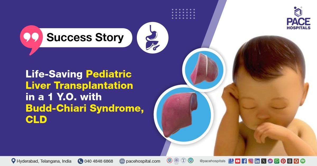
Life-Saving Pediatric Liver Transplantation in a 1 Y.O. with Budd-Chiari Syndrome, CLD
PACE Hospitals
PACE Hospitals' expert Pediatric Liver Transplantation
team successfully performed Pediatric living donor liver transplantation
on a 1-year-old baby with
Chronic
Budd Chiari Syndrome
with CLD (chronic liver disease) and portal hypertension with recurrent UGI (upper gastrointestinal) bleed.
Chief complaints
A 1-year-old baby was presented to the Organ Transplant Department at
PACE Hospitals, Hitech City, Hyderabad, with a history of chronic Budd-Chiari syndrome associated with chronic liver disease (CLD). She had experienced recurrent episodes of upper gastrointestinal (UGI) bleeding. An evaluation was initiated to assess the extent of liver dysfunction and determine the appropriate course of management.
Past Medical History
The baby was a known case of chronic
Budd-Chiari syndrome associated with
end stage liver disease (ESLD), which had led to progressive liver dysfunction and contributed to her recurrent episodes of upper gastrointestinal bleeding.
Diagnosis
Following the admission to PACE Hospitals and understanding the history and physical examination, A comprehensive diagnostic workup was carried out, including a complete blood count, liver function tests, and coagulation profile to assess liver status and bleeding risk. Serum electrolytes and renal function were also evaluated. Imaging studies, such as abdominal ultrasound with Doppler and contrast-enhanced computed tomography (CT) scan, were performed to assess liver morphology and detect venous obstruction.
Upper gastrointestinal endoscopy identified the sources of bleeding, visualized esophageal varices and other mucosal abnormalities, and allowed therapeutic interventions like band ligation to control bleeding and prevent recurrence. Viral hepatitis serology and serum alpha-fetoprotein tests were conducted to rule out infection and malignancy. Blood cultures were also obtained.
Based on the confirmed diagnosis, the baby was advised to undergo chronic Budd-Chiari syndrome Treatment in Hyderabad, India, under the specialized care of the Liver Transplant Department.
Medical Decision Making
After careful observation of the patient’s clinical symptoms and medical condition, the Senior Consultant Surgical Gastroenterologist and Liver Transplant Surgeon Dr. CH. Madhusudhan along with Dr. Prashanth Sanghu Consultant Surgical Gastroenterologist and Liver Transplant Surgeon, and cross consultations include Dr. Pradeep Kiran Panchadi, Dr. Lakshmi Kumar CH, and Dr. Navya Sri Gali recommended a Living Donor Liver Transplant (LDLT) to save the patient.
After a detailed preoperative evaluation, the baby was admitted for
Living Donor Liver Transplantation. Her mother, as a donor, was carefully assessed for suitability, and the risks and benefits of the transplant were thoroughly explained to the family. A detailed pre-anesthesia check-up (PAC) was completed to ensure the patient was fit for surgery and obtained consent.
Surgical Procedure
Following the decision, the patient was scheduled to undergo a Pediatric living donor liver transplantation (LDLT) in Hyderabad at PACE Hospitals, under the expert supervision of the liver transplantation team.
The patient underwent a living donor liver transplantation (LDLT), with her mother as the donor. The procedure was performed in the following steps:
- Donor Hepatectomy: The left lateral segment of the mother's liver was carefully mobilized and removed after ensuring vascular and biliary anatomy suitability.
- Recipient Hepatectomy: The diseased liver of the child was meticulously dissected and removed, preserving major vascular structures for graft implantation.
- Vascular Anastomosis: The donor liver graft was implanted by performing end-to-end anastomoses of the hepatic vein, portal vein, and hepatic artery using fine sutures under magnification.
- Biliary Reconstruction: Biliary continuity was restored using a Roux-en-Y hepaticojejunostomy, appropriate for pediatric LDLT.
- Reperfusion and Hemostasis: The graft was reperfused after completing vascular connections, and hemostasis was achieved. The abdomen was closed in layers over drains, and the patient was shifted to the ICU for postoperative monitoring.
Postoperative Care
The postoperative period was uneventful, and the patient was closely monitored in the ICU to ensure a stable recovery. The patient started on intravenous anticoagulant and antiplatelet therapy, and her recovery remained uneventful. Serial Doppler studies showed normal hepatic artery flow, and both drains were removed as the output gradually decreased. Bilateral conductive breath sounds were noted on respiratory examination and improved with conservative management.
During her hospital stay, she was treated with IV fluids, immunosuppressants, antibiotics, antifungals, PPIs, multivitamins, antiemetics, analgesics, and antipyretics. Liver enzymes showed gradual improvement, and immunosuppressant doses were adjusted accordingly. She was discharged in a hemodynamically stable condition with appropriate medications and follow-up instructions.
Discharge Medications
Upon discharge, the patient was prescribed a combination of oral medications tailored to her postoperative needs. These included antibiotics to prevent or treat infections, proton pump inhibitors (PPIs) to reduce gastric acid and protect the gastrointestinal mucosa, and macrolides for their antimicrobial and anti-inflammatory properties. Corticosteroids were given as part of her immunosuppressive regimen, while antifungal medication was included to prevent opportunistic fungal infections.
Nonsteroidal anti-inflammatory drugs (NSAIDs) were prescribed for pain and inflammation control. Hepatoprotective agents were administered to support liver function, along with nutritional supplements and multivitamins to aid in recovery and overall health. Additionally, antiplatelets and oral anticoagulants were prescribed to reduce the risk of thrombosis and maintain vascular graft patency.
Advice on Discharge
The patient was instructed to apply an ointment over the perianal area to manage excoriation (skin soreness) and promote skin healing. The ointment helped reduce local irritation, provided a protective barrier, and supported the healing of the affected skin.
Emergency Care
The patient was informed to contact the Emergency ward at PACE Hospitals in case of any emergency or development of symptoms like fever, abdominal pain, or vomiting.
Review and Follow-Up
The patient was advised to return for a follow-up visit with Liver Transplant surgeons in Hyderabad at PACE Hospitals after 3 days. This visit will include a comprehensive evaluation, wound assessment, and monitoring of postoperative recovery.
Conclusion
This case demonstrated the effectiveness of living donor liver transplantation (LDLT) as a life-saving treatment for chronic Budd-Chiari syndrome by restoring proper liver function and significantly improving the patient’s overall prognosis.
Significance of ultrasound Doppler in interpretation and management of Budd-Chiari Syndrome (BCS)
Ultrasound Doppler is the first-line imaging modality for diagnosing Budd-Chiari Syndrome (BCS), a rare liver disease caused by hepatic venous outflow obstruction (HVOTO). It can non-invasively detect thrombi in the hepatic veins and assess abnormal venous flow patterns, such as absent, reversed, or "flat" flow. High flow velocities in stenotic segments of the IVC or hepatic veins can also be visualized. With a diagnostic sensitivity and specificity of 85–90%, it is a reliable tool for early detection. In suspected or confirmed cases, referral to a liver transplant specialist is crucial for further management and treatment planning. Early diagnosis and Liver Transplant doctor /specialist involvement can help prevent liver damage and improve long-term outcomes.
Share on
Request an appointment
Fill in the appointment form or call us instantly to book a confirmed appointment with our super specialist at 04048486868

